. 2011 Aug 16;30(20):4323-35.
doi: 10.1038/emboj.2011.292.

C/EBPβ mediates tumour-induced ubiquitin ligase atrogin1/MAFbx upregulation and muscle wasting

Affiliations

C/EBPβ mediates tumour-induced ubiquitin ligase atrogin1/MAFbx upregulation and muscle wasting

Guohua Zhang et al. EMBO J. .

Abstract

Upregulation of ubiquitin ligase atrogin1/MAFbx and muscle wasting are hallmarks of cancer cachexia; however, the underlying mechanism is undefined. Here, we describe a novel signalling pathway through which Lewis lung carcinoma (LLC) induces atrogin1/MAFbx upregulation and muscle wasting. C2C12 myotubes treated with LLC-conditioned medium (LCM) rapidly activates p38 MAPK and AKT while inactivating FoxO1/3, resulting in atrogin1/MAFbx upregulation, myosin heavy chain loss, and myotube atrophy. The p38α/β MAPK inhibitor SB202190 blocks the catabolic effects. Upon activation, p38 associates with C/EBPβ resulting in its phosphorylation and binding to a C/EBPβ-responsive cis-element in the atrogin1/MAFbx gene promoter. The promoter activity is stimulated by LCM via p38β-mediated activation of the C/EBPβ-responsive cis-element, independent of the adjacent FoxO1/3-responsive cis-elements in the promoter. In addition, p38 activation is observed in the muscle of LLC tumour-bearing mice, and SB202190 administration blocks atrogin1/MAFbx upregulation and muscle protein loss. Furthermore, C/EBPβ(-/-) mice are resistant to LLC tumour-induced atrogin1/MAFbx upregulation and muscle wasting. Therefore, activation of the p38β MAPK-C/EBPβ signalling pathway appears a key component of the pathogenesis of LLC tumour-induced cachexia.


Conflict of interest statement

The authors declare that they have no conflict of interest.

Figures
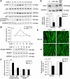
Figure 1
Figure 1
LCM induces atrogin1/MAFbx upregulation and myofibrillar protein loss via p38 MAPK in C2C12 myotubes. (A) LCM activates p38 MAPK and AKT, and inactivates FoxO1/3a. C2C12 myotubes were treated with LCM for indicated time periods as described in Materials and methods. Myotubes were lysed and phosphorylation of p38 MAPK, AKT, and FoxO1/3a were evaluated by western blot analysis with total p38 MAPK, AKT, and GAPDH as controls. (B) LCM upregulates atrogin1/MAFbx expression. The mRNA levels of atrogin1/MAFbx and MuRF1 in C2C12 myotubes treated with LCM for indicated periods were determined by using real-time PCR. *Denotes a difference (P<0.05) from 0 h determined by ANOVA. The protein levels of the ubiquitin ligases were determined by western blot. (C) Upregulation of atrogin1/MAFbx by LCM is mediated by p38 MAPK. C2C12 myotubes were treated with LCM for 4 h with or without the presence of SB202190 (SB, 10 μM). The mRNA level of atrogin1/MAFbx and MuRF1 was determined by real-time PCR. (D) LCM induces MHC loss via p38 MAPK. C2C12 myotubes were treated with LCM for 72 h with or without the presence of SB202190 (10 μM). Western blot analysis was carried out using an antibody against total MHC (MF-20). (E) LCM induces myotube atrophy via p38 MAPK. C2C12 myotubes were treated with LCM as described in (D). Immunofluorescence was performed to stain myotubes with MF-20. Diameter of myotubes was then determined as described previously (Doyle et al, 2011). Bar represents 100 μm. *Denotes a difference (P<0.05) from PBS control (PBS/vehicle) and † denotes a difference from LCM control (LCM/vehicle) determined by ANOVA.
Figure 2
Figure 2
Activated p38 MAPK interacts with and phosphorylates C/EBPβ, activating the binding of C/EBPβ to the atrogin1/MAFbx promoter. (A) The 5′ promoter sequence of the mouse, rat, and human atrogin1/MAFbx gene contains a putative C/EBPβ-binding motif (TTGTGCAA) within 200 bp from the transcription start site. *Indicates conserved sequences. (B) Activation of p38 MAPK by adenovirus-mediated overexpression of MKK6bE. Adenovirus encoding MKK6bE or GFP was used to transduce C2C12 cells as described in Materials and methods. In 48 h, myotubes were lysed. Expression of HA-tagged MKK6bE and activation of p38 MAPK and C/EBPβ level were verified by western blot. C/EBPβ phosphorylation was evaluated by using an antibody specific for Thr-188 phosphorylated C/EBPβ (p-C/EBPβ). (C) Activated p38 MAPK interacts with C/EBPβ. C2C12 myotubes were transduced with adenovirus encoding MKK6bE or GFP. SB202190 (SB, 10 μM) was included in the culture medium as indicated. In 48 h, immunoprecipitation was carried out with antibody against either p38 MAPK or C/EBPβ. Precipitation of the two proteins was analysed by western blot. The input control of the assay is shown in (B). (D) Activation of p38 MAPK results in C/EBPβ phosphorylation. C/EBPβ was immunoprecipitated from cell lysate of C2C12 myotubes expressing MKK6bE or GFP and treated with SB202190 as described above. Phosphorylation of the serine and threonine residues in immunoprecipitated C/EBPβ was analysed by western blot using antibody specific for phosphorylated serine or threonine. (E) Activated p38 MAPK stimulates C/EBPβ binding to the putative C/EBPβ-binding motif identified in the atrogin1/MAFbx promoter. C2C12 myotubes were transduced with adenovirus encoding MKK6bE or GFP as described above. ChIP assay was carried out to evaluate the effect of MKK6bE activation of p38 MAPK on C/EBPβ binding to the putative C/EBPβ-binding motif present in the atrogin1/MAFbx promoter as described in Materials and methods. The inverted image is shown. (F) LCM stimulates the phosphorylation of C/EBPβ. C2C12 myotubes were treated with LCM for indicated time periods. Thr-188 phosphorylation of C/EBPβ (p-C/EBPβ) and total C/EBPβ were determined by western blot analysis of the cell lysates.
Figure 3
Figure 3
The C/EBPβ-binding motif in the atrogin1/MAFbx promoter is a functional cis-element regulated by p38β MAPK. (A) Generation of reporter gene constructs controlled by the atrogin1/MAFbx promoter. Reporter constructs were generated by inserting fragments of the atrogin1/MAFbx promoter into a luciferase reporter vector as described in Materials and methods. The location of the C/EBPβ-binding motif and two previously identified FoxO-responsive cis-elements are as indicated. (B) The C/EBPβ-binding motif mediates MKK6bE stimulation of atrogin1/MAFbx promoter activity. Reporter gene constructs were co-transfected with plasmids encoding the active form of C/EBPβ (LAP) and/or MKK6bE into C2C12 myoblasts as indicated. In 24 h, cells were lysed and luciferase activity was determined. (C) p38β MAPK, but not other isoforms of p38 MAPK, stimulates atrogin1/MAFbx promoter activity in the presence of C/EBPβ. Plasmids encoding an active mutant of p38 MAPK isoform and/or LAP were co-transfected with the pA reporter gene construct into C2C12 myoblasts as indicated. Luciferase activity in myotubes was determined in 24 h. (D) The C/EBPβ-binding motif mediates p38β stimulation of atrogin1/MAFbx promoter activity. Reporter gene constructs were co-transfected with plasmids encoding LAP and/or active p38β MAPK into C2C12 myoblasts as indicated. In 24 h, cells were lysed and luciferase activity was determined. (E) The C/EBPβ-binding motif mediates LCM stimulation of atrogin1/MAFbx promoter activity. Reporter gene constructs were co-transfected with a plasmid encoding LAP into C2C12 myoblasts as indicated. In 24 h, cells were treated with LCM for 3 h. Luciferase activity in lysed cells was then determined. *Indicates difference (P<0.05) from control based on ANOVA.
Figure 4
Figure 4
C/EBPβ mediates upregulation of endogenous atrogin1/MAFbx by p38β MAPK and LCM in C2C12 myotubes. (A) C/EBPβ knockdown by C/EBPβ-specific siRNA. C2C12 myoblasts were transfected with either control or C/EBPβ-specific siRNA. After differentiation for 72 h, C/EBPβ protein level in myotubes was determined by western blot analysis. (B) C/EBPβ mediates p38β MAPK upregulation of the atrogin1/MAFbx gene. In C/EBPβ knockdown myotubes that were co-transfected with a plasmid encoding a constitutively active p38β or transduced with an adenovirus encoding MKK6bE, atrogin1/MAFbx mRNA level was determined by real-time PCR. *Denotes a difference (P<0.05) from control/control siRNA and † denotes a difference from p38β/control siRNA or MKK6bE/control siRNA, correspondently, based on ANOVA. (C) C/EBPβ mediates LLC-induced atrogin1/MAFbx upregulation and MHC loss. Myotubes in which C/EBPβ was knocked down as shown in (A) were treated with control medium or LCM. Atrogin1/MAFbx and MHC levels were determined by western blot in 8 and 72 h, respectively. *Denotes a difference (P<0.05) from control/control siRNA and † denotes a difference from LCM/control siRNA or MKK6bE/control siRNA, correspondently, based on ANOVA.
Figure 5
Figure 5
Inhibition of p38α/β MAPK blocks LLC tumour-induced muscle catabolism. LLC cells or PBS (control) was injected subcutaneously into the right flank of C57BL/6 male mice (8 weeks of age) as described in Materials and methods. SB202190 was i.p. injected daily (5 mg/kg) from day 5 of LLC implant with equal volume of vehicle as control. In 14 days, mice were weighed and euthanized. Tumour and muscle samples were immediately collected and analysed. (A) p38 MAPK is activated in the muscle of LLC tumour-bearing mice. Phosphorylation state of p38 MAPK in TA was analysed by western blot. *Indicates difference (P<0.05) from control based on Student's t-test. (B) C/EBPβ undergoes a p38α/β MAPK-dependent phosphorylation in LLC tumour-bearing mice. C/EBPβ phosphorylation at Thr-188 and total C/EBPβ in the TA of LLC tumour-bearing mice was evaluated by western blot analysis. (C) LLC tumour growth (tumour volume) is not affected by SB202190. Net body weight gain is the difference of body weight between the time of LLC implant and the time of euthanization minus tumour weight. (D) LLC-induced atrogin1/MAFbx upregulation is blocked by SB202190. Atrogin1/MAFbx mRNA isolated from TA was determined by real-time PCR and atrogin1/MAFbx protein levels were determined by western blot analysis. (E) LLC-induced loss of net body weight gain is blocked by SB202190. (F) LLC-induced loss of TA mass is attenuated by SB202190. (G) SB202190 attenuates LLC-induced loss of EDL mass. (H) LLC-induced tyrosine release from EDL is blocked by SB202190. Tyrosine release was determined as described previously (Doyle et al, 2011). *Denotes a difference (P<0.05) from PBS control (PBS/vehicle) and † denotes a difference from LLC control (LLC/vehicle) determined by ANOVA. (I) SB202190 blocks LLC-induced shrinkage in TA fibre cross-sectional area (CSA). CSA of H&E stained TA cross-sections was measured as described in Materials and methods. Bar represents 50 μm.
Figure 6
Figure 6
C/EBPβ is crucial to the development of muscle wasting in LLC tumour-bearing mice. LLC cells or PBS (control) was injected subcutaneously into the right flank of male C/EBPβ−/− and WT mice. In 14 days, mice were weighed and euthanized. Tumour and muscle samples were immediately collected and analysed. (A) LLC tumour growth (tumour volume) in C/EBPβ−/− and WT mice is comparable. (B) LLC tumour-induced atrogin1/MAFbx upregulation is blocked in C/EBPβ−/− mice. Atrogin1/MAFbx levels in TA were determined by real-time PCR and western blot. (C) LLC-induced loss of net body weight gain is attenuated in C/EBPβ−/− mice. Net body weight gain is the difference of body weight between the time of LLC implant and the time of euthanization minus tumour weight. (D) LLC-induced loss of TA mass is abolished in C/EBPβ−/− mice. (E) LLC does not induce loss of EDL mass in C/EBPβ−/− mice. (F) LLC-induced tyrosine release from EDL is blocked in C/EBPβ−/− mice. Tyrosine release was determined as described previously (Doyle et al, 2011). *Denotes a difference (P<0.05) from PBS control (PBS/vehicle) and † denotes a difference from LLC control (LLC/vehicle) determined by ANOVA. (G) LLC-induced shrinkage in TA fibre cross-sectional area (CSA) is blocked in C/EBPβ−/− mice. CSA of H&E stained TA cross-sections was measured as described in Materials and methods. Bar represents 50 μm.
Figure 7
Figure 7
A working model of the signalling mechanism through which LLC induces muscle mass loss.

Similar articles

Cited by

References

    1. Acharyya S, Butchbach ME, Sahenk Z, Wang H, Saji M, Carathers M, Ringel MD, Skipworth RJ, Fearon KC, Hollingsworth MA, Muscarella P, Burghes AH, Rafael-Fortney JA, Guttridge DC (2005) Dystrophin glycoprotein complex dysfunction: a regulatory link between muscular dystrophy and cancer cachexia. Cancer Cell 8: 421–432 - PubMed
    1. Acharyya S, Guttridge DC (2007) Cancer cachexia signaling pathways continue to emerge yet much still points to the proteasome. Clin Cancer Res 13: 1356–1361 - PubMed
    1. Andreyev HJ, Norman AR, Oates J, Cunningham D (1998) Why do patients with weight loss have a worse outcome when undergoing chemotherapy for gastrointestinal malignancies? Eur J Cancer 34: 503–509 - PubMed
    1. Askari N, Beenstock J, Livnah O, Engelberg D (2009) p38 is active in vitro and in vivo when monophosphorylated on Thr180. Biochemistry 48: 2497–2504 - PubMed
    1. Baltgalvis KA, Berger FG, Pena MM, Davis JM, White JP, Carson JA (2009) Muscle wasting and interleukin-6-induced atrogin-I expression in the cachectic Apc (Min/+) mouse. Pflugers Arch 457: 989–1001 - PMC - PubMed

Publication types

MeSH terms