

Vascular stiffness and increased pulse pressure in the aging cardiovascular system
- PMID: 21845218
- PMCID: PMC3154449
- DOI: 10.4061/2011/263585
Vascular stiffness and increased pulse pressure in the aging cardiovascular system
Abstract
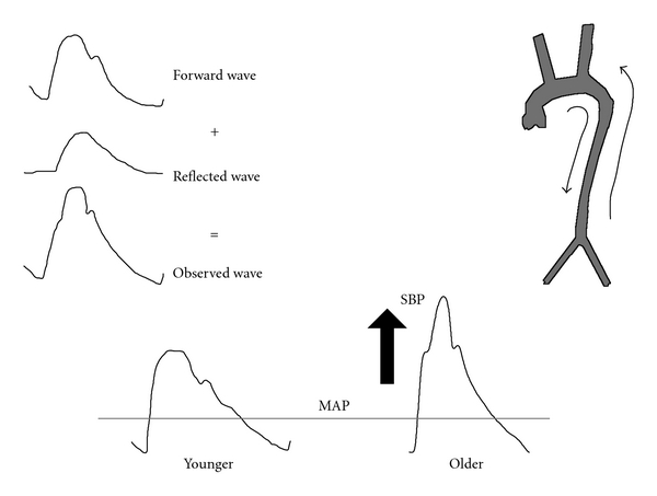
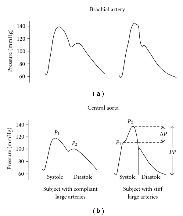
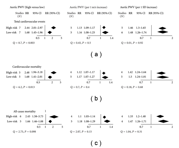
Aging leads to a multitude of changes in the cardiovascular system, including systolic hypertension, increased central vascular stiffness, and increased pulse pressure. In this paper we will review the effects of age-associated increased vascular stiffness on systolic blood pressure, pulse pressure, augmentation index, and cardiac workload. Additionally we will describe pulse wave velocity as a method to measure vascular stiffness and review the impact of increased vascular stiffness as an index of vascular health and as a predictor of adverse cardiovascular outcomes. Furthermore, we will discuss the underlying mechanisms and how these may be modified in order to change the outcomes. A thorough understanding of these concepts is of paramount importance and has therapeutic implications for the increasingly elderly population.
Figures
Similar articles
-
Pathophysiology of hypertension in the elderly.Semin Nephrol. 2002 Jan;22(1):65-70. Semin Nephrol. 2002. PMID: 11785070
-
Impact of Cardiovascular Factors on Pulse Wave Velocity and T otal Vascular Resistance in Different Age Group Patients with Cardiovascular Disorders.Curr Aging Sci. 2019;11(4):261-268. doi: 10.2174/1874609812666190226151500. Curr Aging Sci. 2019. PMID: 30813882 Free PMC article.
-
Impact of cocoa flavanol intake on age-dependent vascular stiffness in healthy men: a randomized, controlled, double-masked trial.Age (Dordr). 2015 Jun;37(3):9794. doi: 10.1007/s11357-015-9794-9. Epub 2015 May 27. Age (Dordr). 2015. PMID: 26013912 Free PMC article. Clinical Trial.
-
Clinical measurement of arterial stiffness obtained from noninvasive pressure waveforms.Am J Hypertens. 2005 Jan;18(1 Pt 2):3S-10S. doi: 10.1016/j.amjhyper.2004.10.009. Am J Hypertens. 2005. PMID: 15683725 Review.
-
Markers of arterial stiffness in peripheral arterial disease.Vasa. 2015 Sep;44(5):341-8. doi: 10.1024/0301-1526/a000452. Vasa. 2015. PMID: 26317253 Review.
Cited by
-
Vascular Aging and COVID-19.Angiology. 2023 Apr;74(4):308-316. doi: 10.1177/00033197221121007. Epub 2022 Aug 28. Angiology. 2023. PMID: 36031949 Free PMC article. Review.
-
Chronic stress and Rosiglitazone increase indices of vascular stiffness in male rats.Physiol Behav. 2017 Apr 1;172:16-23. doi: 10.1016/j.physbeh.2016.03.031. Epub 2016 Mar 31. Physiol Behav. 2017. PMID: 27040922 Free PMC article.
-
Risk Factors and Outcomes With Progressive Mitral Annular Calcification.J Am Heart Assoc. 2023 Sep 19;12(18):e030620. doi: 10.1161/JAHA.123.030620. Epub 2023 Sep 13. J Am Heart Assoc. 2023. PMID: 37702056 Free PMC article.
-
Photoplethysmogram based vascular aging assessment using the deep convolutional neural network.Sci Rep. 2022 Jul 5;12(1):11377. doi: 10.1038/s41598-022-15240-4. Sci Rep. 2022. PMID: 35790836 Free PMC article.
-
Exercise, vascular stiffness, and tissue transglutaminase.J Am Heart Assoc. 2014 Apr 10;3(2):e000599. doi: 10.1161/JAHA.113.000599. J Am Heart Assoc. 2014. PMID: 24721796 Free PMC article.
References
-
- Watanabe M, Sawai T, Nagura H, Suyama K. Age-related alteration of cross-linking amino acids of elastin in human aorta. Tohoku Journal of Experimental Medicine. 1996;180(2):115–130. - PubMed
-
- Cattell MA, Anderson JC, Hasleton PS. Age-related changes in amounts and concentrations of collagen and elastin in normotensive human thoracic aorta. Clinica Chimica Acta. 1996;245(1):73–84. - PubMed
-
- Kass DA. Age-related changes in venticular-arterial coupling: pathophysiologic implications. Heart Failure Reviews. 2002;7(1):51–62. - PubMed
LinkOut - more resources
Full Text Sources
