. 2011 Oct;179(4):2001-15.
doi: 10.1016/j.ajpath.2011.06.025. Epub 2011 Aug 10.

Dendritic degeneration, neurovascular defects, and inflammation precede neuronal loss in a mouse model for tau-mediated neurodegeneration

Affiliations

Dendritic degeneration, neurovascular defects, and inflammation precede neuronal loss in a mouse model for tau-mediated neurodegeneration

Tomasz Jaworski et al. Am J Pathol. 2011 Oct.

Abstract

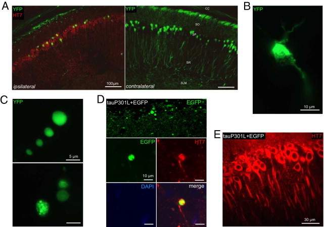
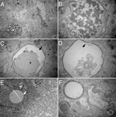
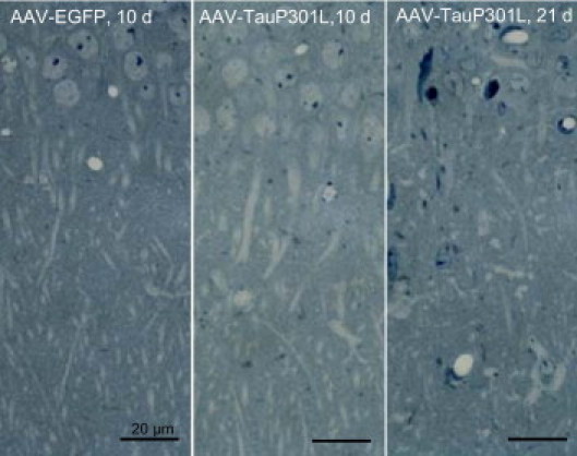
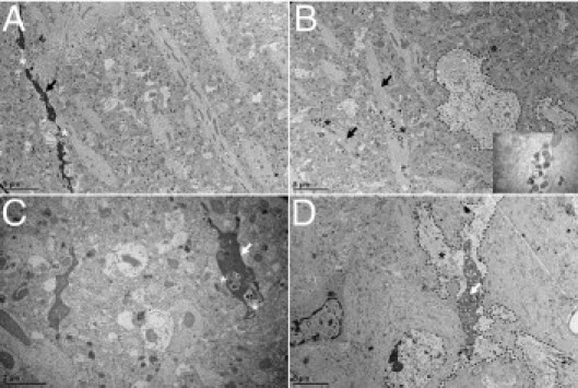
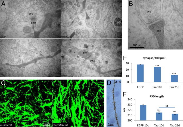
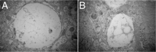
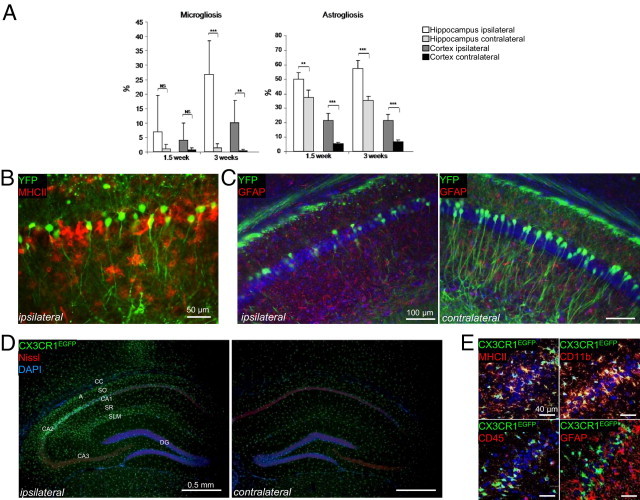
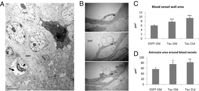
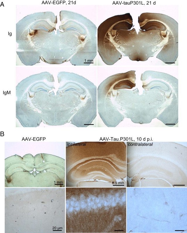
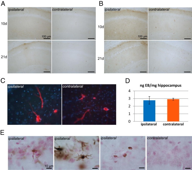
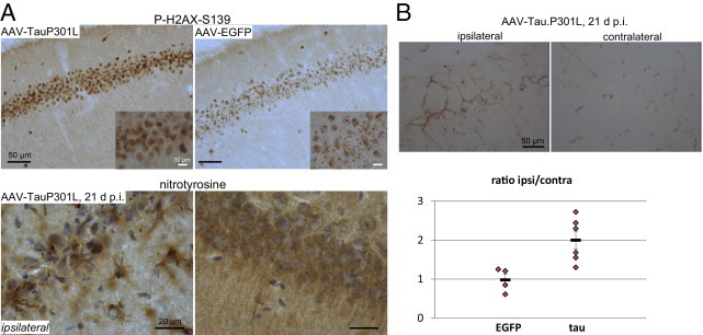
Adeno-associated virus (AAV)-mediated expression of wild-type or mutant P301L protein tau produces massive degeneration of pyramidal neurons without protein tau aggregation. We probed this novel model for genetic and structural factors and early parameters of pyramidal neurodegeneration. In yellow fluorescent protein-expressing transgenic mice, intracerebral injection of AAV-tauP301L revealed early damage to apical dendrites of CA1 pyramidal neurons, whereas their somata remained normal. Ultrastructurally, more and enlarged autophagic vacuoles were contained in degenerating dendrites and manifested as dark, discontinuous, vacuolated processes surrounded by activated astrocytes. Dendritic spines were lost in AAV-tauP301L-injected yellow fluorescent protein-expressing transgenic mice, and ultrastructurally, spines appeared dark and degenerating. In CX3CR1(EGFP/EGFP)-deficient mice, microglia were recruited early to neurons expressing human tau. The inflammatory response was accompanied by extravasation of plasma immunoglobulins. α2-Macroglobulin, but neither albumin nor transferrin, became lodged in the brain parenchyma. Large proteins, but not Evans blue, entered the brain of mice injected with AAV-tauP301L. Ultrastructurally, brain capillaries were constricted and surrounded by swollen astrocytes with extensions that contacted degenerating dendrites and axons. Together, these data corroborate the hypothesis that neuroinflammation participates essentially in tau-mediated neurodegeneration, and the model recapitulates early dendritic defects reminiscent of "dendritic amputation" in Alzheimer's disease.


Figures

Figure 1
Figure 1
Tau damages dendrites and axons of CA1 neurons. A: YFP-expressing transgenic mice (n = 6) were analyzed to visualize degenerating neuritic processes. At 21 days after infection, AAV-tauP301L induced loss of apical and proximal dendrites and a significant decrease in the number of pyramidal neurons. Immunofluorescent staining was performed using HT7-bio and Strepta-Alexa 594 (red). A, alveus; SLM, stratum lacunosum moleculare; SO, stratum oriens; SP, stratum pyramidale; SR, stratum radiatum. B: Degenerating neuron in the CA1 of mouse injected with AAV-tauP301L. Note shrinkage and fragmentation of cytoplasmic YFP signal. C: Examples of YFP-positive dilatations in the alveus in YFP-expressing mice injected with AAV-tauP301L. YFP signal appeared as punctate (upper panel) or smooth (lower panel). D: WT mice were co-injected with AAV-tauP301L (10E8 t.u.) and AAV-EGFP (10E8 t.u.) (n = 5) or injected only with AAV-EGFP (10E8 t.u.) (n = 4). Images were obtained in the CA1 from 40-μm thick sections using a microscope (Leica Microsystems GmbH, Wetzlar, Germany). Tau-induced degeneration was associated with deposition of EGFP in dilatations in alveus similarly as in YFP-expressing mice. E: Neurons co-injected with tauP301L and EGFP express tau (HT7 staining) in the somatodendritic compartment.
Figure 2
Figure 2
Axonal disease. Ultrastructural aspects of axonal degeneration representing various stages. A: Accumulation of autophagic vacuoles (asterisk). B: Example of digested axoplasm (asterisk). C and D: Accumulation and detachment (arrows) of dark axoplasm (asterisk). E: Extremely swollen degenerated axon is almost completely electron-lucent (arrow) next to a capillary (asterisk). F: Axon with split myelin sheath.
Figure 3
Figure 3
Dendritic changes on neurodegeneration. Thin sections of brain from AAV-EGFP-injected mice at 10 days after infection (n = 3), AAV-tauP301L–injected mice at 10 days (n = 4) and 21 days (n = 4), stained with toluidine blue, to compare projecting apical dendrites of CA1 pyramidal neurons into the stratum radiatum.
Figure 4
Figure 4
Ultrastructure of dendritic degeneration in CA1 stratum radiatum. Representative examples of the ultrastructure of the CA1 stratum radiatum reveal various stages of dendritic degeneration. A: Lack of continuity (asterisks) of dark degenerating dendrite (arrow). B: Healthy-looking dendrites (arrows) accumulating autophagic vacuoles (asterisks). Inset: Adjacent astrocyte is delineated (dashed line). C: Autophagic vacuoles (asterisks) in a dark dendrite (arrow). D: Dark perforated dendrite (arrow) surrounded by white astrocytic process (asterisk). Astrocyte is delineated (dashed line).
Figure 5
Figure 5
Synaptic degeneration. A: Representative examples of the ultrastructure of synapses in CA1 demonstrate dark synaptic degeneration. Presynaptic and postsynaptic compartments are denoted. B: Higher magnification of the upper right panel in A demonstrates degenerating dark synapse. C: Confocal microscopy of the CA1 stratum radiatum in brain from AAV-tauP301L–injected YFP-expressing transgenic mice (n = 6) at 21 days after infection to compare the extent of degenerating dendritic shafts and spines in injected and noninjected hippocampi. D: A single dendrite from CA1 stratum radiatum of AAV-tauP301L–injected WT mouse positive for AT8. Accumulation of phospho-tau along dendritic process and in postsynaptic spines is apparent. E: Quantification of synaptic density (PSD/100 μm2). Values are given as mean ± SEM. *P < 0.001 (one-way analysis of variance). AAV-EGFP–injected mice (n = 3) were compared with AAV-tauP301L–injected mice (n = 4) at 10 days after infection and with AAV-tauP301L–injected mice (n = 4) at 21 days after infection. Total number of synapses for EGFP-injected mice was 15,796 in area of 55,781.83 μm2, for tauP301L-injected mice at 10 days was 17,779 in area of 72,944.06 μm2, and for tauP301L-injected mice at 21 days was 9189 in area of 81,023.39 μm2. F: Quantification of PSD length is given in nanometers. Values are given as mean ± SEM. *P < 0.001 (one-way analysis of variance). Mice injected with AAV-EGFP at 10 days (n = 3) were compared with those injected with AAV-tauP301L at 10 days (n = 4) and AAV-tauP301L at 21 days (n = 4) after infection. Total number of nonperforated synapses for EGFP-injected mice was 1547, with total length of 354,257 μm; for tauP301L-injected mice at 10 days was 2079, with total length of 447,194 μm; and for tauP301L-injected mice at 21 days was 2053, with total length of 436,814 μm.
Figure 6
Figure 6
Spongiform degeneration. Examples of ultrastructure of neuropil illustrate vacuolization and spongiform appearance of CA1 stratum radiatum in hippocampus of AAV-tauP301L–injected WT mice at 21 days after infection. A shows a large empty vacuole. B shows a vacuole filled with flocculent material.
Figure 7
Figure 7
Tau expression causes neuronal loss intimately related to inflammation. A: Timeline of AAV-tauP301L–mediated inflammation. WT mice were intracerebrally injected with AAV-tauP301L and analyzed for GFAP and MHCII at 1½ weeks (n = 4) and 3 weeks (n = 6) after infection. Quantification of GFAP- and MHCII-positive signal in the ipsilateral (white bars) and contralateral (light gray bars) hippocampus and ipsilateral (gray bars) and contralateral (black bars) cortex. Values are given as mean ± SD. B: IHC for MHCII in YFP-expressing mice (n = 6) intracerebrally injected with AAV-tauP301L and analyzed at 21 days after infection. Degenerating neurons are in close proximity to activated microglial cells. Anti-MHCII antibody was followed by rabbit anti-rat biotinylated secondary antibody and Strepta-Alexa 594 (red). YFP-positive neurons are shown in green. C: IHC for GFAP in YFP-expressing mice (n = 6) intracerebrally injected with AAV-tauP301L and analyzed at 21 days after infection. Degenerating neurons are in close proximity to astroglial cells. Anti-GFAP antibody was followed by anti-rabbit biotinylated secondary antibody and Strepta-Alexa 594 (red). YFP-positive neurons are shown in green. Nuclei stained with DAPI are shown in blue. D: In AAV-tauP301L–injected CX3CR1EGFP/EGFP mice (n = 4), the fractalkine receptor–deficient EGFP-positive microglia are recruited to or by the degenerating neurons. Sections were stained for cytoplasmic Nissl substance (red) and nuclei (DAPI, blue). EGFP-positive microglia are shown in green. CA, cornu ammonis; DG, dentate gyrus; SLM, stratum lacunosum moleculare; SO, stratum oriens; SR, stratum radiatum. E: Confocal microscopy of glial markers in AAV-tauP301L–injected CX3CR1EGFP/EGFP mice (n = 4). Microglial markers MHCII and CD11b, unlike astroglial marker GFAP, co-localize with CX3CR1EGFP/EGFP-positive microglia. MHCII, CD11b, CD45, and GFAP were immunodetected using secondary antibody coupled to Alexa 594 (red). EGFP-positive microglia are shown in green, and nuclei stained with DAPI are shown in blue.
Figure 8
Figure 8
Inflammation is linked to vascular defects. A: Ultrastructure of degenerating CA1 region shows a dark neuron (asterisk) in proximity to a capillary (arrowhead), surrounded by a swollen astrocytic process (arrow) that likely is causing compression of the vessel. Den, apical dendrite; nuc, nucleus of CA1 pyramidal neuron. B: Ultrastructure of capillary wall in the brain of AAV-tauP301L–injected WT mice at 21 days after infection. Both the capillary wall linings and the tight junctions (asterisk) appeared normal and intact. Nevertheless, some membrane blebbing was evident at the luminal side (lum) of the endothelial cells in some capillaries. C: Quantification of the area occupied by the wall of capillaries in the brain of WT transgenic mice injected with AAV-EGFP at 10 days after infection (n = 3), with AAV-tauP301L at 10 days (n = 4), and at 21 days (n = 4). The number of capillaries analyzed for each condition was 75, 74, and 111, respectively. Data are given as mean ± SEM. *P < 0.05, **P < 0.01. D: Quantification of the astrocytic processes around blood vessels in the same mice as analyzed in C. *P < 0.05, **P < 0.01.
Figure 9
Figure 9
Selective extravasation of plasma proteins IgG, IgM, and α2-macroglobulin. A: IHC for mouse immunoglobulins (Ig) or mouse IgM on brain sections from AAV-tauP301L–injected WT mice (n = 6) at 21 days after infection compared with AAV-EGFP–injected WT mice (n = 4), as indicated. IHC was performed using specific horseradish peroxidase–labeled secondary antibodies only. B: Immunohistochemical detection of α2-macroglobulin in brain sections from AAV-tauP301L–injected WT mice at 10 days after infection (n = 4) compared with AAV-EGFP–injected WT mice (n = 4), as indicated. The lower panels are higher magnifications of pyramidal neurons in the CA1 region in the corresponding upper panels.
Figure 10
Figure 10
Selective extravasation. IHC for albumin (A) and transferrin (B) in sections of brain from AAV-tauP301L–injected mice at 10 days (n = 4) and 21 days (n = 6) after infection. C: WT mice were injected with AAV-tauP301L, and at 21 days after infection, 3 hours before euthanasia, were injected i.p. with 25 mg/kg Evans blue solution. The nonperfused brains were fixed in 4% paraformaldehyde and cut into 40-μm sections for microscopic analysis for Evans blue dye fluorescence. Note the positive reaction in blood vessels and the negative reaction in surrounding parenchyma. D: Mice (n = 3) were euthanized under anesthesia via perfusion with silane solution. The hippocampi were dissected and processed for Evans blue dye extraction (see Materials and Methods). Absorbance was measured at 620 nm, and Evans blue dye was quantified by comparison with a standard curve of various Evans blue dye concentrations. Data are expressed relative to the hippocampal weight in nanograms Evans blue dye per milligram tissue. E: Perls Prussian iron staining on sections of brain from AAV-tauP301L–injected mice (n = 3) at 21 days after infection. Nuclei were visualized using nuclear fast red. The stratum radiatum and stratum oriens are shown at the ipsilateral site (middle panels), and CA1 pyramidal regions ipsilateral versus contralateral (right and left panels).
Figure 11
Figure 11
Oxidative stress and increased expression of PECAM-1. A: Upper panel: IHC for phosphorylated H2AX (S139) on sections of brain from AAV-tauP301L–injected mice (n = 4) at 10 days after infection compared with AAV-EGFP–injected mice (n = 4). Inset shows increased size and number of phosphorylated H2AX–positive foci in AAV-tauP301L–injected mice. Lower panel: IHC for nitrotyrosine on sections of brain from AAV-tauP301L–injected mice (n = 6) at 21 days after infection. Comparison of ipsilateral versus contralateral sites reveals increased nitrotyrosine levels in glial cells. B: Upper panel: IHC for PECAM-1 (CD31) on sections of brain from AAV-tauP301L–injected mice at 21 days after infection. Lower panel: Quantification of PECAM-1–positive blood-vessels in brain of AAV-tauP301L–injected mice (n = 6) and AAV-EGFP–injected mice (n = 4) at 21 days after infection, expressed as mean ± SEM ratio of the ipsilateral versus contralateral sides.

Similar articles

Cited by

References

    1. Jaworski T., Dewachter I., Lechat B., Croes S., Termont A., Demedts D., Borghgraef P., Devijver H., Filipkowski R.K., Kaczmarek L., Kügler S., Van Leuven F. AAV-tau mediates pyramidal neurodegeneration by cell-cycle re-entry without neurofibrillary tangle formation in WT mice. PLoS One. 2009;4:e7280. - PMC - PubMed
    1. Braak E., Braak H. Alzheimer's disease: transiently developing dendritic changes in pyramidal cells of sector CA1 of the Ammon's horn. Acta Neuropathol. 1997;93:323–325. - PubMed
    1. Terwel D., Muyllaert D., Dewachter I., Borghgraef P., Croes S., Devijver H., Van Leuven F. Amyloid activates GSK-3beta to aggravate neuronal tauopathy in bigenic mice. Am J Pathol. 2008;172:786–798. - PMC - PubMed
    1. Jaworski T., Kügler S., Van Leuven F. Modeling of tau-mediated synaptic and neuronal degeneration in Alzheimer's disease. Int J Alzheimer Dis. 2010 pii:573138. - PMC - PubMed
    1. Jaworski T., Dewachter I., Seymour C.M., Borghgraef P., Devijver H., Kügler S., Van Leuven F. Alzheimer's disease: old problem, new views from transgenic and viral models. Biochim Biophys Acta. 2010;1802:808–818. - PubMed

Publication types

MeSH terms