. 2011 Aug 16;108(33):13588-93.
doi: 10.1073/pnas.1108472108. Epub 2011 Aug 8.

The aneurogenic limb identifies developmental cell interactions underlying vertebrate limb regeneration

Affiliations

The aneurogenic limb identifies developmental cell interactions underlying vertebrate limb regeneration

Anoop Kumar et al. Proc Natl Acad Sci U S A. .

Abstract

The removal of the neural tube in salamander embryos allows the development of nerve-free aneurogenic limbs. Limb regeneration is normally nerve-dependent, but the aneurogenic limb regenerates without nerves and becomes nerve-dependent after innervation. The molecular basis for these tissue interactions is unclear. Anterior Gradient (AG) protein, previously shown to rescue regeneration of denervated limbs and to act as a growth factor for cultured limb blastemal cells, is expressed throughout the larval limb epidermis and is down-regulated by innervation. In an aneurogenic limb, the level of AG protein remains high in the epidermis throughout development and regeneration, but decreases after innervation following transplantation to a normal host. Aneurogenic epidermis also shows a fivefold difference in secretory gland cells, which express AG protein. The persistently high expression of AG in the epithelial cells of an aneurogenic limb ensures that regeneration is independent of the nerve. These findings provide an explanation for this classical problem, and identify regulation of the epidermal niche by innervation as a distinctive developmental mechanism that initiates the nerve dependence of limb regeneration. The absence of this regulation during anuran limb development might suggest that it evolved in relation to limb regeneration.


Conflict of interest statement

The authors declare no conflict of interest.

Figures
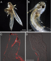
Fig. 1.
Fig. 1.
Generation of aneurogenic larvae. The extent of innervation was analyzed by the reactivity of neuron-specific tubulin antibody. (A) Parabiosis was performed at stage 24 and the neural tube was removed from the larva on the left at stage 29. The forelimb of both aneurogenic (Left) and normal host (Right) larvae is indicated by an arrow. The parabiotic pair has reached developmental stage 46. (B) An aneurogenic larva (stage 46) generated by complete removal of the neural tube at stage 28. The arrow identifies the three-digit forelimb. (C) Longitudinal section of the forelimb of a normal host larva showing the distribution of nerve (red). Note the extensive innervation to the larval skin. (D) A section of the forelimb of an aneurogenic larva that lacks innervation. The images (C and D) are a composite overlay of fluorescence with differential interference contrast (DIC). N, nerve. [Scale bar, 200 μm (C and D).]
Fig. 2.
Fig. 2.
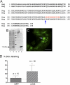
Characterization of A. maculatum Anterior Gradient protein and its biological activity. (A) Protein sequence alignment of newt (nAG) and A. maculatum (mAG) Anterior Gradient proteins. The red letters indicate the peptide (14 amino acids) in nAG that has been used to generate a rabbit antibody referred to as 224. The peptide differs at one amino acid (arrow) from the respective sequence in mAG. (B) An immunoblot showing that mAG is a secreted protein. Cos 7 cells were cotransfected with plasmids expressing mAG or GFP. The culture medium and the cell lysate were collected at 48 h after transfection and analyzed by Western blotting. The mAG protein is present in the medium (lane 5) and is found at a lower level in the cell lysate (lane 2). Lanes 1 and 4 are cell lysate and medium, respectively, from parallel cultures where the Cos 7 cells were transfected with GFP alone. Lane 3, molecular weight marker. (C) A cross-section of the small intestine of A. maculatum showing the specific reactivity of the peptide antibody 224 with the secretory goblet cells (arrow). (D) Activity of secreted mAG on cultured newt blastemal cells. Blastemal cells growing in microwells were incubated in the presence of 0.05% serum-containing medium, concentrated medium from control protein (GFP) transfections, or concentrated medium from mAG transfection of Cos 7 cells. The cells were labeled with BrdU for 72 h, fixed, and analyzed for S-phase reentry by staining with anti-BrdU antibody. The baseline S-phase reentry level with 0.05% serum medium is indicated by a dotted line. The blastemal cells incubated with mAG show a significant increase in S-phase reentry. (Scale bar, 100 μm.) Error bars, SD. *P < 0.05.
Fig. 3.
Fig. 3.
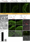
Developmental regulation of mAG protein. Expression of mAG protein in tissue sections was detected by reactivity with 224 antibody. (A) Normal innervated limb. (B) Section of a limb 10 d after denervation. (C) Aneurogenic limb. Note that expression is high throughout the larval epidermis. The epidermis is arrowed in AC. (D) Electron micrograph showing gland cells (arrows) in an aneurogenic larva at stage 43–44. The unicellular gland cells have large inclusions and are distributed throughout the larval skin. (E) Immunogold labeling of mAG protein in gland cells of an aneurogenic larva. The gold particles are localized to the rough endoplasmic reticulum (arrows). (F) Quantitative distribution of gland cells in aneurogenic and control epidermis. The number of cells with glandular morphology was counted from an entire larval epidermis. The total number of cells was tabulated from two independent samples (stage 43–44). (G) Morphology of the larva at stage 38. The limb bud is indicated by an arrow. (H) mAG expression in larval skin of the limb bud. (I) Expression in an aneurogenic limb bud. (J) Morphology of the stage 40 larva. The limb bud is indicated by an arrow. (K) Section of the limb at stage 40. Arrows point to an area of the epidermis that shows down-regulation by innervation. (L) Aneurogenic epidermis at stage 40 retains a higher level of mAG. (M and N) Neuron-specific antibody staining of K and L with DIC overlay. (O) Section through the distal tip of a normal limb bud at stage 40 showing BrdU incorporation. Note the BrdU-positive cells (green) in the epidermis. The image is an overlay of DIC with fluorescence. (P) BrdU-labeled cells in an aneurogenic limb bud at stage 40. (Q) Apoptotic cells were detected by TUNEL assay in the aneurogenic larval epidermis at stage 40 (arrows). (R) DIC overlay showing the localization within gland cells. E, epidermis; ER, endoplasmic reticulum; M, mesenchyme; N, nerve. [Scale bars, 100 μm (AC); 5 μm (D); 0.2 μm (E); and 50 μm (H, I, and KR).]
Fig. 4.
Fig. 4.
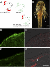
Transplantation of aneurogenic limb. (A) Schematic representation of the transplantation experiment. The left limb from an aneurogenic larva at stage 45 was transplanted to the left side of a normal host larva after removal of its corresponding limb. The larval salamander was maintained for 17 d after transplantation of the limb. The right reference limb from the aneurogenic larva was removed at the time of transplantation and processed for immunolabeling with mAG antibody. (B) A host larva with the aneurogenic limb transplant (Left; arrow). (C) The right reference limb of the aneurogenic larva showing expression of mAG in the larval skin. (D) Staining with AAT antibody showing the absence of innervation. (E) Transplanted aneurogenic limb showing down-regulation of mAG protein (compare with C) after reinnervation of the aneurogenic limb from the host larva. (F) Staining with AAT shows reinnervation of the aneurogenic limb from the host larva. Both D and F are composite overlays of AAT antibody (red) with DIC. [Scale bar, 50 μm (CF).]
Fig. 5.
Fig. 5.
Regeneration of normal and aneurogenic limbs. The upper panels are schematic representations of the limbs before amputation (A, normal with nerves; B, aneurogenic). The images of the limb blastemas are at 3 d after amputation of the limb. The amputation plane is shown with a white dotted line. (A) Normal limb blastema. The gland cells (arrows) are proximal to the amputation plane and express mAG, whereas the wound epithelium of the regenerate does not show detectable reactivity. The nerve sheath in the stump tissue shows strong reactivity with mAG protein. (Inset) AAT staining of the nerve in the limb coinciding with the sheath staining. (B) An aneurogenic limb blastema. The wound epithelium shows strong reactivity with mAG protein. Gland cells are present within the wound epithelium (arrows) as well as proximal to the amputation plane. (C and D) Electron micrograph showing the morphology of the 3-d limb blastemas. The amputation plane is represented with a red dotted line. (C) The wound epithelium in a control limb regenerate has two layers of epithelial cells that migrate over the cartilage of the stump tissue. (D) The aneurogenic limb blastema has a well-formed wound epithelium. A gland cell (arrow) is visible at the amputation plane. Note the difference in the extent of the limb blastemas in C and D. C, cartilage; M, mesenchyme; N, nerve; WE, wound epidermis. [Scale bars, 100 μm (A, B, and D); and 20 μm (C).]

Similar articles

Cited by

References

    1. Hall SM. Mechanisms of repair after traumatic injury. In: Dyck PJ, Thomas PK, editors. Peripheral Neuropathy. 4th Ed. Philadelphia: Elsevier; 2005. pp. 1403–1434.
    1. Parrinello S, et al. EphB signaling directs peripheral nerve regeneration through Sox2-dependent Schwann cell sorting. Cell. 2010;143:145–155. - PMC - PubMed
    1. Singer M. The influence of the nerve in regeneration of the amphibian extremity. Q Rev Biol. 1952;27:169–200. - PubMed
    1. Goldhamer DJ, Tomlinson BL, Tassava RA. Ganglia implantation as a means of supplying neurotrophic stimulation to the newt regeneration blastema: Cell-cycle effects in innervated and denervated limbs. J Exp Zool. 1992;262:71–80. - PubMed
    1. Kintner CR, Brockes JP. Monoclonal antibodies to the cells of a regenerating limb. J Embryol Exp Morphol. 1985;89:37–55. - PubMed

Publication types

Associated data

LinkOut - more resources