. 2011 Dec 20;108(51):20317-24.
doi: 10.1073/pnas.1100278108. Epub 2011 Aug 5.

Retrotransposons that maintain chromosome ends

Affiliations

Retrotransposons that maintain chromosome ends

Mary-Lou Pardue et al. Proc Natl Acad Sci U S A. .

Abstract

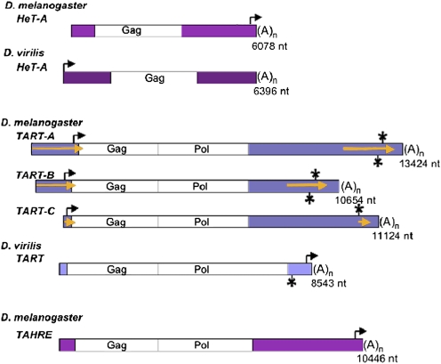
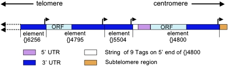
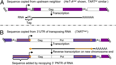
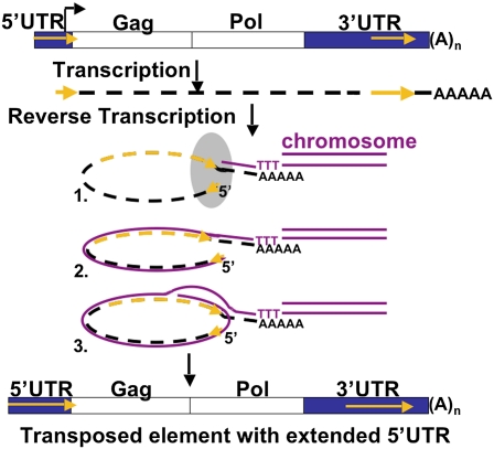
Reverse transcriptases have shaped genomes in many ways. A remarkable example of this shaping is found on telomeres of the genus Drosophila, where retrotransposons have a vital role in chromosome structure. Drosophila lacks telomerase; instead, three telomere-specific retrotransposons maintain chromosome ends. Repeated transpositions to chromosome ends produce long head to tail arrays of these elements. In both form and function, these arrays are analogous to the arrays of repeats added by telomerase to chromosomes in other organisms. Distantly related Drosophila exhibit this variant mechanism of telomere maintenance, which was established before the separation of extant Drosophila species. Nevertheless, the telomere-specific elements still have the hallmarks that characterize non-long terminal repeat (non-LTR) retrotransposons; they have also acquired characteristics associated with their roles at telomeres. These telomeric retrotransposons have shaped the Drosophila genome, but they have also been shaped by the genome. Here, we discuss ways in which these three telomere-specific retrotransposons have been modified for their roles in Drosophila chromosomes.


Conflict of interest statement

The authors declare no conflict of interest.

Figures

Fig. 1.
Fig. 1.
Telomeric retrotransposons from D. melanogaster and D. virilis (approximately to scale). Magenta, 5′ and 3′ UTRs of HeT-A and TAHRE; blue, 5′ and 3′ UTRs of TART; white, Gag and Pol ORFs; gold arrows in 5′ and 3′ UTRs of TARTmel elements (see Figs. 3B and 4 and accompanying text), PNTRs; (A)n, 3′ oligoA; bent arrows, transcription start sites for full-length sense-strand RNA (note that, for HeT-Amel and TARTvir, the element transcribed is immediately downstream of the element shown); asterisks above TART elements, start site for short sense-strand RNA; asterisks below TARTs, start site for nearly full-length antisense RNA (not determined for TART-C). The length of 5′ UTRs in TARTmel elements is extremely variable. Those lengths shown here representing the three subfamilies were the first to be sequenced; other members of these subfamilies have shorter or longer 5′ UTRs. 5′ PNTR extends to the 5′ end of the element; thus, the length of any element’s 3′ PNTR is defined by the length of its 5′ PNTR. Although the TARTvir 3′ UTR is much shorter than the 3′ UTR of the other elements, it is more than two times the length of the 3′ UTR of nontelomeric jockey clade elements that we have analyzed. TARTvir Pol ORF also has a 3′ extension of ∼1.2 kb, with no obvious motifs to indicate its function (14). This sequence is not seen in the other elements and might do double duty as 3′ UTR when the element forms telomere DNA. Elements shown are HeT-Amel, U06920, nucleotides 1,015–7,097; HeT-Avir, AY369259, nucleotides 7,211–13,612; TARTmel -A, AY561850; TARTmel -B, U14101; TARTmel -C, AY600955; TARTvir, AY219709, nucleotides 4,665–13,208; and TAHRE, AJ542581.
Fig. 2.
Fig. 2.
The four most proximal elements in the sequenced array from the XL telomere drawn approximately to scale. All elements are HeT-A, and each element is joined by its 3′ oligoA to its proximal neighbor. The most proximal element, HeT-A {}4,800, is complete, two elements are truncated in the 3′ UTR, and one element is truncated in the ORF. Elements are identified by FlyBase identifier number. Dark blue, 3′ UTR; light blue, ORF; magenta, 5′ UTR; white, string of Tags on the 5′ end of complete element {}4,800; gold, beginning of the subtelomere region; bent arrows, transcription start sites (each arrow indicates a cluster of three closely spaced sites at the 3' end of the element). The start site on {}5,504 initiates transcription of {}4,800, a transposition-competent element. Other starts will not produce productive transcripts. Physical mapping of BACs from this stock indicates that this telomere extends >100 kb further to the left (51), but no sequence is available.
Fig. 3.
Fig. 3.
Mechanisms for adding buffering 5′ sequence. (A) Sequence copied from upstream neighbor. Used by HeT-Amel and TARTvir. Telomere segment with a complete HeT-Amel flanked by other HeT-As. Transcription starts at the bent arrow in the upstream element and continues through the complete element. The resulting RNA (black line) has a Tag of the last nucleotides of the upstream element. On transposition, this Tag will become the 5′ end of the new element, undergo erosion, and if transposed again, be internalized into the string of variably eroded Tags indicated by the gray box at the 5′ end of the complete element. (B) Sequence copied from the 3′ UTR of transposing RNA. Used by TARTmel. Telomere segment with a complete TARTmel flanked by distal TART and proximal HeT-A. (A)n, 3′ oligoA in DNA; AAAAAA, polyA tail on RNA; gold arrows, PNTRs; other annotation as in Fig. 1. Transcription starts at the bent arrow and produces RNA with a very short 5′ UTR. When this is reverse-transcribed onto the chromosome end, the RT jumps back to the 3′ UTR and copies sequence to extend the 5′ UTR (Fig. 4).
Fig. 4.
Fig. 4.
Proposed mechanism for extending the 5′ end of D. melanogaster TART. Transcription starts (bent arrow) near the ATG of ORF 1 (gag), producing a transposition intermediate RNA (dashed black line) lacking most of the 5′ UTR. This RNA has a small piece of the parent element’s 5′ PNTR (short gold arrow) and a complete 3′ PNTR (long gold arrow). Steps 1–3 show the RNA as it is reverse-transcribed into DNA on the chromosome end. (Step 1) The polyA tail associates with the chromosomal DNA (magenta), and RT begins to copy the RNA. The gray oval represents proteins proposed to hold the RNA in a conformation that brings the 5′ PNTR sequence into proximity to the 3′ end of the 3′ PNTR (omitted for clarity in later steps). (Step 2) When RT reaches the 5′ end of the transcript, it makes a template jump back to the matching 3′ end of the 3′ PNTR. (Step 3) RT dissociates the RNA–DNA complex and recopies some or all of the 3′ PNTR. As a result, the transposed element will have more 5′ UTR sequence than the RNA did and possibly more sequence and longer PNTRs than the element from which it was derived.
Fig. 5.
Fig. 5.
Evolution of HeT-A sequences in the centromere region of the Y chromosome, deduced by Mendez-Lago et al. (45) (not to scale). The bottom diagram shows telomere sequence transposed into the Y chromosome: eight HeT-A elements (orange arrows) and one partial TART (#4; yellow arrow). Elements 1, 2, 3, and 5 are truncated HeT-As, and elements 6–9 are complete HeT-As. The top diagram shows 159 kb cloned in the sequenced BAC. The partial elements underwent complex amplifications to make up the 18HT satellite, which is partially represented by pentagons and black arrows on the left and is not further considered here. The end result of the several amplifications of the initially complete elements is shown on the right (numbering retained from ref. to indicate origin of different parts of the sequence). Elements with two numbers result from amplifications of parts of two elements. Triangles, nontelomeric retrotransposons (copia, mdg1, diver, F, and 1731) that inserted at various times during the sequential amplifications of this DNA; green boxes, segment of autosomal region 42A transposed into element 8 and later duplicated. [Based on figure 7 in the work by Mendez-Lago et al. (45) and reproduced with permission from Oxford University Press.]

Similar articles

Cited by

References

    1. Blackburn EH. Switching and signaling at the telomere. Cell. 2001;106:661–673. - PubMed
    1. Greider CW. Telomerase activity, cell proliferation, and cancer. Proc Natl Acad Sci USA. 1998;95:90–92. - PMC - PubMed
    1. Epel ES, et al. Accelerated telomere shortening in response to life stress. Proc Natl Acad Sci USA. 2004;101:17312–17315. - PMC - PubMed
    1. Pardue ML, DeBaryshe PG. Retrotransposons provide an evolutionarily robust non-telomerase mechanism to maintain telomeres. Annu Rev Genet. 2003;37:485–511. - PubMed
    1. Pardue ML, DeBaryshe PG. Adapting to life at the end of the line. How Drosophila telomeric retrotransposons cope with their job. Mobile Genetic Elements. 2011;1:128–134. - PMC - PubMed

Publication types

LinkOut - more resources