

Clinical diagnosis and staging of cholangiocarcinoma
- PMID: 21808282
- PMCID: PMC3331791
- DOI: 10.1038/nrgastro.2011.131
Clinical diagnosis and staging of cholangiocarcinoma
Abstract
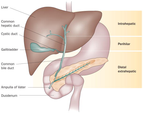
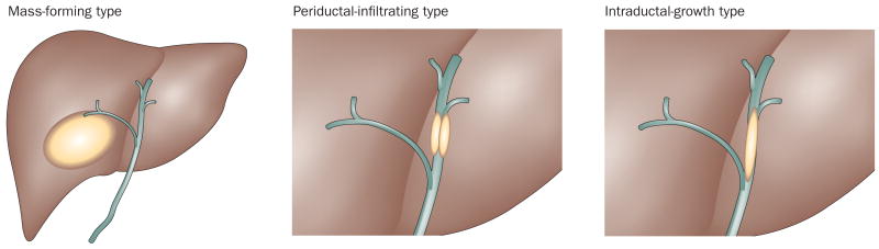
Cholangiocarcinoma is the most frequent biliary malignancy. It is difficult to diagnose owing to its anatomic location, growth patterns and lack of definite diagnostic criteria. Currently, cholangiocarcinoma is classified into the following types according to its anatomic location along the biliary tree: intrahepatic, perihilar or distal extrahepatic cholangiocarcinoma. These cholangiocarcinoma types differ in their biological behavior and management. The appropriate stratification of patients with regard to the anatomic location and stage of cholangiocarcinoma is a key determinate in their management. Staging systems can guide this stratification and provide prognostic information. In addition, staging systems are essential in order to compare and contrast the outcomes of different therapeutic approaches. A number of staging systems exist for cholangiocarcinoma-several early ones have been updated, and new ones are being developed. We discuss the emerging diagnostic criteria as well as the different staging systems for cholangiocarcinoma, and provide a critical appraisal regarding these advances in biliary tract malignancies.
Conflict of interest statement
Figures
Similar articles
-
Cholangiocarcinoma.Surg Pathol Clin. 2018 Jun;11(2):403-429. doi: 10.1016/j.path.2018.02.005. Surg Pathol Clin. 2018. PMID: 29751883 Review.
-
Imaging bile duct tumors: staging.Abdom Imaging. 2013 Oct;38(5):1071-81. doi: 10.1007/s00261-013-0021-9. Abdom Imaging. 2013. PMID: 23793410 Review.
-
Cholangiocarcinoma.Surg Clin North Am. 2019 Apr;99(2):315-335. doi: 10.1016/j.suc.2018.12.004. Epub 2019 Feb 10. Surg Clin North Am. 2019. PMID: 30846037 Review.
-
Evaluation and management of intrahepatic and extrahepatic cholangiocarcinoma.Cancer. 2016 May 1;122(9):1349-69. doi: 10.1002/cncr.29692. Epub 2016 Jan 22. Cancer. 2016. PMID: 26799932 Review.
-
Current approaches to the diagnosis and treatment of cholangiocarcinoma.Curr Gastroenterol Rep. 2006 Feb;8(1):30-7. doi: 10.1007/s11894-006-0061-1. Curr Gastroenterol Rep. 2006. PMID: 16510032 Review.
Cited by
-
Apolipoprotein B Is Associated With the Microenvironment of Cholangiocarcinoma.Front Oncol. 2021 Mar 31;11:654689. doi: 10.3389/fonc.2021.654689. eCollection 2021. Front Oncol. 2021. PMID: 33954113 Free PMC article.
-
The role of extracellular vesicles in cholangiocarcinoma tumor microenvironment.Front Pharmacol. 2024 Jan 10;14:1336685. doi: 10.3389/fphar.2023.1336685. eCollection 2023. Front Pharmacol. 2024. PMID: 38269274 Free PMC article. Review.
-
Distal extrahepatic cholangiocarcinoma mimicking groove pancreatitis: A case report and literature review.Front Oncol. 2022 Sep 27;12:948799. doi: 10.3389/fonc.2022.948799. eCollection 2022. Front Oncol. 2022. PMID: 36237325 Free PMC article.
-
Defining the optimal bilirubin level before hepatectomy for hilar cholangiocarcinoma.BMC Cancer. 2020 Sep 23;20(1):914. doi: 10.1186/s12885-020-07385-0. BMC Cancer. 2020. PMID: 32967634 Free PMC article.
-
The Importance of Imaging Techniques in the Assessment of Biliary Tract Cancer.Curr Health Sci J. 2017 Jul-Sep;43(3):201-208. doi: 10.12865/CHSJ.43.03.03. Epub 2017 Sep 28. Curr Health Sci J. 2017. PMID: 30595876 Free PMC article.
References
-
- Everhart JE, Ruhl CE. Burden of digestive diseases in the United States Part III: liver, biliary tract, and pancreas. Gastroenterology. 2009;136:1134–1144. - PubMed
-
- DeOliveira ML, et al. New staging system and a registry for perihilar cholangiocarcinoma. Hepatology. 2011;53:1363–1371. - PubMed
-
- Welzel TM, McGlynn KA, Hsing AW, O'Brien TR, Pfeiffer RM. Impact of classification of hilar cholangiocarcinomas (Klatskin tumors) on the incidence of intra- and extrahepatic cholangiocarcinoma in the United States. J Natl Cancer Inst. 2006;98:873–875. - PubMed
-
- Burak K, et al. Incidence and risk factors for cholangiocarcinoma in primary sclerosing cholangitis. Am J Gastroenterol. 2004;99:523–526. - PubMed
Publication types
MeSH terms
Grants and funding
LinkOut - more resources
Full Text Sources
Other Literature Sources
Medical
