

Viral weapons of membrane destruction: variable modes of membrane penetration by non-enveloped viruses
- PMID: 21804909
- PMCID: PMC3144554
- DOI: 10.1016/j.coviro.2011.05.002
Viral weapons of membrane destruction: variable modes of membrane penetration by non-enveloped viruses
Abstract
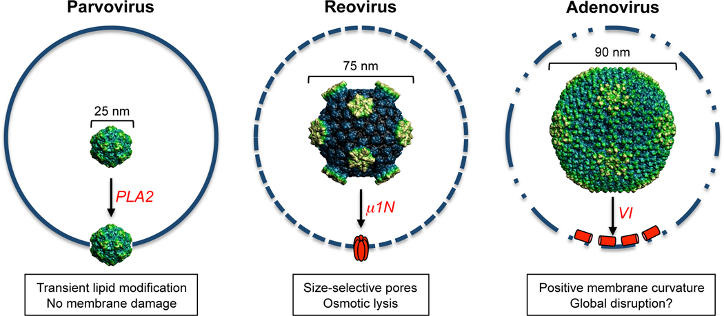
Significant progress has recently been obtained in our understanding of cellular entry by nonenveloped viruses (NEVs). A key step in the entry process involves the disruption or remodeling of the limiting cell membrane allowing the virus to gain access to the cellular replication machinery. Biochemical, genetic and structural data from diverse virus groups have shed light on the process of membrane penetration thereby revealing both the conservation and divergence of the mechanisms and principles governing this process. In general, membrane breach is achieved via the highly regulated spatiotemporal exposure of a virally encoded membrane lytic factor, resulting in the transfer of the viral genome or nucleocapsid into the cytosol.
Figures
Similar articles
-
Breach: Host Membrane Penetration and Entry by Nonenveloped Viruses.Trends Microbiol. 2018 Jun;26(6):525-537. doi: 10.1016/j.tim.2017.09.010. Epub 2017 Oct 25. Trends Microbiol. 2018. PMID: 29079499 Review.
-
Penetration of nonenveloped viruses into the cytoplasm.Annu Rev Cell Dev Biol. 2007;23:23-43. doi: 10.1146/annurev.cellbio.23.090506.123454. Annu Rev Cell Dev Biol. 2007. PMID: 17456018 Review.
-
Non-Enveloped Virus Entry: Structural Determinants and Mechanism of Functioning of a Viral Lytic Peptide.J Mol Biol. 2016 Aug 28;428(17):3540-56. doi: 10.1016/j.jmb.2016.06.006. Epub 2016 Jun 16. J Mol Biol. 2016. PMID: 27320388
-
Cell entry of enveloped viruses.Adv Genet. 2011;73:121-83. doi: 10.1016/B978-0-12-380860-8.00004-5. Adv Genet. 2011. PMID: 21310296 Free PMC article. Review.
-
Vaccinia virus A25 and A26 proteins are fusion suppressors for mature virions and determine strain-specific virus entry pathways into HeLa, CHO-K1, and L cells.J Virol. 2010 Sep;84(17):8422-32. doi: 10.1128/JVI.00599-10. Epub 2010 Jun 10. J Virol. 2010. PMID: 20538855 Free PMC article.
Cited by
-
TBK1 is part of a galectin 8 dependent membrane damage recognition complex and drives autophagy upon Adenovirus endosomal escape.PLoS Pathog. 2022 Jul 20;18(7):e1010736. doi: 10.1371/journal.ppat.1010736. eCollection 2022 Jul. PLoS Pathog. 2022. PMID: 35857795 Free PMC article.
-
"Repair Me if You Can": Membrane Damage, Response, and Control from the Viral Perspective.Cells. 2020 Sep 7;9(9):2042. doi: 10.3390/cells9092042. Cells. 2020. PMID: 32906744 Free PMC article. Review.
-
Ceramide formation mediated by acid sphingomyelinase facilitates endosomal escape of caliciviruses.Virology. 2015 Sep;483:218-28. doi: 10.1016/j.virol.2015.04.022. Epub 2015 May 15. Virology. 2015. PMID: 25985440 Free PMC article.
-
Membranotropic peptides mediating viral entry.Pept Sci (Hoboken). 2018 Sep;110(5):e24040. doi: 10.1002/pep2.24040. Epub 2018 Feb 13. Pept Sci (Hoboken). 2018. PMID: 32328541 Free PMC article. Review.
-
Poxvirus membrane biogenesis: rupture not disruption.Cell Microbiol. 2013 Feb;15(2):190-9. doi: 10.1111/cmi.12072. Epub 2012 Dec 16. Cell Microbiol. 2013. PMID: 23168015 Free PMC article. Review.
References
-
- Bong DT, Steinem C, Janshoff A, Johnson JE, Reza Ghadiri M. A highly membrane-active peptide in flock house virus: Implications for the mechanism of nodavirus infection. Chem Biol. 1999;6:473–481. - PubMed
-
- Gastaldelli M, Imelli N, Boucke K, Amstutz B, Meier O, Greber UF. Infectious adenovirus type 2 transport through early but not late endosomes. Traffic. 2008;9:2265–2278. - PubMed
Publication types
MeSH terms
Substances
Grants and funding
LinkOut - more resources
Full Text Sources
Medical
