. 2011 Sep;25(9):1636-49.
doi: 10.1210/me.2011-0035. Epub 2011 Jul 28.

IGF-I stimulates cooperative interaction between the IGF-I receptor and CSK homologous kinase that regulates SHPS-1 phosphorylation in vascular smooth muscle cells

Affiliations

IGF-I stimulates cooperative interaction between the IGF-I receptor and CSK homologous kinase that regulates SHPS-1 phosphorylation in vascular smooth muscle cells

Yashwanth Radhakrishnan et al. Mol Endocrinol. 2011 Sep.

Abstract

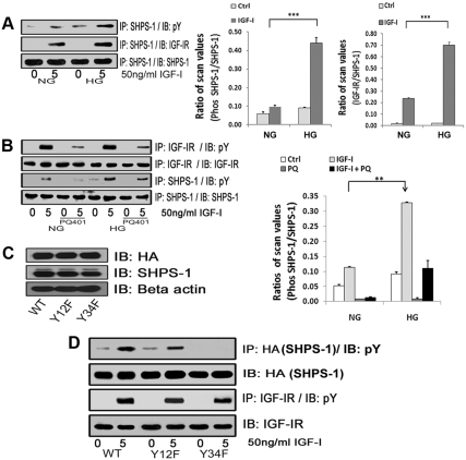
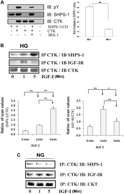
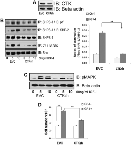
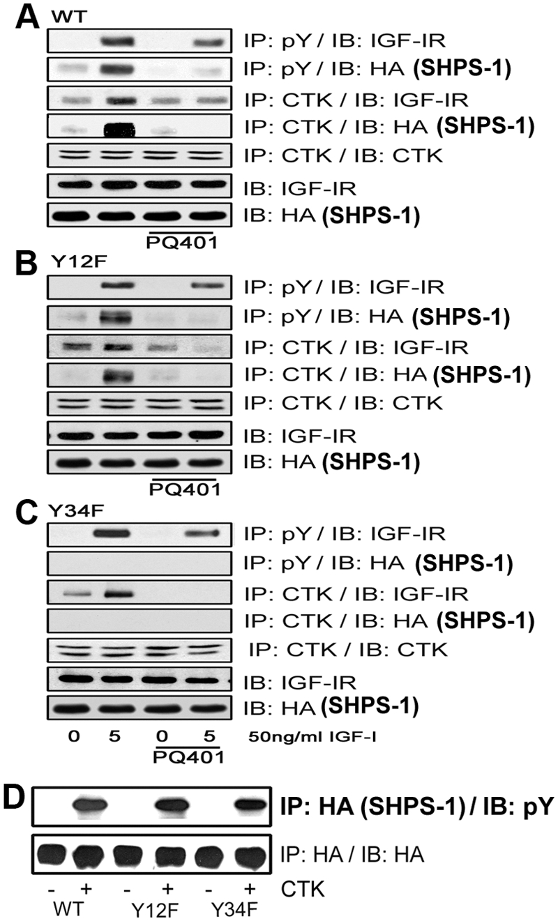
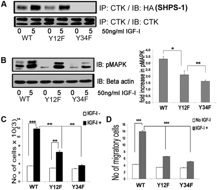
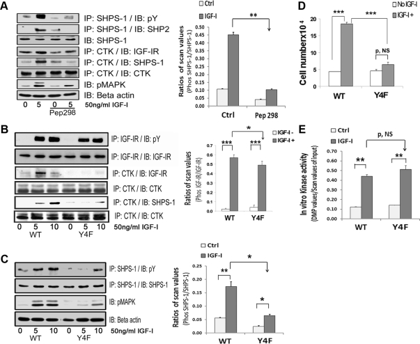
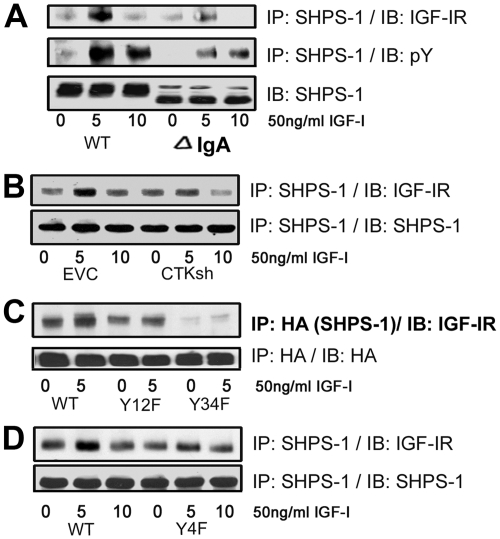
IGF-I plays an important role in smooth muscle cell proliferation and migration. In vascular smooth muscle cells cultured in 25 mm glucose, IGF-I stimulated a significant increase in Src homology 2 domain containing protein tyrosine phosphatase substrate-1 (SHPS-1) phosphorylation compared with 5 mm glucose and this increase was required for smooth muscle cell proliferation. A proteome-wide screen revealed that carboxyl-terminal SRC kinase homologous kinase (CTK) bound directly to phosphotyrosines in the SHPS-1 cytoplasmic domain. Because the kinase(s) that phosphorylates these tyrosines in response to IGF-I is unknown, we determined the roles of IGF-I receptor (IGF-IR) and CTK in mediating SHPS-1 phosphorylation. After IGF-I stimulation, CTK was recruited to IGF-IR and subsequently to phospho-SHPS-1. Expression of an IGF-IR mutant that eliminated CTK binding reduced CTK transfer to SHPS-1, SHPS-1 phosphorylation, and cell proliferation. IGF-IR phosphorylated SHPS-1, which provided a binding site for CTK. CTK recruitment to SHPS-1 resulted in a further enhancement of SHPS-1 phosphorylation. CTK knockdown also impaired IGF-I-stimulated SHPS-1 phosphorylation and downstream signaling. Analysis of specific tyrosines showed that mutation of tyrosines 428/452 in SHPS-1 to phenylalanine reduced SHPS-1 phosphorylation but allowed CTK binding. In contrast, the mutation of tyrosines 469/495 inhibited IGF-IR-mediated the phosphorylation of SHPS-1 and CTK binding, suggesting that IGF-IR phosphorylated Y469/495, allowing CTK binding, and that CTK subsequently phosphorylated Y428/452. Based on the above findings, we conclude that after IGF-I stimulation, CTK is recruited to IGF-IR and its recruitment facilitates CTK's subsequent association with phospho-SHPS-1. This results in the enhanced CTK transfer to SHPS-1, and the two kinases then fully phosphorylate SHPS-1, which is necessary for IGF-I stimulated cellular proliferation.


Figures

Fig. 1.
Fig. 1.
Effects of high glucose on IGF-I-stimulated SHPS-1 phosphorylation and IGF-IR/SHPS-1 association. A, Quiescent VSMC cultures were incubated in SFM containing 5 (DMEM-NG) or 25 (DMEM-HG) mm glucose overnight. Cells were stimulated with IGF-I (50 ng/ml) for 5 min. The extent of SHPS-1 tyrosine phosphorylation was determined by immunoprecipitating (IP) cell lysates with an anti-SHPS-1 antibody and then immunoblotting (IB) with an antiphosphotyrosine (pY) antibody (upper panel). Similarly the extent of the SHPS-1 association with IGF-IR was determined by IP using an anti-SHPS-1 antibody and then IB with an anti IGF-IR antibody (middle panel). As a loading control, the cell lysates were immunoprecipitated and immunoblotted with an anti-SHPS-1 antibody (lower panel). The bar graphs show pooled results from at least three independent experiments. The error bars represent mean ± se. ***, P < 0.001 when amount of SHPS-1 phosphorylation or its association with IGF-IR at 5 min in response to IGF-I is compared between DMEM-NG and DMEM-HG cells. B, Quiescent VSMC cultured and maintained in DMEM-NG or DMEM-HG were serum starved and then incubated with or without the IGF-IR tyrosine kinase inhibitor, PQ401 (10 μm) for 1–2 h. IGF-I was added for 5 min. Cell lysates were immunoprecipitated with an anti-IGF-IR antibody (first panel) or with an anti-SHPS-1 antibody (third panel) and immunoblotted for pY. For loading controls, IP and IB of cell lysates were performed with anti-IGF-IR (second panel) or anti-SHPS-1 antibody (fourth panel). The bar graphs show pooled results from at least three independent experiments. Error bars represent mean ± se. **, P < 0.01 when amount of SHPS-1 phosphorylation at 5 min in response to IGF-I is compared between DMEM-NG and DMEM-HG cells. C, VSMC expressing WT SHPS-1 (SHPS1-WT), VSMC expressing Y428F/Y452F mutant (Y12F), and VSMC expressing Y469F/Y495F mutant (Y34F) were serum starved in DMEM-HG and analyzed for recombinant protein expression. Cell lysates were immunoblotted using an anti-HA antibody (top panel), anti-SHPS-1 antibody (middle panel), and anti-β-actin antibodies (bottom panel). D, Confluent cells expressing SHPS1-WT or the SHPS-1-mutants (Y12F and Y34F) were serum starved for 16 h in DMEM-HG and then exposed to IGF-I for 5 min. The extent of HA-tagged SHPS-1 and IGF-IR phosphorylation was determined by IP HA (first panel) or IGF-IR (third panel) and then immunoblotted with an anti-pY antibody. An equal amount of protein from cell lysates from the same experiment was used to immunoblot for total HA (second panel) and total IGF-IR (fourth panel).
Fig. 2.
Fig. 2.
In vitro phosphorylation of SHPS-1 CD by CTK and CTK association with SHPS-1 and IGF-IR A, The cytoplasmic domain of the SHPS-1 (SHPS-1/CD) generated by in vitro translation using wheat germ extract was incubated in vitro (denoted by the + sign) with and without known amount of CTK and IRS-1 as described in Materials and Methods. The reaction mix was analyzed by immunoblotting (IB) for phosphotyrosine (upper panel) or SHPS-1 (middle panel) and CTK (bottom panel). The relative fold increase in the SHPS-1/CD phosphorylation and suppression by IRS-1 is shown in the bar graph. The error bars represent mean ± se. **, P < 0.01. B and C, Quiescent VSMC cultures were incubated in SFM containing DMEM-HG (B) or DMEM-NG (C) overnight. Cells were stimulated with IGF-I (50 ng/ml) for the periods of time indicated. The extent of the CTK association with SHPS-1 and IGF-IR was determined by immunoprecipitating (IP) using an anti-CTK antibody and then immunoblotted with an anti SHPS-1(first panel) or anti-IGF-IR antibody (second panel). Cell lysates from the same experiment were used to immunoprecipitate and immunoblot with anti-CTK antibody (third panel). The bar graphs show pooled results from at least three independent experiments. Error bars represent mean ± se. **, P < 0.01 when the amount of CTK associated with SHPS-1 at 5 min or IGF-IR at 1 min in response to IGF-I is compared with no stimulation or when the amount at 1 min is compared with 5 min. *, P < 0.05 when the amount of CTK associated with IGF-IR at 5 min is compared with no stimulation.
Fig. 3.
Fig. 3.
Silencing CTK impairs IGF-I induced SHPS-1 phosphorylation. A, Confluent VSMC expressing an empty control vector (EVC) or CTK shRNA maintained in DMEM-HG and serum starved for 18 h in HG were analyzed for CTK protein expression (top panel) or for β-actin (lower panel). B, Confluent EVC and CTK shRNA cell cultures were serum starved for 18 h and IGF-I (50 ng/ml) was added for 5 min. Cell lysates were immunoprecipitated (IP) with an anti-SHPS-1 antibody and then immunoblotted (IB) for pY (first panel) or SHP-2 (second panel) or with an anti-SHPS-1 antibody (third panel). Cell lysates from similar experiments were immunoprecipitated with an anti-pY antibody and immunoblotted with an anti-Shc antibody (fourth panel). Twenty micrograms of protein from the same whole cell lysate were directly immunoblotted with anti-Shc antibody (fifth panel). The bar graphs show pooled results from at least three independent experiments. Error bars represent mean ± se. **, P < 0.01 when amount of SHPS-1 phosphorylation at 5 min in response to IGF-I is compared between EVC and CTK shRNA cells. C, Lysates containing 20 μg of protein from the same or similar experiment were directly immunoblotted with an anti-pMAPK antibody or an anti β-actin antibody. D, EVC and CTK shRNA cells were plated (3 × 10⁴) in DMEM-HG with 2% FBS before exposure to IGF-I (50 ng/ml) in DMEM-HG with 0.2% platelet-poor plasma. Forty-eight hours after the addition of IGF-I, cell number was determined by tryptan blue staining and counting. The bar graphs show pooled results from at least three independent experiments. The error bars represent mean ± se. ***, P < 0.001 when cell number in response to IGF-I is compared between EVC and CTK shRNA cells.
Fig. 4.
Fig. 4.
IGF-IR and CTK phosphorylate specific tyrosines on SHPS-1. A–C, Quiescent VSMC expressing SHPS1-WT (A), Y12F (B), and Y34F (C) were cultured and maintained in DMEM-HG and then serum starved overnight before being incubated with or without the IGF-IR tyrosine kinase inhibitor, PQ401 (10 μm), for 1–2 h. IGF-I was added for 5 min. Cell lysates were immunoprecipitated (IP) with an anti-pY antibody and then immunoblotted (IB) for IGF-IR (first panel) or HA (second panel). Similarly, cell lysates from the same experiments were immunoprecipitated with an anti-CTK antibody and then immunoblotted with anti-IGF-IR (third panel), anti-HA (fourth panel), or anti-CTK antibodies (fifth panel). Twenty micrograms of protein from the same whole-cell lysate were directly immunoblotted with an anti-IGF-IR antibody (sixth panel) or with an anti-HA antibody (seventh panel). D, Confluent SHPS-WT, Y12F, and Y34F cells were serum starved for 16 h in DMEM-HG, and the cell lysates were immunoprecipitated with anti-HA antiserum. The immunoprecipitates were incubated with or without CTK to determine HA-tagged SHPS-1 phosphorylation in vitro. After centrifugation the resultant supernatants were immunoblotted using a pY antibody (upper panel) or an HA antibody as a loading control (lower panel).
Fig. 5.
Fig. 5.
Substitution for specific tyrosines on SHPS-1 alters CTK association and IGF-I signaling-stimulated actions. A, Confluent VSMC expressing SHPS1-WT and SHPS1 (Y12F and Y34F) mutant cells were serum starved for 16 h in DMEM-HG and then exposed to IGF-I for 5 min. Cell lysates were immunoprecipitated (IP) using anti-CTK antiserum then immunoblotted (IB) for HA-tagged SHPS-1. An equal amount of protein from cell lysates was used for IP/IB to estimate total CTK. B, Twenty micrograms of protein from the same cell lysates were directly immunoblotted with anti-pMAPK or anti-β-actin. Error bars represent mean ± se. *, P < 0.05 when phosphorylation of MAPK at 5 min in response to IGF-I is compared between WT and Y12F; **, P < 0.01 when Y12F and Y34F are compared. C, SHPS-1-WT, Y12F, and Y34F cells were plated (3 × 10⁴) in DMEM-HG with 2% FBS before exposure to IGF-I (50 ng/ml) in DMEM-HG with 0.2% platelet-poor plasma. Forty-eight hours after the addition of IGF-I, the cell number was determined by tryptan blue staining and counting. The bar graphs show pooled results from at least three independent experiments. Error bars represent mean ± se. ***, P < 0.001 when the increase in cell number cell number in response to IGF-I in WT cells is compared with control; **, P < 0.01 when increase in cell number in response to IGF-I in Y12F cells is compared with control; **, P < 0.01 when the increases in cell number between WT and Y12F cells or between Y12F and Y34F cells are compared. D, SHPS-1 WT, Y12F, and Y34F cells were grown to confluent density in six-well plates in DMEM-HG containing 10% FBS. After wounding, they were allowed to migrate with or without IGF-I in medium containing 0.2% FBS for 48 h. The total number of cells migrating past the wound edge in five predetermined areas was quantified. ***, P < 0.001 when the number of WT cell migration in response to IGF-I is compared with control or with the number of migration of Y12F or Y34F cells.
Fig. 6.
Fig. 6.
CTK transfer to IGF-IR facilitates SHPS-1 phosphorylation. A, VSMC maintained in DMEM-HG were serum starved and then incubated with or without cell-permeable peptide 298 at a concentration of 10 μg/ml for 2 h. IGF-I was added for 5 min with and without the peptide. Cell lysates were immunoprecipitated (IP) with an anti-CTK antibody and immunoblotted (IB) with anti-IGF-IR (fourth panel), anti-SHPS1 (fifth panel), or anti-CTK antibodies (sixth panel). The cell lysates were also immunoprecipitated with anti-SHPS-1 antibody and immunoblotted for pY (first panel) or SHP-2 (second panel). Twenty micrograms of protein from the same or similar cell lysates were directly immunoblotted with anti-SHPS-1 (third panel), pMAPK (seventh panel) or anti-β-actin (eighth panel) antibodies. The bar graph shows the relative decrease in SHPS-1 phosphorylation for at least three independent experiments. The error bars represent mean ± se. **, P < 0.001 when the increase in SHPS-1 phosphorylation in response to IGF-I is compared with or without peptide 298 (Pep298). Ctrl, Control. B, Confluent VSMC expressing IGF-IR-WT and IGF-IR-Y4F mutant cells were serum starved for 16 h in DMEM-HG and then exposed to IGF-I for 5 or 10 min. Cell lysates were immunoprecipitated with an anti-IGF-IR antibody and immunoblotted for pY (first panel) or for total IGF-IR (second panel). Cell lysates from the same experiments were immunoprecipitated with an anti-CTK antibody and immunoblotted using an anti-IGF-IR antibody (third panel), an anti-SHPS1 antibody (fifth panel), or an anti-CTK antibody (fourth and sixth panels). C, Lysates from an experiment similar to that shown in B were immunoprecipitated with an anti-SHPS-1 antibody and immunoblotted for pY (first panel) or total SHPS-1 (second panel). Twenty micrograms of total protein from the above cell lysates were directly immunoblotted with anti-pMAPK (third panel) or anti-β-actin (fourth panel). The bar graph represents the pooled results from three independent experiments and shows the relative decrease in SHPS-1 phosphorylation. The error bars represent mean ± se. **, P < 0.01; * P < 0.05 indicate the significant differences between two treatments or two cell types. D, IGF-I-R-WT and IGF-I-R-Y4F cells were plated (3 × 10⁴) in DMEM-HG with 2% FBS before exposure to IGF-I (50 ng/ml) in DMEM-HG with 0.2% platelet-poor plasma. Forty-eight hours after the addition of IGF-I, cell number was determined by tryptan blue staining and counting. The bar graphs show pooled results from at least three independent experiments. Error bars represent mean ± se. ***, P < 0.001 when number of proliferating cells is compared between WT and Y4F cells in response to IGF-I. P, NS indicates no significant difference between two treatments. E, Confluent VSMC expressing IGF-IR-WT and IGF-IR-Y4F mutant cells were serum starved for 16 h in DMEM-HG and then exposed to IGF-I for 1 min. As indicated, cell lysates were precleared with protein A beads and then immunoprecipitated with an anti-IGF-IR antibody. Immunoprecipitates were used in an in vitro kinase reaction as described in Materials and Methods. The relative kinase activity is depicted in the bar graph. The error bars represent mean ± se. P = NS. **, P < 0.01 indicates a significant difference between two treatments. P, NS indicates no significant difference.
Fig. 7.
Fig. 7.
IGF-IR/SHPS-1 association and IGF-IR-stimulated SHPS-1 phosphorylation is mediated in part by CTK. A, Confluent VSMC expressing SHPS-1WT or SHPS-1 IgA deletion mutant were serum starved for 16 h in DMEM-HG and then exposed to IGF-I for the times indicated. Cell lysates were immunoprecipitated (IP) with an anti-SHPS-1 antibody and then immunoblotted (IB) for IGF-IR (top panel) or with an anti-pY antibody (middle panel). Twenty micrograms of protein from the same or similar whole-cell lysate were directly IB with an anti-SHPS-1 antibody (lower panel). B, Confluent empty control vector (EVC), and CTK shRNA cell cultures were serum starved for 16 h and IGF-I (50 ng/ml) was added for times indicated. The cell lysates were immunoprecipitated with an anti-SHPS-1 antibody and then immunoblotted for IGF-IR or using an anti-SHPS-1 antibody. C, Confluent VSMC-expressing SHPS-1-WT and SHPS-1 (Y12F and Y34F) mutant cells were serum starved for 16 h in DMEM-HG and then exposed to IGF-I for 5 min. The extent of HA-tagged SHPS-1 association with IGF-IR was determined by immunoprecipitating HA and immunoblotting for IGF-IR or with an anti-HA antibody as a loading control. D, Confluent VSMC-expressing IGF-IR-WT and IGF-IR-Y4F mutant cells were serum starved for 16 h in DMEM-HG and then exposed to IGF-I for the indicated period of time. Cell lysates were immunoprecipitated with an anti-SHPS-1 antibody and immunoblotted using an anti-IGF-IR antibody or an anti-SHPS-1 antibody.

Similar articles

Cited by

References

    1. Arnqvist HJ, Bornfeldt KE, Chen Y, Lindström T. 1995. The insulin-like growth factor system in vascular smooth muscle: interaction with insulin and growth factors. Metabolism 44:58–66 - PubMed
    1. Joslin E, Kahn CR. 2005. Joslin's diabetes mellitus. 14th ed. Boston: Lippincott Williams, Wilkins
    1. Radhakrishnan Y, Busby WH, Jr, Shen X, Maile LA, Clemmons DR. 2010. Insulin-like growth factor-I-stimulated insulin receptor substrate-1 negatively regulates Src homology 2 domain-containing protein-tyrosine phosphatase substrate-1 function in vascular smooth muscle cells. J Biol Chem 285:15682–15695 - PMC - PubMed
    1. Shen X, Xi G, Radhakrishnan Y, Clemmons DR. 2009. Identification of novel SHPS-1-associated proteins and their roles in regulation of insulin-like growth factor-dependent responses in vascular smooth muscle cells. Mol Cell Proteomics 8:1539–1551 - PMC - PubMed
    1. Ling Y, Maile LA, Lieskovska J, Badley-Clarke J, Clemmons DR. 2005. Role of SHPS-1 in the regulation of insulin-like growth factor I-stimulated Shc and mitogen-activated protein kinase activation in vascular smooth muscle cells. Mol Biol Cell 16:3353–3364 - PMC - PubMed

Publication types

MeSH terms