. 2011 Jul 26;30(17):3647-61.
doi: 10.1038/emboj.2011.245.

Vaccinia extracellular virions enter cells by macropinocytosis and acid-activated membrane rupture

Affiliations

Vaccinia extracellular virions enter cells by macropinocytosis and acid-activated membrane rupture

Florian Ingo Schmidt et al. EMBO J. .

Abstract

Vaccinia virus (VACV), the model poxvirus, produces two types of infectious particles: mature virions (MVs) and extracellular virions (EVs). EV particles possess two membranes and therefore require an unusual cellular entry mechanism. By a combination of fluorescence and electron microscopy as well as flow cytometry, we investigated the cellular processes that EVs required to infect HeLa cells. We found that EV particles were endocytosed, and that internalization and infection depended on actin rearrangements, activity of Na(+)/H(+) exchangers, and signalling events typical for the macropinocytic mechanism of endocytosis. To promote their internalization, EVs were capable of actively triggering macropinocytosis. EV infection also required vacuolar acidification, and acid exposure in endocytic vacuoles was needed to disrupt the outer EV membrane. Once exposed, the underlying MV-like particle presumably fused its single membrane with the limiting vacuolar membrane. Release of the viral core into the host cell cytosol allowed for productive infection.


Conflict of interest statement

The authors declare that they have no conflict of interest.

Figures

Figure 1
Figure 1
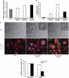
Quality of EV particles. (A) EV quality—infectious particles. Clarified supernatants of RK13 cells infected with VACV IHD-J, WR, or WR ΔA34R were titrated on BSC-40 cells after incubation with or without Mab 7D11. As a control, purified MVs of the same strains were subjected to the same treatment and titration. Plaque numbers were normalized to untreated virus samples. Results represent the average of three independent experiments (mean±s.e.m.). (B) EV quality—fluorescent particles. Clarified supernatants of RK13 cells infected with IHD-J mCherry–A5 F13–GFP were incubated with 7D11, bound to cover slips, and fixed. 7D11 was visualized with AF 647 goat anti-mouse and images recorded by confocal microscopy. Virus particles were detected by their fluorescent core and assigned to the following categories: MV−7D11 (mCherry), MV+7D11 (mCherry and 7D11 staining), EV−7D11 (mCherry, GFP), or EV+7D11 (mCherry, GFP, and 7D11 staining). Average values of three independent experiments are shown±s.e.m. (C) Fluorescent EVs. Representative images from (B). Examples of a neutralized MV (MV+7D11, open arrowhead), an intact EV (EV−7D11, arrow), and a disrupted EV (EV+7D11, closed arrowhead) are highlighted.
Figure 2
Figure 2
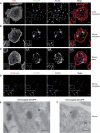
EV internalization by fluorescence and electron microscopy. (AD) IHD-J F13–GFP EVs (AC) or IHD-J mCherry–A5 F13–GFP EVs (D) were bound to HeLa cells on ice for 1 h (MOI 15). EV particles were stained with VMC-20 (anti-B5) directly (A) or after incubation at 37°C for 30 min (BD). VMC-20 staining was performed either under non-permeabilizing (non-perm.) conditions to visualize bound virions (A, B, D) or under permeabilizing (perm.) conditions to visualize bound and internalized virions (C). Images were recorded by confocal microscopy and representative maximum projections of Z-stacks are shown. Arrows in the inset of B highlight F13–GFP-containing EV membranes not accessible to VMC-20 staining. In the inset of (D), EV particles (arrows) and an EV membrane without a core (open arrowhead), all not accessible to VMC-20 staining, as well as a virus core or MV without an EV membrane (closed arrowhead), are highlighted. Scale bars=10 μm. (E, F) IHD-J F13–GFP EVs were bound to HeLa cells on ice for 1 h (MOI 300). Cells were incubated at 37°C for 30 min, fixed, and subjected to cryosectioning and immunolabelling with anti-GFP and protein A-gold. Images were recorded by transmission electron microscopy and representative pictures are shown. Arrows in (F) highlight MV and EV membranes. Mi, mitochondrion; Nu, nucleus. Scale bars=200 nm.
Figure 3
Figure 3
EV internalization by flow cytometry. IHD-J F13–GFP EVs (MOI 175) were bound to HeLa cells on ice for 1 h and cells incubated at 0 or 37°C for 30 min. To detect internalized EVs (int.), bound virions were removed and cells were detached with trypsin; to measure total cell-associated virions (total), cells were detached with EDTA. Cells were fixed, and green fluorescence quantified by flow cytometry. Representative histograms of untreated samples are shown in (A); green fluorescence intensity from three independent experiments was quantified and the average of measured geometric means of internalized (B) and total (C) EVs is displayed±s.e.m.
Figure 4
Figure 4
Cellular factors required for EV infection. (AD) HeLa cells were left untreated or treated with DMSO (highest used concentration), staurosporine (Stau), genistein (Geni), wortmannin (Wort), calphostin C (CalC), rottlerin (Rott), 3-indolepropionic acid (IPA-3), Iressa (Ires), chlorpromazine (Chlo), ML-7 (all 30 min), cytochalasin D (Cyto), jasplakinolide (Jasp), or ethylisopropyl amiloride (EIPA) (all 15 min). MVs or EVs of IHD-J GFP (A), WR GFP (C), or WR ΔA34R GFP (D) were preincubated in the presence of drugs and in the case of EVs with 7D11 (MOI 2). Cells were infected and green fluorescent cells quantified 4 h p.i. by flow cytometry. Infection levels were normalized to untreated samples. (B) EV (IHD-J F13–GFP) and MV (IHD-J EGFP–A5) internalization in the presence of various drugs was quantified as described in Figure 3. The geometric mean of green fluorescence intensity of 0°C samples was substracted from 37°C samples and internalization normalized to untreated samples. All experiments were performed three times independently, mean values±s.e.m. are shown.
Figure 5
Figure 5
EVs and cellular phenotypes of macropinocytosis. (A) VAVC-induced fluid-phase uptake. WR ΔA34R EVs and WR wt MVs were bound to serum-starved HeLa cells on ice for 1.5 h and cells pulsed for 10 min at 37°C with 10 kDa dextran-AF 488. Cells were washed, harvested, fixed, and geometric means of green fluorescence quantified by flow cytometry. Dextran uptake was normalized to uptake by unstimulated cells and mean values±s.e.m. of three independent experiments are shown. (B, C) VAVC-induced cell blebbing. WR ΔA34R EGFP–A5 MVs or concentrated EVs were bound to HeLa cells at room temperature (RT). Cells were incubated at 37°C for 40 min, fixed, stained, and analysed by DIC and wide-field fluorescence microscopy. The number of total and blebbing cells from three independent experiments (∼250 cells per condition and experiment) was counted and mean±s.e.m. is shown (B). Representative images are shown in (C). Scale bars=20 μm. (D) WR ΔA34R infection—ANX5. Exposed PS of concentrated EVs or MVs of WR ΔA34R GFP (MOI 4) was masked with ANX5. Subsequently, virions were incubated at 37°C for 1 h (in case of EVs in the presence of 7D11) and used for infection experiments as described in Figure 4. Experiments were performed three times independently, mean values±s.e.m. are shown.
Figure 6
Figure 6
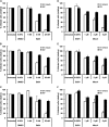
EV infection and acidification of endocytic vacuoles. Experimental setup as in Figure 4; HeLa cells were pretreated with BafA (AC) or MonA (DF) for 1 h; controls with solvents at the highest used concentrations were included. Infection with IHD-J GFP (A, D), WR GFP (B, E), or WR ΔA34R GFP (C, F) in the presence of drugs was quantified by flow cytometry; mean±s.e.m. of three independent, normalized experiments are shown.
Figure 7
Figure 7
EV internalization and acid-mediated EV membrane disruption. (A) IHD-J—internalization. EV (IHD-J F13–GFP) and MV (IHD-J EGFP–A5) internalization in the presence of BafA, MonA, and EIPA was quantified as described in Figure 4B. (B) In vitro EV membrane disruption. IHD-J, WR, and WR ΔA34R EVs were incubated at pH 5.0 or pH 7.4 and 37°C for 5 min. EVs were titrated with or without 7D11 at pH 7.4 as described in Figure 1A. The percentage of intact EVs was calculated by normalizing plaque numbers after 7D11 neutralization to plaque numbers in untreated samples; mean values±s.e.m. of three independent experiments are presented. (C) EV infection—acid bypass of BafA block. HeLa cells were left untreated or treated with BafA for 1 h. EVs of IHD-J GFP, WR GFP, or WR ΔA34R GFP were preincubated in the presence of drugs and 7D11 for 1 h at 37°C (MOI 2). Virus particles were bound to cells on ice. Cells were shifted to 37°C in pH 7.4 or pH 4.5 medium for 5 min and then incubated in full medium with drugs for 4 h. Infected cells were quantified by flow cytometry. The percentage of infected cells was normalized to the untreated samples. All experiments were performed three times independently, mean values±s.e.m. are shown, asterisks mark significant differences (P<0.05).
Figure 8
Figure 8
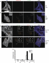
EV accumulation and core release in the presence of BafA. (AE) HeLa cells were infected with IHD-J mCherry–A5 F13–GFP EVs (MOI 25) (A, B) or 7D11-treated IHD-J EGFP–A5 EVs (MOI 8 after 7D11 incubation) (CE) in the presence of 5 μg/ml ActD and 25 nM BafA where indicated. Particles were bound to pretreated cells on ice and incubated in full DMEM with drugs at 37°C for 3 h. Samples were fixed and stained for actin (A, B) or L1 (Mab 7D11) and actin (C, D). (AD) Images were recorded by confocal microscopy and representative maximum projections of Z-stacks are shown. Arrows in the insets of (B) highlight EVs, the arrowhead marks a free EV membrane. Arrows in the insets of (C, D) highlight released viral cores, arrowheads point at viral particles that were stained for the MV-membrane marker L1. Scale bars=10 μm. (E) The percentage of released cores after infection with 7D11-treated EVs, MVs, and 7D11-treated MVs in the presence of the indicated drugs was quantified in three independent experiments. Controls with bound virus (0 h) were included. Core release from 7D11-neutralized MVs was only quantified in the presence of ActD. Mean values±s.e.m. are shown.
Figure 9
Figure 9
Mechanism of VACV EV entry. VACV EVs induce fluid-phase uptake by macropinocytosis and are internalized with the bulk fluid. The pH in macropinosomes decreases in the course of their maturation, which triggers disruption of the outer EV membrane. Exposed EFCs in the membrane of MV-like particles catalyse or regulate fusion with limiting endocytic membranes and thereby release virus cores with the genome into the cytosol allowing for successful replication.

Similar articles

Cited by

References

    1. Aldaz-Carroll L, Whitbeck JC, Ponce de Leon M, Lou H, Hirao L, Isaacs SN, Moss B, Eisenberg RJ, Cohen GH (2005) Epitope-mapping studies define two major neutralization sites on the vaccinia virus extracellular enveloped virus glycoprotein B5R. J Virol 79: 6260–6271 - PMC - PubMed
    1. Amstutz B, Gastaldelli M, Kalin S, Imelli N, Boucke K, Wandeler E, Mercer J, Hemmi S, Greber UF (2008) Subversion of CtBP1-controlled macropinocytosis by human adenovirus serotype 3. EMBO J 27: 956–969 - PMC - PubMed
    1. Banfield BW, Leduc Y, Esford L, Schubert K, Tufaro F (1995) Sequential isolation of proteoglycan synthesis mutants by using herpes simplex virus as a selective agent: evidence for a proteoglycan-independent virus entry pathway. J Virol 69: 3290–3298 - PMC - PubMed
    1. Benhnia MR, McCausland MM, Moyron J, Laudenslager J, Granger S, Rickert S, Koriazova L, Kubo R, Kato S, Crotty S (2009) Vaccinia virus extracellular enveloped virion neutralization in vitro and protection in vivo depend on complement. J Virol 83: 1201–1215 - PMC - PubMed
    1. Chakrabarti S, Sisler JR, Moss B (1997) Compact, synthetic, vaccinia virus early/late promoter for protein expression. Biotechniques 23: 1094–1097 - PubMed

Publication types