. 2011 Jul 25;194(2):229-43.
doi: 10.1083/jcb.201012017. Epub 2011 Jul 18.

HJURP is a CENP-A chromatin assembly factor sufficient to form a functional de novo kinetochore

Affiliations

HJURP is a CENP-A chromatin assembly factor sufficient to form a functional de novo kinetochore

Meghan C Barnhart et al. J Cell Biol. .

Abstract

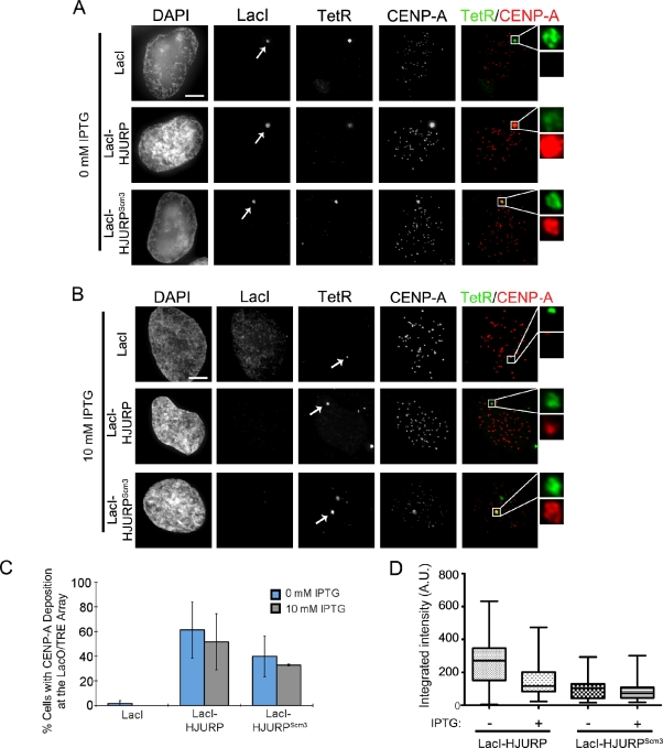
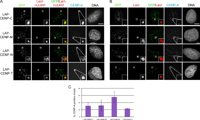
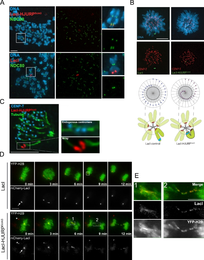
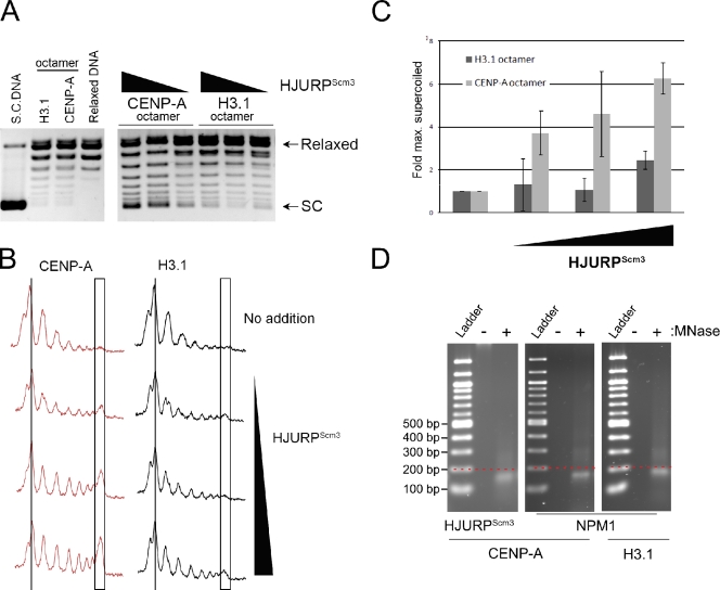
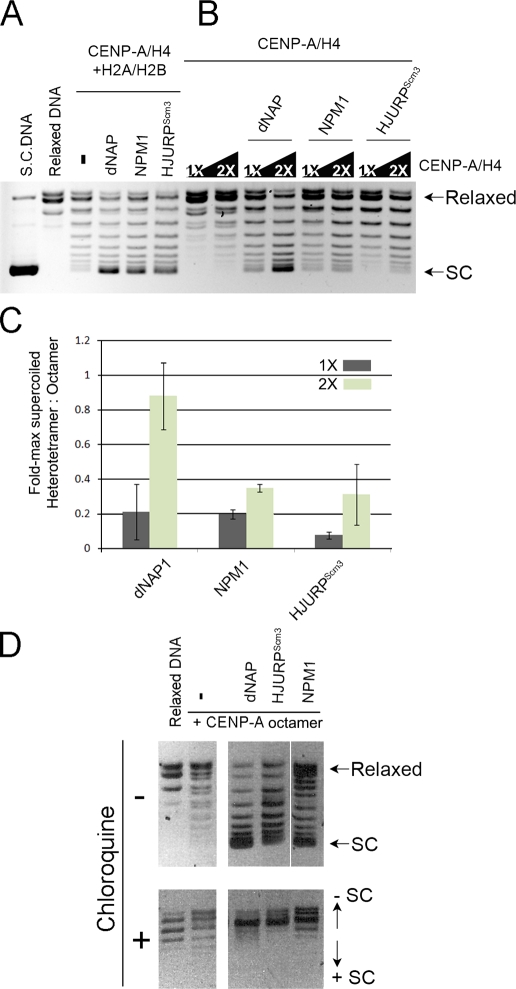
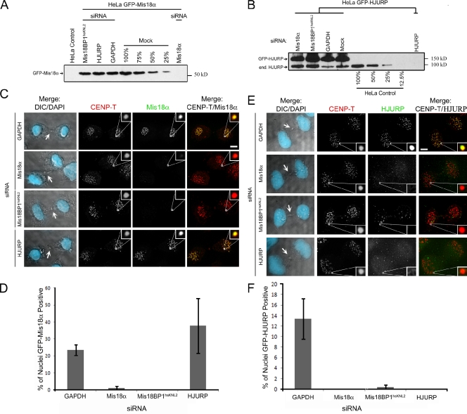
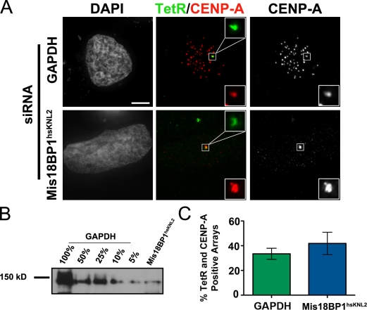
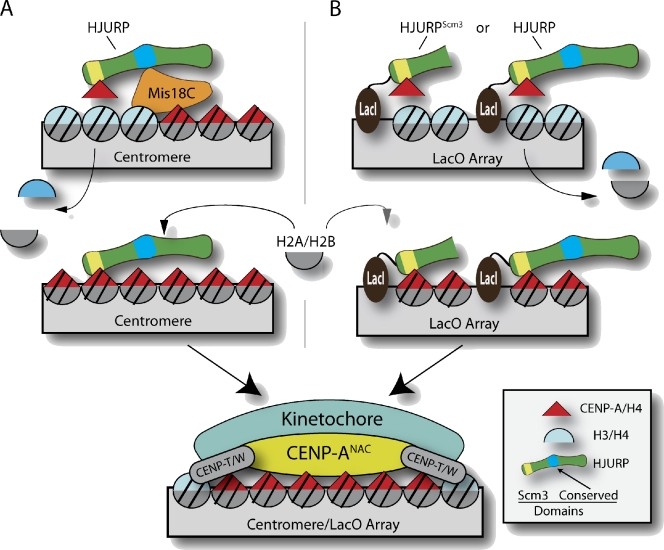
Centromeres of higher eukaryotes are epigenetically marked by the centromere-specific CENP-A nucleosome. New CENP-A recruitment requires the CENP-A histone chaperone HJURP. In this paper, we show that a LacI (Lac repressor) fusion of HJURP drove the stable recruitment of CENP-A to a LacO (Lac operon) array at a noncentromeric locus. Ectopically targeted CENP-A chromatin at the LacO array was sufficient to direct the assembly of a functional centromere as indicated by the recruitment of the constitutive centromere-associated network proteins, the microtubule-binding protein NDC80, and the formation of stable kinetochore-microtubule attachments. An amino-terminal fragment of HJURP was able to assemble CENP-A nucleosomes in vitro, demonstrating that HJURP is a chromatin assembly factor. Furthermore, HJURP recruitment to endogenous centromeres required the Mis18 complex. Together, these data suggest that the role of the Mis18 complex in CENP-A deposition is to recruit HJURP and that the CENP-A nucleosome assembly activity of HJURP is responsible for centromeric chromatin assembly to maintain the epigenetic mark.


Figures

Figure 1.
Figure 1.
HJURP-dependent CENP-A recruitment and incorporation into the LacO/TRE array. (A and B) Recruitment of endogenous CENP-A to the LacO/TRE array in the presence of LacI-HJURP or LacI-HJURPScm3. All LacI constructs have an N-terminal mCherry tag. Representative images of preextracted cells treated with 0 mM IPTG (A) or 10 mM IPTG (B) for 1 h before fixation. Endogenous CENP-A was detected using a monoclonal anti–CENP-A antibody. mCherry-LacI fusions of HJURP or HJURPScm3 and GFP-TetR markers are transiently transfected at equal ratios, and DNA is visualized using DAPI. Cells are fixed at 48 h after transfection. Arrows indicate the array. Insets show magnified views of boxed regions. (C) Quantification of CENP-A staining at the LacO/TRE array. Error bars represent the standard deviations between two experiments. At least 30 cells per condition were analyzed; n = 2. In the case of IPTG treatment, cells in which the residual mCherry signal was still visible at the array were excluded. (D) Quantification of the amount of CENP-A at the array in LacI-HJURP– and LacI-HJURPScm3–transfected cells with and without treatment with IPTG (>28 cells/condition). Middle lines in each box represent the mean integrated intensity for each condition, and whiskers represent the maximum and minimum intensities observed. A.U., arbitrary unit. Bars, 5 µm.
Figure 2.
Figure 2.
HJURP-deposited CENP-A recruits constitutive centromere proteins. (A) LacO/TRE U2OS cells were transiently transfected with LacI-HJURP and constructs expressing LAP (GFP localization and purification)-tagged CENP-C, CENP-M, CENP-N, or CENP-T. Cells were preextracted and fixed 72 h after transfection. The presence of CENP-A was assessed using antibodies against endogenous CENP-A. Insets show the arrays at a higher magnification. (B) LacI control images for LAP-CENP-C–T, indicating that recruitment is never observed in the absence of CENP-A. (C) Graph showing the percentage of doubly transfected (GFP and LacI-HJURP) U2OS LacO/TRE cells with endogenous CENP-A present at the array, which also recruited the indicated constitutive centromere proteins (≥30 cells per condition; error bars represent standard deviation). Bars, 5 µm.
Figure 3.
Figure 3.
Recruitment of CENP-A by HJURPScm3 mediates kinetochore formation at the LacO array. (A) Mitotic chromosome spreads from U20S-LacO/TRE cells transfected with LacI or LacI-HJURPScm3, arrested in nocodazole, and stained with antibodies for NDC80. 40% of LacI-HJURP arrays recruited NDC80. (B) Monastrol-arrested cells transfected with LacI or LacI-HJURPScm3 and immunostained for centromere marker CENP-T. Radial distribution plots describe the mean centromere position (black circle) in the cells measured (>26 cells per condition) relative to the center of the DNA mass. The array position is diagrammed relative to the center of the DNA mass as blue triangles (LacI) or red diamonds (LacI-HJURPScm3). The gray circle represents one standard deviation from the mean centromere position. The LacI-HJURPScm3 array falls within the centromere region in 69% of transfected cells versus 15% for LacI controls. (C) Selective stabilization of kinetochore-bound microtubules through cold treatment demonstrates the LacI-HJURPScm3 arrays form stable microtubule interactions similar to endogenous centromeres. Insets show magnified views of the boxed region. (D) LacO-SceI-TRE NIH3T3 cells were transfected with YFP–histone H2B and followed by live-cell imaging as they progress through mitosis. Times are given relative to the last frame when cells were in metaphase. Arrows indicate the array, and asterisks indicate nonchromatin-bound unspecific LacI staining. (E) Insets taken from images in D show the behavior of the array (red in merge) and YFP-H2B (green in merge) for (1 and 2) LacI-HJURPScm3 at 6 and 9 min into anaphase, respectively. Bars: (A–D) 5 µm; (E) 2 µm.
Figure 4.
Figure 4.
HJURPScm3 is sufficient to assemble CENP-A nucleosomes in vitro. (A) Plasmid supercoiling assays were conducted using recombinant MBP-tagged HJURPScm3 and recombinant CENP-A octamer (including histones H4, H2A, and H2B) or histone H3.1 octamer to assess the relative ability of HJURP to assemble CENP-A– and H3.1-containing nucleosomes. The relaxed DNA lane contains topoisomerase-treated supercoiled (S.C.) plasmid DNA. HJURPScm3 induced supercoiling more efficiently in the presence of CENP-A relative to H3.1. (B) Line scans across topoisomers within conditions presented in A. Lines indicate the least supercoiled topoisomers. Boxes indicate the location of the maximally assembled topoisomers. (C) Assembly reactions from A containing H3.1 and CENP-A are graphed here as fold intensity over reactions containing no HJURPScm3. Error bars show standard deviations. (D) Assembly reactions in A (using HJURPScm3) or assembly reactions using NPM1 digested with micrococcal nuclease to show DNA protection of the assembled species. A dotted line was drawn to indicate the migration of a 200-bp fragment.
Figure 5.
Figure 5.
HJURPScm3-assembled CENP-A nucleosomes are negatively supercoiled and contain H2A and H2B. (A and B) Supercoiling assay comparing assembly efficiencies of chaperones dNAP, NPM1, and HJURPScm3 with CENP-A histone octamers (CENP-A–H4 and H2A/H2B) in A or with CENP-A–H4 alone in B. CENP-A–H4 levels added to the reactions were varied from 1 to 2× compared with the amount of CENP-A–H4 present in the reactions in A. Line scans are presented in Fig. S3. SC, supercoiled. (C) Integrated intensities of maximally supercoiled populations were measured from reactions in A and B. Values are graphed as fold-maximally supercoiled heterotetramer to octamer. Error bars show standard deviations. (D) Supercoiling assay showing assembly activities (top) for dNAP, HJURPScm3, and NPM1. Supercoiled DNA was separated by agarose gel electrophoresis with (bottom) or without (top) the DNA intercalating agent chloroquine to distinguish negatively and positively supercoiled DNA. The minus signs indicate no addition of chaperone. The white line indicates that intervening lanes have been spliced out.
Figure 6.
Figure 6.
Recruitment of HJURP to centromeres requires the Mis18 complex. (A and B) Cellular extracts from siRNA-treated and control cell lines were analyzed by Western blotting using anti-GFP (A) or anti-HJURP antibodies (B). Each lane contains lysate from 105 cells. Dilution series were generated from mock-treated HeLa GFP-Mis18α (A) or parental HeLa (B) cells. (C) Stable GFP-Mis18α cells lines were treated with siRNA against Mis18α, Mis18BP1hsKNL2, HJURP, or GAPDH (control). Representative images of siRNA-treated GFP-Mis18α cells were selected in which a midbody was clearly present (differential interference contrast [DIC], arrows) to show the cell was in early G1. DAPI staining was overlaid onto the differential interference contrast image. Cells were stained with anti–CENP-T. (D) Mean percentage of GFP-Mis18α centromere-positive nuclei from a population of ≥57 cells in each siRNA treatment from two experiments. (E) Similar image acquisition as in C. Here, stable HeLa GFP-HJURP cells were treated with the same siRNAs. (F) Mean percentage of GFP-HJURP centromere-positive nuclei from a population of ≥135 cells in each siRNA treatment from two experiments. Error bars show standard deviations. Insets show magnified views of boxed regions. Bars, 5 µm.
Figure 7.
Figure 7.
The Mis18 complex is not required for CENP-A deposition at the LacO/TRE array. (A) Representative images of endogenous CENP-A recruitment in U2OS-LacO cells treated with 15 mM IPTG after 72 h of either GAPDH or Mis18BP1hsKNL2 siRNA treatment. Cells had been transiently transfected with LacI-HJURPScm3 and GFP-TetR 48 h before fixation. Cells were transfected after an initial 24 h siRNA treatment to ensure Mis18BP1hsKNL2 depletion before CENP-A establishment at the array. Insets show magnified views of boxed regions. Bar, 5 µm. (B) Cellular extracts from GAPDH and Mis18BP1hsKNL2 siRNA-treated cells were analyzed by Western blotting using an anti-Mis18BP1hsKNL2 antibody. Each lane contains lysate from 107 cells. (C) Quantification of CENP-A staining at the LacO/TRE array marked by GFP-TetR after 72 h of GAPDH or Mis18BP1hsKNL2 siRNA treatment and 1 h of 15 mM IPTG treatment. At least 30 cells per condition were analyzed; n = 2. Error bars represent the standard deviation between the two experiments. The p-value between GAPDH and Mis18BP1hsKNL2 is 0.3609.
Figure 8.
Figure 8.
HJURP recruitment determines centromere position. (A) HJURP is recruited to centromeres through the action of Mis18. During G1, HJURP directly assembles CENP-A nucleosomes at centromeres along with histones H2A and H2B. The recruitment of HJURP is the critical step in determining the site of the centromere. (B) Redirecting HJURP to an integrated LacO array bypasses the requirement for Mis18, results in deposition of CENP-A, and is sufficient to form a functional kinetochore.

Similar articles

Cited by

References

    1. Allshire R.C., Karpen G.H. 2008. Epigenetic regulation of centromeric chromatin: old dogs, new tricks? Nat. Rev. Genet. 9:923–937 10.1038/nrg2466 - DOI - PMC - PubMed
    1. Bergmann J.H., Rodríguez M.G., Martins N.M., Kimura H., Kelly D.A., Masumoto H., Larionov V., Jansen L.E., Earnshaw W.C. 2011. Epigenetic engineering shows H3K4me2 is required for HJURP targeting and CENP-A assembly on a synthetic human kinetochore. EMBO J. 30:328–340 10.1038/emboj.2010.329 - DOI - PMC - PubMed
    1. Bernad R., Sánchez P., Rivera T., Rodríguez-Corsino M., Boyarchuk E., Vassias I., Ray-Gallet D., Arnaoutov A., Dasso M., Almouzni G., Losada A. 2011. Xenopus HJURP and condensin II are required for CENP-A assembly. J. Cell Biol. 192:569–582 10.1083/jcb.201005136 - DOI - PMC - PubMed
    1. Black B.E., Foltz D.R., Chakravarthy S., Luger K., Woods V.L., Jr, Cleveland D.W. 2004. Structural determinants for generating centromeric chromatin. Nature. 430:578–582 10.1038/nature02766 - DOI - PubMed
    1. Black B.E., Brock M.A., Bédard S., Woods V.L., Jr, Cleveland D.W. 2007. An epigenetic mark generated by the incorporation of CENP-A into centromeric nucleosomes. Proc. Natl. Acad. Sci. USA. 104:5008–5013 10.1073/pnas.0700390104 - DOI - PMC - PubMed

Publication types

LinkOut - more resources