

doi: 10.1038/nsmb.2086.
A RING E3-substrate complex poised for ubiquitin-like protein transfer: structural insights into cullin-RING ligases
Affiliations
- PMID: 21765416
- PMCID: PMC3245743
- DOI: 10.1038/nsmb.2086
Item in Clipboard
A RING E3-substrate complex poised for ubiquitin-like protein transfer: structural insights into cullin-RING ligases
Nat Struct Mol Biol.
.
Abstract
How RING E3 ligases mediate E2-to-substrate ubiquitin-like protein (UBL) transfer remains unknown. Here we address how the RING E3 RBX1 positions NEDD8's E2 (UBC12) and substrate (CUL1). We find that existing structures are incompatible with CUL1 NEDD8ylation and report a new conformation of RBX1 that places UBC12 adjacent to CUL1. We propose RING domain rotation as a general mechanism for UBL transfer for the largest family of E3s.
Figures

UBC12–RBX1 interactions. (a) WT and mutant UBC12-mediated CUL1 NEDD8ylation in vitro. Mutated residues are orange on UBC12 (otherwise cyan)–RBX1 (pink) model. (b) 2D HSQC [15N-1H] spectra of 15N-UBC12core in the absence (red) and presence (blue) of equimolar RBX1RING. A subset of shifted resonances are labeled in the spectra. Residues with chemical shift differences greater than one standard deviation above the mean are labeled and colored orange on UBC12 (otherwise cyan)–RBX1 (pink) model.
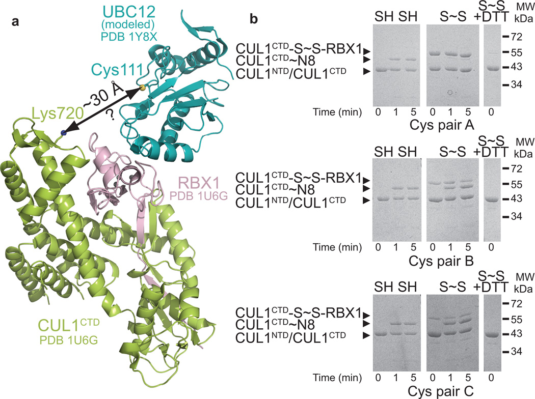
Models from prior structures reveal an E2-to-substrate gap. (a) Structural model of CUL1CTD–RBX1–UBC12core (colored light green, pink, cyan respectively) complex based on previous CUL1–RBX1 and RING–E2 structures–,, (Supplementary Fig. 1). CUL1 NEDD8 modification site and UBC12 catalytic cysteine are indicated by blue and yellow spheres respectively. (b) In vitro CUL1 NEDD8ylation assays using disulfide-linked (S~S) and unlinked (SH SH) split’n’coexpress CUL1–RBX1 (ref. 7). Pairs A–C refer to three distinct combinations of engineered cysteines (Supplementary Fig. 2).
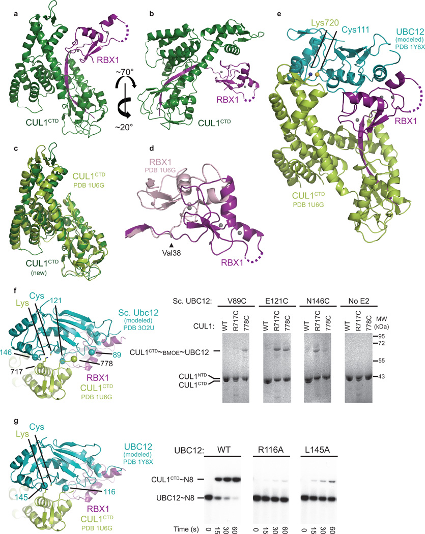
Structure of CUL1CTD–RBX1 in new conformation. (a) One copy of CUL1CTD (dark green) –RBX1 (purple) from the asymmetric unit. (b) Same as (a), but rotated ~70° in y and ~20° in x. (c) Structural overlay with previous CUL1CTD (light green) oriented similarly to (a),. (d) Structural overlay with previous RBX1 (pink) in same orientation as (b). (e) Structural model of CUL1CTD–RBX1–UBC12core (light green, purple, cyan respectively) complex based on the new RBX1 conformation. (f) Bis-Maleimidoethane (BMOE)-crosslinking between engineered cysteine mutants of Ubc12 and CUL1CTD. Residues mutated to cysteine are numbered on the model (left, view rotated by ~70° and ~40° about the×and z-axes relative to panel e) and products of crosslinking reactions are shown for all possible Ubc12+CUL1–RBX1 combinations (right). Note residue numbering for yeast Ubc12. For reference, Ubc12 catalytic cysteine and CUL1 NEDD8ylation site are shown as sticks. (g) In vitro CUL1 NEDD8ylation for UBC12 mutants at the predicted interface. Mutated residues are shown on the model (left).
Comment in
-
Conformational flexibility and rotation of the RING domain in activation of cullin-RING ligases.Nat Struct Mol Biol. 2011 Aug 3;18(8):863-5. doi: 10.1038/nsmb.2117. Nat Struct Mol Biol. 2011. PMID: 21811311 No abstract available.
Similar articles
-
Structure of a RING E3 trapped in action reveals ligation mechanism for the ubiquitin-like protein NEDD8.Cell. 2014 Jun 19;157(7):1671-84. doi: 10.1016/j.cell.2014.04.037. Cell. 2014. PMID: 24949976 Free PMC article.
-
A dual E3 mechanism for Rub1 ligation to Cdc53.Mol Cell. 2010 Sep 10;39(5):784-96. doi: 10.1016/j.molcel.2010.08.030. Mol Cell. 2010. PMID: 20832729 Free PMC article.
-
Structural insights into NEDD8 activation of cullin-RING ligases: conformational control of conjugation.Cell. 2008 Sep 19;134(6):995-1006. doi: 10.1016/j.cell.2008.07.022. Cell. 2008. PMID: 18805092 Free PMC article.
-
NEDD8 and ubiquitin ligation by cullin-RING E3 ligases.Curr Opin Struct Biol. 2021 Apr;67:101-109. doi: 10.1016/j.sbi.2020.10.007. Epub 2020 Nov 5. Curr Opin Struct Biol. 2021. PMID: 33160249 Free PMC article. Review.
-
Genetically engineered mouse models for functional studies of SKP1-CUL1-F-box-protein (SCF) E3 ubiquitin ligases.Cell Res. 2013 May;23(5):599-619. doi: 10.1038/cr.2013.44. Epub 2013 Mar 26. Cell Res. 2013. PMID: 23528706 Free PMC article. Review.
Cited by
-
Structural basis for a reciprocal regulation between SCF and CSN.Cell Rep. 2012 Sep 27;2(3):616-27. doi: 10.1016/j.celrep.2012.08.019. Epub 2012 Sep 6. Cell Rep. 2012. PMID: 22959436 Free PMC article.
-
Mechanism of Lysine 48 Selectivity during Polyubiquitin Chain Formation by the Ube2R1/2 Ubiquitin-Conjugating Enzyme.Mol Cell Biol. 2016 May 16;36(11):1720-32. doi: 10.1128/MCB.00097-16. Print 2016 Jun 1. Mol Cell Biol. 2016. PMID: 27044868 Free PMC article.
-
Characterization of an A3G-VifHIV-1-CRL5-CBFβ Structure Using a Cross-linking Mass Spectrometry Pipeline for Integrative Modeling of Host-Pathogen Complexes.Mol Cell Proteomics. 2021;20:100132. doi: 10.1016/j.mcpro.2021.100132. Epub 2021 Aug 11. Mol Cell Proteomics. 2021. PMID: 34389466 Free PMC article.
-
Twists and turns in ubiquitin-like protein conjugation cascades.Protein Sci. 2011 Dec;20(12):1941-54. doi: 10.1002/pro.750. Epub 2011 Nov 9. Protein Sci. 2011. PMID: 22012881 Free PMC article. Review.
-
RNF111-dependent neddylation activates DNA damage-induced ubiquitination.Mol Cell. 2013 Mar 7;49(5):897-907. doi: 10.1016/j.molcel.2013.01.006. Epub 2013 Feb 7. Mol Cell. 2013. PMID: 23394999 Free PMC article.
References
Publication types
MeSH terms
Substances
Associated data
- Actions
Grants and funding
- R01 CA082491-10/CA/NCI NIH HHS/United States
- R01 GM069530-10/GM/NIGMS NIH HHS/United States
- P41 RR015301/RR/NCRR NIH HHS/United States
- P30 CA021765-33S1/CA/NCI NIH HHS/United States
- R01 GM069530-09/GM/NIGMS NIH HHS/United States
- RR-15301/RR/NCRR NIH HHS/United States
- R01CA082491/CA/NCI NIH HHS/United States
- R01 CA082491-08/CA/NCI NIH HHS/United States
- P30 CA021765/CA/NCI NIH HHS/United States
- P30 CA021765-31/CA/NCI NIH HHS/United States
- R01 CA082491/CA/NCI NIH HHS/United States
- HHMI_/Howard Hughes Medical Institute/United States
- P30 CA021765-32/CA/NCI NIH HHS/United States
- R01 GM069530/GM/NIGMS NIH HHS/United States
- R01 GM069530-08/GM/NIGMS NIH HHS/United States
- R01GM069530/GM/NIGMS NIH HHS/United States
- R01 CA082491-09/CA/NCI NIH HHS/United States
LinkOut - more resources
Full Text Sources
Molecular Biology Databases
Miscellaneous
