. 2011 Sep;22(17):3218-30.
doi: 10.1091/mbc.E10-11-0874. Epub 2011 Jul 14.

MARCH ubiquitin ligases alter the itinerary of clathrin-independent cargo from recycling to degradation

Affiliations

MARCH ubiquitin ligases alter the itinerary of clathrin-independent cargo from recycling to degradation

Craig A Eyster et al. Mol Biol Cell. 2011 Sep.

Abstract

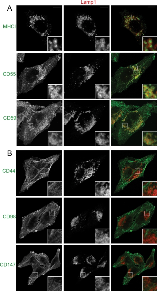
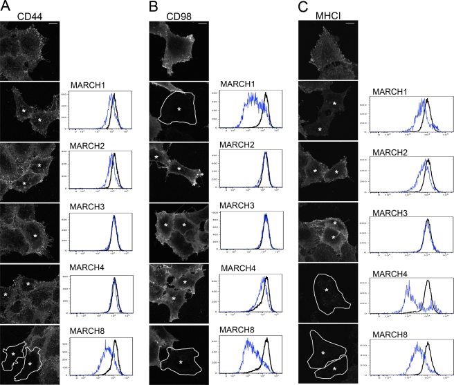
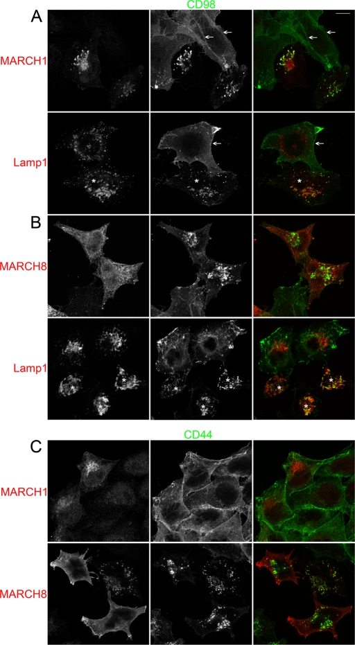
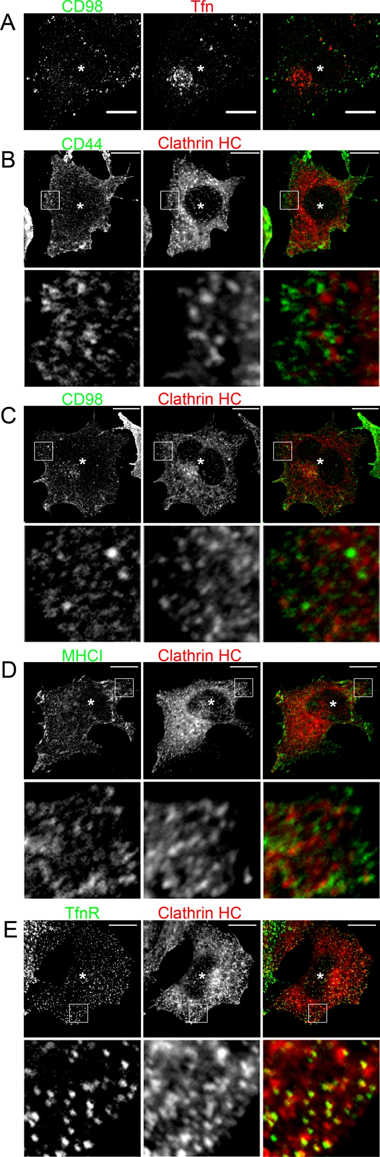
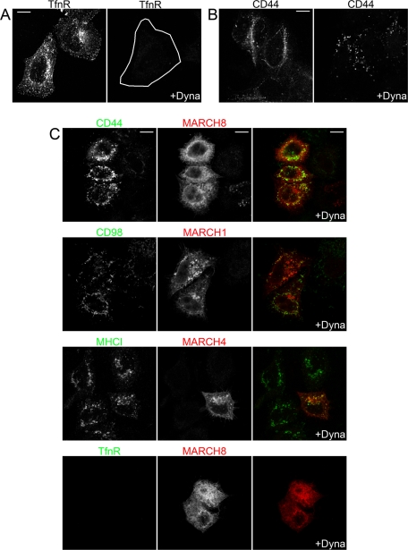
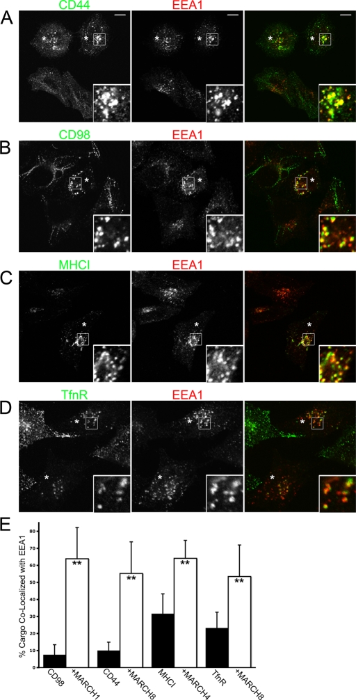
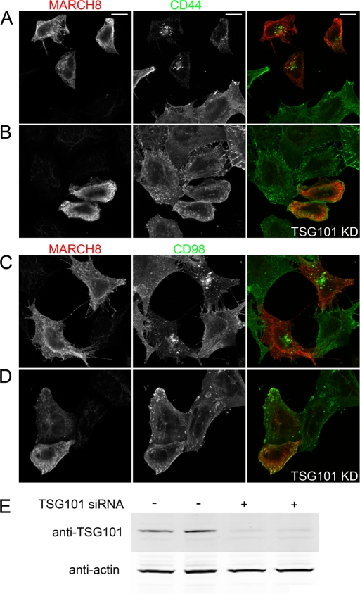
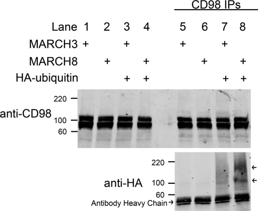
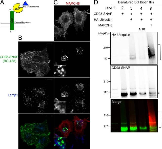
Following endocytosis, internalized plasma membrane proteins can be recycled back to the cell surface or trafficked to late endosomes/lysosomes for degradation. Here we report on the trafficking of multiple proteins that enter cells by clathrin-independent endocytosis (CIE) and determine that a set of proteins (CD44, CD98, and CD147) found primarily in recycling tubules largely failed to reach late endosomes in HeLa cells, whereas other CIE cargo proteins, including major histocompatibility complex class I protein (MHCI), trafficked to both early endosome antigen 1 (EEA1) and late endosomal compartments in addition to recycling tubules. Expression of the membrane-associated RING-CH 8 (MARCH8) E3 ubiquitin ligase completely shifted the trafficking of CD44 and CD98 proteins away from recycling tubules to EEA1 compartments and late endosomes, resulting in reduced surface levels. Cargo affected by MARCH expression, including CD44, CD98, and MHCI, still entered cells by CIE, suggesting that the routing of ubiquitinated cargo occurs after endocytosis. MARCH8 expression led to direct ubiquitination of CD98 and routing of CD98 to late endosomes/lysosomes.


Figures

FIGURE 1:
FIGURE 1:
Clathrin-independent cargo proteins show differential trafficking to late endosomes. HeLa cells were incubated with antibodies to MHCI, CD55, or CD59 (A) and CD44, CD98, or CD147 (B) for 1 h to allow endocytosis to occur. Cells were washed with media and then transferred to media containing 25 mM NH4Cl for 24 h. Cells were then fixed as described in Materials and Methods, and the internalized antibodies were detected with Alexa 488 (green) goat anti–mouse secondary antibodies. Lamp1 was immunolabeled with a rabbit antibody and an Alexa 594 (red) goat anti–rabbit secondary antibody to detect late endosomal and lysosomal structures. Images were acquired as described in Materials and Methods. Bars, 10 μm.
FIGURE 2:
FIGURE 2:
CD98 does not accumulate in late endosomes over similar time periods as MHCI. Primary antibodies to CD98 or MHCI were added to HeLa cells to allow internalization for 1 h. Cells were washed and transferred to fresh media without (top) or with 25 mM NH4Cl (bottom). After 2 (A), 6 (B), 13 (C), and 24 (D) h cells were washed and fixed. A rabbit antibody to Lamp1 was used with an Alexa 633 (blue) secondary antibody to detect LE. Isotype-specific secondary antibodies to IgG1 488 (green) and to IgG2a 594 (red) were used to detect CD98 and MHCI, respectively. Bars, 10 μm.
FIGURE 3:
FIGURE 3:
Effect of MARCH protein expression on surface levels of CD44, CD98, and MHCI. HeLa cells were transfected with the indicated MARCH-FLAG construct as described. After 18 h, the surface of the cells was labeled with mouse antibodies to CD44, CD98, and MHCI and processed for either immunofluorescence or flow cytometry. In fluorescence images, MARCH-transfected cells (as detected by rabbit anti-FLAG staining; not shown) are indicated by an asterisk and outlined in some cases. Graphs show the Alexa 488 staining intensity (x-axis) versus cell number (y-axis) for surface CD44 (A), CD98 (B), or MHCI (C) in control cells (black line) vs. indicated MARCH-expressing cells (blue line) from flow cytometry analysis. Bars, 10 μm.
FIGURE 4:
FIGURE 4:
MARCH expression increases CIE cargo delivery to late endosomes. HeLa cells were transfected with either MARCH1-FLAG (A, C) or MARCH8-FLAG (B, C) constructs for 18 h. Primary antibodies to CD44 (C) or CD98 (A, B) were added to cells and allowed to internalize for 1 h. Cells were washed and transferred to media containing 25 mM NH4Cl for 2 h. Mouse antibodies to CD44 and CD98 were detected with anti–mouse Alexa 488 (green) and rabbit anti-FLAG or rabbit anti-Lamp1 with anti–rabbit Alexa 594 (red). Cells indicated with an asterisk in A and B, bottom, are assumed to be MARCH1-FLAG and MARCH8-FLAG transfected, respectively, due to the large change in CD98 localization only observed in MARCH1-FLAG– or MARCH8-FLAG–transfected cells as shown at the top. Cell surface and recycling tubule staining for CD98 are indicated with arrows in A. Bars, 10 μm.
FIGURE 5:
FIGURE 5:
CIE cargo proteins do not internalize with Tfn or colocalize with surface clathrin in MARCH-expressing cells. (A) HeLa cells were transfected with MARCH8-FLAG. After 18 h, CD98 antibody and Tfn633 were added to cells and allowed to internalize for 5 min. Surface CD98 antibody and Tfn633 were removed with acid wash and fixed. MARCH8-FLAG was stained with a rabbit anti-FLAG antibody. CD98 and FLAG antibodies were detected with Alexa 488 anti–mouse and Alexa 594 anti–rabbit secondary antibodies, respectively. Tfn633 is pseudo-colored red, and asterisk denotes a MARCH8-FLAG–stained cell. In B–E, HeLa cells were transfected with plasmids encoding MARCH1-FLAG (C), MARCH8-FLAG (B, E), or MARCH4-FLAG (D). After 18 h, cells were fixed and incubated with primary antibodies to CD44 (B), CD98 (C), MHCI (D), and TfnR (E) in the absence of saponin to label surface antigen. Clathrin heavy chain (HC) was detected using a rabbit antibody in the presence of saponin. Surface antibodies were detected with Alexa 488 anti–mouse secondary and rabbit antibodies to clathrin HC with an Alexa 594 anti–rabbit secondary. Asterisk denotes MARCH-expressing cells identified as in Figure 4. Bars, 10 μm.
FIGURE 6:
FIGURE 6:
Dynasore inhibits internalization of CDE but not CIE cargo proteins in MARCH-expressing cells. (A, B) Untransfected HeLa cells were pretreated with 80 μM Dynasore (+Dyna) or vehicle control for 1 h, and then monoclonal antibodies to TfnR (A) or CD44 (B) were added and allowed to internalize for 30 min in the continued presence of Dynasore or vehicle control. Surface antibody to TfnR and CD44 was removed by acid wash, cells were then fixed, and internalized CD44 and TfnR antibodies were detected with Alexa 594 anti–mouse secondary antibodies. (C) HeLa cells transfected with indicated MARCH-FLAG constructs were allowed to internalize monoclonal antibodies to indicated CIE cargos or TfnR in the presence of Dynasore as before. Surface antibodies were removed with acid wash, and cells were fixed and stained with a rabbit anti-FLAG antibody. Internalized antibodies to cargos and the rabbit anti-FLAG antibody were detected with Alexa 488 anti–mouse secondary and Alexa 594 anti–rabbit secondary antibodies. Bars, 10 μm.
FIGURE 7:
FIGURE 7:
CD44, CD98, MHCI, and TfnR show increased localization with EEA1 in MARCH-expressing cells. HeLa cells transfected with MARCH1-FLAG (B), MARCH4-FLAG (C), or MARCH8-FLAG (A, D) were identified as in Figure 4 and are indicated with an asterisk. Primary antibodies to CD44 (A), CD98 (B), MHCI (C), or TfnR (D) were added to cells and allowed to internalize for 30 min. Mouse antibodies to CD44, CD98, MHCI, and TfnR were detected with anti–mouse Alexa 488 (green) and rabbit anti-EEA1 with anti–rabbit Alexa 594 (red). Bar, 10 μm. (E) The percentage of each cargo colocalizing with EEA1 in the absence (black bars) or presence (clear bars) of the indicated MARCH protein was quantified using Metamorph software. **Statistical significance at p < 0.001 between indicated cargo in control (black bars) and MARCH-expressing cells (clear bars). Bars, 10 μm.
FIGURE 8:
FIGURE 8:
TSG101 knockdown reverses the effect of MARCH expression on CIE cargo proteins. HeLa cells were mock transfected (A, C) or transfected with an siRNA to TSG101 for 24 h (B, D). Cells were subsequently transfected with MARCH8-FLAG for 18 h (A–D). Total CD44 (A, B) or CD98 (C, D) distribution was detected with mouse antibodies and MARCH8-FLAG transfectants with a rabbit anti-FLAG antibody. Primary antibodies were detected with Alexa 488 (green) anti–mouse secondary and Alexa 594 anti–rabbit secondary, respectively, in mock (A, C) and TSG101 knockdown cells (B, D). (E) Western blot of mock and TSG101 knockdown lysates from replicate cultures. TSG101 protein levels (relative to actin) were reduced to 25% of control levels in siRNA-treated cells. Bars, 10 μm.
FIGURE 9:
FIGURE 9:
Ubiquitin is present in immunoprecipitated endogenous CD98 complexes in MARCH8-expressing cells. HeLa cells were transfected with MARCH3-FLAG or MARCH8-FLAG with or without HA-ubiquitin. After 18 h, CD98 was immunoprecipitated from whole-cell lysates as described in Materials and Methods. Ten percent of whole-cell lysates were immunoblotted with anti-CD98 antibodies (lanes 1–4). Molecular weights are identified in kDa. CD98 immunoreactive bands were detected at ∼90 and 110 kDA. Twenty percent of recovered CD98 immunoprecipitates were immunoblotted with anti-CD98 or anti-HA antibodies (lanes 5–8). Two HA-reactive bands were present in the MARCH8-expressing CD98 immunoprecipitates (lane 8 arrows).
FIGURE 10:
FIGURE 10:
CD98-SNAP traffics as CD98 does and is directly ubiquitinated in MARCH8-expressing cells. (A) Schematic diagram of CD98-SNAP interaction with BG ligands. Binding of BG to SNAP produces a covalent bond between the SNAP and BG. HeLa cells expressing CD98-SNAP with (C) or without (B) MARCH8-FLAG were labeled with BG-488 for 1 h and chased in NH4Cl for 2 h. A rabbit anti-Lamp1 and mouse anti-FLAG were used with Alexa 633 anti–rabbit and Alexa 594 anti–mouse secondary antibodies to visualize late endosomes and MARCH-transfected cells respectively. Bars, 10 μm. (D) BG-biotin–labeled CD98-SNAP was immunoprecipitated from denatured lysates of cells expressing the indicated proteins and separated on SDS–PAGE and immunoblotted as described in Materials and Methods. HA-ubiquitin was detected with mouse anti-HA and an Alexa 680 anti–mouse secondary antibody (red in merged blot). The biotin-labeled CD98-SNAP was detected with NeutrAvidin, DyLight-800 Conjugated (green in merged blot). Lane 4 was loaded with 1/10 of the total protein of lane 3, which is equivalent to the amount of CD98-SNAP in lane 5 (in MARCH8-expressing cells), to directly compare the ubiquitination levels for equivalent amounts of CD98-SNAP with and without MARCH8 expression. The arrow shows CD98-SNAP, and brackets indicate ubiquitinated forms of CD98-SNAP.

Similar articles

Cited by

References

    1. Acconcia F, Sigismund S, Polo S. Ubiquitin in trafficking: the network at work. Exp Cell Res. 2009;315:1610–1618. - PubMed
    1. Arvan P, Zhao X, Ramos-Castaneda J, Chang A. Secretory pathway quality control operating in Golgi, plasmalemmal, and endosomal systems. Traffic. 2002;3:771–780. - PubMed
    1. Bartee E, Eyster CA, Viswanathan K, Mansouri M, Donaldson JG, Fruh K. Membrane-associated RING-CH proteins associate with Bap31 and target CD81 and CD44 to lysosomes. PLoS One. 2010;5:e151325. - PMC - PubMed
    1. Bartee E, Mansouri M, Hovey Nerenberg BT, Gouveia K, Fruh K. Downregulation of major histocompatibility complex class I by human ubiquitin ligases related to viral immune evasion proteins. J Virol. 2004;78:1109–1120. - PMC - PubMed
    1. Bartee E, McCormack A, Fruh K. Quantitative membrane proteomics reveals new cellular targets of viral immune modulators. PLoS Pathog. 2006;2:e107. - PMC - PubMed

Publication types

MeSH terms

Substances

LinkOut - more resources