Bacteria-specific neutrophil dysfunction associated with interferon-stimulated gene expression in the acute respiratory distress syndrome
- PMID: 21755013
- PMCID: PMC3130788
- DOI: 10.1371/journal.pone.0021958
Bacteria-specific neutrophil dysfunction associated with interferon-stimulated gene expression in the acute respiratory distress syndrome
Abstract
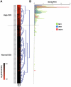
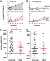
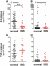
Acute respiratory distress syndrome (ARDS) is a poorly understood condition with greater than 30% mortality. Massive recruitment of neutrophils to the lung occurs in the initial stages of the ARDS. Significant variability in the severity and duration of ARDS-associated pulmonary inflammation could be linked to heterogeneity in the inflammatory capacity of neutrophils. Interferon-stimulated genes (ISGs) are a broad gene family induced by Type I interferons. While ISGs are central to anti-viral immunity, the potential exists for these genes to evoke extensive modification in cellular response in other clinical settings. In this prospective study, we sought to determine if ISG expression in circulating neutrophils from ARDS patients is associated with changes in neutrophil function. Circulating neutrophil RNA was isolated, and hierarchical clustering ranked patients' expression of three ISGs. Neutrophil response to pathogenic bacteria was compared between normal and high ISG-expressing neutrophils. High neutrophil ISG expression was found in 25 of 95 (26%) of ARDS patients and was associated with reduced migration toward interleukin-8, and altered responses to Staphylococcus aureus, but not Pseudomonas aeruginosa, which included decreased p38 MAP kinase phosphorylation, superoxide anion release, interleukin-8 release, and a shift from necrotic to apoptotic cell death. These alterations in response were reflected in a decreased capacity to kill S. aureus, but not P. aeruginosa. Therefore, the ISG expression signature is associated with an altered circulating neutrophil response phenotype in ARDS that may predispose a large subgroup of patients to increased risk of specific bacterial infections.
Trial registration: ClinicalTrials.gov NCT00548795.
Conflict of interest statement
Figures


Similar articles
-
Extremes of Interferon-Stimulated Gene Expression Associate with Worse Outcomes in the Acute Respiratory Distress Syndrome.PLoS One. 2016 Sep 8;11(9):e0162490. doi: 10.1371/journal.pone.0162490. eCollection 2016. PLoS One. 2016. PMID: 27606687 Free PMC article.
-
Differential type I interferon response and primary airway neutrophil extracellular trap release in children with acute respiratory distress syndrome.Sci Rep. 2020 Nov 4;10(1):19049. doi: 10.1038/s41598-020-76122-1. Sci Rep. 2020. PMID: 33149247 Free PMC article.
-
Increased neutrophil migratory activity after major trauma: a factor in the etiology of acute respiratory distress syndrome?Crit Care Med. 2002 Aug;30(8):1717-21. doi: 10.1097/00003246-200208000-00007. Crit Care Med. 2002. PMID: 12163782
-
The mercurial nature of neutrophils: still an enigma in ARDS?Am J Physiol Lung Cell Mol Physiol. 2014 Feb;306(3):L217-30. doi: 10.1152/ajplung.00311.2013. Epub 2013 Dec 6. Am J Physiol Lung Cell Mol Physiol. 2014. PMID: 24318116 Free PMC article. Review.
-
Understanding the role of neutrophils in acute respiratory distress syndrome.Biomed J. 2021 Aug;44(4):439-446. doi: 10.1016/j.bj.2020.09.001. Epub 2020 Sep 10. Biomed J. 2021. PMID: 33087299 Free PMC article. Review.
Cited by
-
Does Type I Interferon Limit Protective Neutrophil Responses during Pulmonary Francisella Tularensis Infection?Front Immunol. 2014 Jul 23;5:355. doi: 10.3389/fimmu.2014.00355. eCollection 2014. Front Immunol. 2014. PMID: 25101094 Free PMC article. Review. No abstract available.
-
A double-inactivated severe acute respiratory syndrome coronavirus vaccine provides incomplete protection in mice and induces increased eosinophilic proinflammatory pulmonary response upon challenge.J Virol. 2011 Dec;85(23):12201-15. doi: 10.1128/JVI.06048-11. Epub 2011 Sep 21. J Virol. 2011. PMID: 21937658 Free PMC article.
-
Molecular pathology of emerging coronavirus infections.J Pathol. 2015 Jan;235(2):185-95. doi: 10.1002/path.4454. J Pathol. 2015. PMID: 25270030 Free PMC article. Review.
-
Extremes of Interferon-Stimulated Gene Expression Associate with Worse Outcomes in the Acute Respiratory Distress Syndrome.PLoS One. 2016 Sep 8;11(9):e0162490. doi: 10.1371/journal.pone.0162490. eCollection 2016. PLoS One. 2016. PMID: 27606687 Free PMC article.
-
Alternative pre-mRNA splicing of Toll-like receptor signaling components in peripheral blood mononuclear cells from patients with ARDS.Am J Physiol Lung Cell Mol Physiol. 2017 Nov 1;313(5):L930-L939. doi: 10.1152/ajplung.00247.2017. Epub 2017 Aug 3. Am J Physiol Lung Cell Mol Physiol. 2017. PMID: 28775099 Free PMC article.
References
-
- Gong MN, Thompson BT, Williams P, Pothier L, Boyce PD, et al. Clinical predictors of and mortality in acute respiratory distress syndrome: potential role of red cell transfusion. Crit Care Med. 2005;33:1191–1198. - PubMed
-
- Ely EW, Wheeler AP, Thompson BT, Ancukiewicz M, Steinberg KP, et al. Recovery rate and prognosis in older persons who develop acute lung injury and the acute respiratory distress syndrome. Ann Intern Med. 2002;136:25–36. - PubMed
-
- Moss M, Guidot DM, Steinberg KP, Duhon GF, Treece P, et al. Diabetic patients have a decreased incidence of acute respiratory distress syndrome. Crit Care Med. 2000;28:2187–2192. - PubMed
-
- Tsangaris I, Tsantes A, Bonovas S, Lignos M, Kopterides P, et al. The impact of the PAI-1 4G/5G polymorphism on the outcome of patients with ALI/ARDS. Thromb Res. 2009;123:832–836. - PubMed
Publication types
MeSH terms
Substances
Associated data
Grants and funding
LinkOut - more resources
Full Text Sources
Other Literature Sources

