Review
. 2011 Sep;63(3):772-810.
doi: 10.1124/pr.110.004135. Epub 2011 Jul 13.

Exploring the neuroimmunopharmacology of opioids: an integrative review of mechanisms of central immune signaling and their implications for opioid analgesia

Affiliations
Review

Exploring the neuroimmunopharmacology of opioids: an integrative review of mechanisms of central immune signaling and their implications for opioid analgesia

Mark R Hutchinson et al. Pharmacol Rev. 2011 Sep.

Abstract

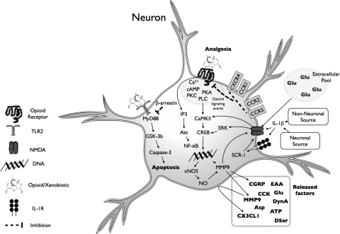
Vastly stimulated by the discovery of opioid receptors in the early 1970s, preclinical and clinical research was directed at the study of stereoselective neuronal actions of opioids, especially those played in their crucial analgesic role. However, during the past decade, a new appreciation of the non-neuronal actions of opioids has emerged from preclinical research, with specific appreciation for the nonclassic and nonstereoselective sites of action. Opioid activity at Toll-like receptors, newly recognized innate immune pattern recognition receptors, adds substantially to this unfolding story. It is now apparent from molecular and rodent data that these newly identified signaling events significantly modify the pharmacodynamics of opioids by eliciting proinflammatory reactivity from glia, the immunocompetent cells of the central nervous system. These central immune signaling events, including the release of cytokines and chemokines and the associated disruption of glutamate homeostasis, cause elevated neuronal excitability, which subsequently decreases opioid analgesic efficacy and leads to heightened pain states. This review will examine the current preclinical literature of opioid-induced central immune signaling mediated by classic and nonclassic opioid receptors. A unification of the preclinical pharmacology, neuroscience, and immunology of opioids now provides new insights into common mechanisms of chronic pain, naive tolerance, analgesic tolerance, opioid-induced hyperalgesia, and allodynia. Novel pharmacological targets for future drug development are discussed in the hope that disease-modifying chronic pain treatments arising from the appreciation of opioid-induced central immune signaling may become practical.


Figures

Fig. 1.
Fig. 1.
TLR4 signaling cascade and evidence for modulation by neuropathic pain and opioids. TLR4 signaling occurs via a cascade of events. The classic TLR4 ligand is Gram-negative bacterial LPS (hexagon), which is transported to the cell via LPS-binding protein and transfers LPS to CD14 on the cell membrane. This leads to intracellular activation of acid sphingomyelinase, which generates ceramide. Ceramide induces the generation of a lipid raft containing the coreceptor MD2, TLR4, and the 70- and 90-kDa heat shock proteins (HSP), among other elements. Ceramide also activates NADPH oxidase, which leads to peroxynitrite formation. CD14 transfers LPS to MD2, leading to both MD2-TLR4 heterodimerization and then homodimerization of MD2-TLR4 pairs. Recent evidence also suggests that opioids, such as morphine, can interact with MD2, causing a similar activation of a functional TLR4 signaling unit. Ensuing intracellular signaling occurs through toll-interleukin 1 receptor domain containing adaptor protein (TIRAP) to at least three parallel pathways: cell motility and cell survival/apoptosis occur through the phosphatidylinositol 3-kinase (PI3K)/Akt pathway, and proinflammatory products such as cytokines result from activation of the NF-κB and MAPK pathways. The gray boxes provide summaries of the converging evidence that neuropathic pain and opioids interact with the TLR4 signaling cascade. IRAK, IL-1-receptor-associated kinase; UEV1A, ubiquitin-conjugating enzyme E2 variant 1A; RIP2, receptor-interacting protein 2; TAK1, transforming growth factor-β-activated kinase-1; TAB3, transforming growth factor-β-activated kinase 1/MAP3K7 binding protein 3; MKK, MAP kinase kinase; IKK, IκB kinase complex; IκBα, inhibitor of nuclear factor-κB α; NEMO, NF-κB essential modifier. [Adapted from Watkins LR, Hutchinson MR, Rice KC, and Maier SF (2009) The “toll” of opioid-induced glial activation: improving the clinical efficacy of opioids by targeting glia. Trends Pharmacol Sci 30:581–591. Copyright © 2009 Elsevier Science. Used with permission.]
Fig. 2.
Fig. 2.
Stereoselective and nonstereoselective binding of a labeled opioid active (−)-ligand (filled reverse L) and the displacement of its binding from various types of sites in tissue by excess unlabeled opioid inactive (+)-ligand (open L) or excess unlabeled (−)-ligand (open reverse L). A, the binding of the labeled (−)-ligand that will participate in all the possible kinds of binding interactions. B, binding of excess unlabeled (+)-ligand and labeled (−)-ligand will result in blockade of labeled (−)-ligand entering the nonstereoselective but saturable binding sites. Thus, the difference of A minus B represents nonstereoselective but saturable binding. C, binding of excess unlabeled (−)-ligand will result in blockade of labeled (−)-ligand entering the saturable by nonstereoselective and stereoselective binding sites. Thus, the difference of B minus C represents saturable stereoselective binding. [Adapted from Goldstein A, Lowney LI, and Pal BK (1971) Stereospecific and nonspecific interactions of the morphine congener levorphanol in subcellular fractions of mouse brain. Proc Natl Acad Sci USA 68:1742–1747. Copyright © 1971 United States National Academy of Sciences. Used with permission.]
Fig. 3.
Fig. 3.
Consequences of opioid-induced central immune signaling in astrocytes. Opioid-induced central immune signaling can directly and indirectly modify astrocyte function. Opioid exposure is associated with increased expression of astrocyte chemokine receptors and activation markers, as well as the release of inflammatory mediators. It is noteworthy that there is also disruption of the astrocyte control of extracellular glutamate homeostasis owing to decreased glutamate uptake by astrocyte glutamate transporters, creating an environment of neuroexcitability (see section III.C for details).
Fig. 4.
Fig. 4.
Consequences of opioid-induced central immune signaling in microglia. Opioid-induced microglial responses occur via opioid engagement of classic opioid receptors expressed by microglia and via activation of TLR4-dependent signaling. The activation of these signaling events leads to the release of many inflammatory mediators, increased cellular motility and the up-regulation of cell surface receptors that enhance the microglial sentinel role. These classic and nonclassic actions of opioids at microglia result in a profound proinflammatory central immune signaling response that can influence neuronal and non-neuronal cells. If these microglial-dependent events occur in nociceptive control centers of the CNS, such as the dorsal spinal cord, a profound effect on the pharmacodynamics of the administered opioid can be expected (see section III.C for details).
Fig. 5.
Fig. 5.
Consequences of opioid-induced central immune signaling in neurons. Neuronal systems can be profoundly modulated and can substantially contribute to central immune signaling. Opioids can act to induce central immune signaling from neurons via direct activation of classic opioid receptors resulting in the release of a myriad of factors that can contribute to neuroexcitability. Moreover, opioids engage with neuronally expressed TLR2 in a nonclassic opioid fashion to induce neuronal apoptosis. Central immune signaling via chemokine receptor induced heterologous desensitization can inhibit classic neuronal opioid signaling. Central immune signals, such as interleukin-1β, from either neuronal or non-neuronal sources can directly and indirectly increase NMDA receptor activity, also contributing to neuroexcitation (see section III.C for details).
Fig. 6.
Fig. 6.
In silico MD2 docking of opioid ligands predicts in vitro TLR4 signaling activity. A, Hutchinson et al. (2010c) examined the in silico docking results of several opioid ligands to human MD2. These results were used to build an in silico docking to in vitro TLR4 signaling response prediction model (gray circles). This was subsequently tested on seven opioid and three nonopioid ligands, with the predicted (open squares) and actual (closed squares) in vitro scores displayed. The data indicate that the simulated interactions of these ligands with MD2 represented an excellent predictor of in vitro TLR4 signaling activity. It is noteworthy that in this case, not only the efficacy but also the direction of response (agonist versus antagonist) was modeled. B, Hutchinson et al. (2010c) subsequently compiled a modified complete 20-ligand in-silico-to-in-vitro model and retested the prediction ability of the model on the structurally disparate glial attenuator ibudilast. The data suggest that the actions of ibudilast in this human embryonic kidney 293-TLR4 in vitro model were due to ibudilast action at MD2. [Reproduced from Hutchinson MR, Zhang Y, Shridhar M, Evans JH, Buchanan MM, Zhao TX, Slivka PF, Coats BD, Rezvani N, Wieseler J, Hughes TS, Landgraf KE, Chan S, Fong S, Phipps S, Falke JJ, Leinwand LA, Maier SF, Yin H, Rice KC, and Watkins LR (2010c) Evidence that opioids may have toll-like receptor 4 and MD-2 effects. Brain Behav Immun 24:83–95. Copyright © 2010 Academic Press. Used with permission.]
Fig. 7.
Fig. 7.
Computer modeling of opioid interaction with the dimerized TLR4-MD2 complex. In silico docking of (−)-morphine (black stick and ball) to the 3D crystalline structure of the human MD2 (dark gray) and TLR4 (light) complex (Protein Data Bank ID 3FXI) demonstrates that the preferred docking location of (−)-morphine is to the LPS binding domain of MD2.

Similar articles

Cited by

References

    1. Abbadie C, Bhangoo S, De Koninck Y, Malcangio M, Melik-Parsadaniantz S, White FA. (2009) Chemokines and pain mechanisms. Brain Res Rev 60:125–134 - PMC - PubMed
    1. Abbadie C, Lindia JA, Cumiskey AM, Peterson LB, Mudgett JS, Bayne EK, DeMartino JA, MacIntyre DE, Forrest MJ. (2003) Impaired neuropathic pain responses in mice lacking the chemokine receptor CCR2. Proc Natl Acad Sci USA 100:7947–7952 - PMC - PubMed
    1. Adler MW, Rogers TJ. (2005) Are chemokines the third major system in the brain? J Leukoc Biol 78:1204–1209 - PubMed
    1. Agostini S, Eutamene H, Cartier C, Broccardo M, Improta G, Houdeau E, Petrella C, Ferrier L, Theodorou V, Bueno L. (2010) Evidence of central and peripheral sensitization in a rat model of narcotic bowel-like syndrome. Gastroenterology 139:553–563, 563.e551–555 - PubMed
    1. Alonso E, Garrido E, Díez-Fernández C, Pérez-García C, Herradón G, Ezquerra L, Deuel TF, Alguacil LF. (2007) Yohimbine prevents morphine-induced changes of glial fibrillary acidic protein in brainstem and alpha2-adrenoceptor gene expression in hippocampus. Neurosci Lett 412:163–167 - PubMed

Publication types

MeSH terms