. 2011;6(7):e21876.
doi: 10.1371/journal.pone.0021876. Epub 2011 Jul 5.

Loss of PTB or negative regulation of Notch mRNA reveals distinct zones of Notch and actin protein accumulation in Drosophila embryo

Affiliations

Loss of PTB or negative regulation of Notch mRNA reveals distinct zones of Notch and actin protein accumulation in Drosophila embryo

Cedric S Wesley et al. PLoS One. 2011.

Abstract

Polypyrimidine Tract Binding (PTB) protein is a regulator of mRNA processing and translation. Genetic screens and studies of wing and bristle development during the post-embryonic stages of Drosophila suggest that it is a negative regulator of the Notch pathway. How PTB regulates the Notch pathway is unknown. Our studies of Drosophila embryogenesis indicate that (1) the Notch mRNA is a potential target of PTB, (2) PTB and Notch functions in the dorso-lateral regions of the Drosophila embryo are linked to actin regulation but not their functions in the ventral region, and (3) the actin-related Notch activity in the dorso-lateral regions might require a Notch activity at or near the cell surface that is different from the nuclear Notch activity involved in cell fate specification in the ventral region. These data raise the possibility that the Drosophila embryo is divided into zones of different PTB and Notch activities based on whether or not they are linked to actin regulation. They also provide clues to the almost forgotten role of Notch in cell adhesion and reveal a role for the Notch pathway in cell fusions.


Conflict of interest statement

Competing Interests: The authors have declared that no competing interests exist.

Figures

Figure 1
Figure 1. Mutant heph embryos manifest suppression of the CNS development in the ventral region, a phenotype linked to excess canonical Notch signaling phenotype.
Expression of Hunchback, a well-known neuronal marker, was used to assess the CNS development. Hunchback expression in Notch null and gain-of-canonical Notch signaling (Nintra/NICD) embryos is shown in Figure S2. Suppressed neurogenesis phenotype became apparent as early as stage 9 (about four hours of embryogenesis) and was severe at stage 14–15 (about 14–16 hours of embryogenesis). Variation in the suppression of neurogenesis in heph embryos was observed, which is presumably due to variable maternal contribution. All embryos shown were from the same experiment and were processed identically.
Figure 2
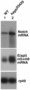
Figure 2. RNA of Notch and E(spl) genes m5 and m8, the latter targets of canonical Notch signaling, were over- expressed in heph03429 embryos.
RNAs used on northern blots were extracted from 3 to 6 hours old embryos that manifest peak canonical Notch signaling activity related to the CNS development. rp49 is the loading control.
Figure 3
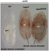
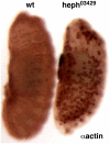
Figure 3. Dorsal closure process is blocked in mutant heph embryos.
While in the wild type embryo the dorsal region was closed and the extra-embryonic amnioserosa (AS) was completely eliminated by about 22 hours after egg laying (left), in heph03429 and heph2 embryos the dorsal closure process was incomplete and AS persisted even after 48 hours. Embryos were labeled using the actin antibody. All embryos/larva shown were from the same experiment and were processed identically.
Figure 4
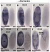
Figure 4. Pericardial cells are in excess and mislocalized in heph03429 embryos indicating that cardiogenesis is disrupted in these embryos.
AC. The same heph03429 and wild type embryos imaged from different perspectives. D. Pericardin was present in unusual places in heph03429 embryos, such as the head and the tail. Note the large amnioserosal region (as) that persisted in heph03429 embryos. E. The ventral epidermis was reasonably developed in heph03429 embryos indicating that these embryos produced sufficient amounts of canonical Notch signaling in the ventral region. F. Pericardial cells continued to be in excess and cardiogenesis blocked in heph03429 embryos at the time when embryogenesis and dorsal vessel formation was complete in wild type embryos (stage 17). All embryos shown were from the same experiment and were processed identically.
Figure 5
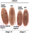
Figure 5. Notch protein level is different in different regions of heph03429 embryos.
A. Notch protein accumulated in the dorso-lateral region of stage 15 heph03429 embryos but not in the ventral region. B. Accumulation of Notch protein in the dorso-lateral regions, and depletion in the ventral region, of heph03429 embryos became apparent as early as Stage 9. All embryos shown were from the same experiment and were processed identically.
Figure 6
Figure 6. Actin level was high and disorganized in the dorso-lateral regions of heph03429 embryos.
A, B, and C: the same set of embryos shown from different perspectives. Absence of CNS labeling in the ventral region of the heph03429 embryo is due to the absence of neuronal cells. Embryos are at stage 15. All embryos shown were from the same experiment and were processed identically.
Figure 7
Figure 7. Apoptosis is not increased in heph03429 and Nnd1-dse mutant embryos.
Normal apoptosis observed in the CNS of wild type embryo was not observed in either heph03429 or Nnd1-dse embryos as the CNS development in the latter embryos was suppressed. Embryos are at stage 14. The brightness of yolk is due to autofluorescence.
Figure 8
Figure 8. Notch and actin accumulated near the cell surfaces in the dorso-lateral regions of heph03429 embryos.
A, B. Low magnification images from DeltaVision restoration microscopy showing that Notch and actin expression overlap. C, D. Two sets of high magnification images from DeltaVision microscopy showing that Notch and actin accumulate near the surfaces of fused cells and not in the nucleus (marked by DAPI).
Figure 9
Figure 9. Embryos of the gain-of-Function Notch allele, Nnd1-dse, manifest heph03429-like phenotypes.
A. Actin accumulated in the dorso-lateral regions of Nnd1-dse embryos. B. The CNS development is suppressed in the ventral region of Nnd1-dse embryos. C. Dorsal closure was blocked in Nnd1-dse embryos. Please compare Nnd1-dse images in this figure with those of heph03429 embryos in Figures 3 and 6. Embryos in A and B are at stage 14; the embryos in C are at stage 17 when embryogenesis ends in wild type embryos. All embryos were from the same experiment and were processed identically.
Figure 10
Figure 10. Nnd1-dse embryos show Notch protein accumulation pattern that is similar to that observed in heph03429 embryos.
While the Notch protein accumulated in the dorso-lateral region, it was depleted in the ventral region. Depletion was more clearly discernible in stage 9 embryos that have not yet produced the notochord (that tends to obscure the depletion in images of embryos at later stages). Please compare Nnd1-dse images in this figure with those of heph03429 embryos in Figure 5. All embryos shown were from the same experiment and were processed identically.
Figure 11
Figure 11. Reduction of Notch activity in heph03429 embryos suppresses actin accumulation in the dorso-lateral regions.
N55e11 is a null allele of Notch. heph03429/+; N55e11/Y and heph03429 ; N55e11/Y embryos were obtained from the same cross. +; N55e11/Y embryos are mostly composed of neuronal cells (with very few if any epidermal cells) and therefore show a higher level of actin compared to wild type embryos of similar stages. Suppression of actin level in the ventral region of heph03429 ; N55e11/Y was due to rescue of epidermal tissue (a consequence of increased canonical Notch signaling). Actin accumulation was not observed in the dorso-lateral regions of these embryos.
Figure 12
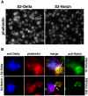
Figure 12. Enrichment of Notch receptor near the cell surface results in F-actin enrichment also near the cell surface.
A. F-actin levels in S2 cells expressing Notch (S2-Notch cells) and S2 cells expressing ligand Delta (S2-Delta cells) that were not mixed together. B. F-actin levels in S2-Notch and S2-Delta cells brought together by Notch and Delta binding. Notch and Delta binding results in Notch receptor enrichment (clustering) at contact points between the two cell types (Delta does not show this response [27]). Notch enrichment diminishes over time, possibly due to production of Nintra/NICD or reduction in Notch synthesis.
Figure 13
Figure 13. Phalloidin labeling indicates that actin that accumulates in the dorso-lateral regions of heph03429 and Nnd1-dse embryos is F-actin.
A. Phalloidin labeling pattern in heph03429 and Nnd1-dse embryos resembled the actin antibody staining pattern (shown in Figures 6 and 9). Mutant and yw embryos were placed next to one another in a multi-well plate and imaged together. Arrowheads point to the ‘cable-like’ structures in heph03429 and wild type embryos. B. F-actin accumulation in the dorso-lateral regions of heph03429 embryos presented ‘cable-like’ patterns. Embryos were mounted individually and imaged under identical settings.
Figure 14
Figure 14. Large cells are produced in live clone-8 (cl-8) and Delta (actDelta) cell aggregates.
A. Samples of cl-8 cells treated with S2 or actDelta cells at three million cells per milliliter density. B. Samples of cl-8 cells treated with S2 or actDelta cells at one million cells per milliliter density. Please note that Notch and Delta mediated cell aggregations are apparent only in cl-8+actDelta samples. Arrows point to large cells that formed within aggregates. Cell densities above one million per milliliter (required for the formation of cell aggregates) had no effect on the frequency of large cells.
Figure 15
Figure 15. Large cells in live cl-8+actDelta cell aggregates are products of cell fusions.
CellTracker Red labeled cl-8 cells and CellTracker Green labeled actDelta cells were used in the cell aggregation assay. These CellTracker dyes are confined to the treated cells or to their progeny upon division. The two rows represent samples from two independent assays. Arrowheads point to some fused cells. Note that other cells are either red or green and that there is no bleed-through between the red and the green channels.
Figure 16
Figure 16. The pattern of actin accumulation in heph03429 embryos resembles the pattern of normal actin levels in wild type embryos.
The image of actin labeled wild type and heph03429 embryos was contrast-enhanced to reveal the faint actin patterns in the wild type embryo.

Similar articles

Cited by

References

    1. Le Sommer C, Lesimple M, Mereau A, Menoret S, Allo M-R, et al. PTB regulates the processing of a 3′-terminal exon by repressing both splicing and polyadenylation. Mol. Cell. Biol. 2005;25:9595–9607. - PMC - PubMed
    1. Castelo-Branco P, Furger A, Wollerton M, Smith C, Moreira A, et al. Polypyrimidine tract binding protein modulates efficiency of polyadenylation. Mol. Cell. Biol. 2004;24:4174–4183. - PMC - PubMed
    1. Wilusz CJ, Wormington M, Peltz SW. The cap-to-tail guide to mRNA turnover. Nat. Rev. Mol. Cell Biol. 2001;2:237–246. - PubMed
    1. Wilusz CJ, Gao M, Jones CL, Wilusz J, Peltz SW. Poly(A)-binding proteins regulate both mRNA deadenylation and decapping in yeast cytoplasmic extracts. RNA. 2001;7:1416–1424. - PMC - PubMed
    1. Bentley DL. Rules of engagement: co-transcriptional recruitment of pre-mRNA processing factors. Curr. Opin. Cell Biol. 2005;17:251–256. - PubMed

Publication types

MeSH terms

LinkOut - more resources