Cell proliferation in human epiretinal membranes: characterization of cell types and correlation with disease condition and duration
- PMID: 21750605
- PMCID: PMC3133557
Cell proliferation in human epiretinal membranes: characterization of cell types and correlation with disease condition and duration
Abstract
Purpose: To quantify the extent of cellular proliferation and immunohistochemically characterize the proliferating cell types in epiretinal membranes (ERMS) from four different conditions: proliferative vitreoretinopathy (PVR), proliferative diabetic retinopathy, post-retinal detachment, and idiopathic ERM.
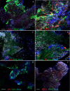
Methods: Forty-six ERMs were removed from patients undergoing vitrectomy and immediately fixed in paraformaldehyde. The membranes were processed whole and immunolabeled with either anti-MIB-1 or anti-SP6 to detect the K(i)-67 protein in proliferating cells, in combination with anti-glial fibrillary acidic protein or anti-vimentin to identify glia, anti-ezrin to identify retinal pigment epithelial cells, Ricinus communis to identify immune cells, and Hoechst to label nuclei. Digital images were collected using a laser scanning confocal microscope. The cell types were identified, their combined proliferative indices were tabulated as the average number of anti-K(i)-67-positive cells/mm(2) of tissue, and the number of dividing cells was related to the specific ocular condition and estimated disease duration.
Results: ERMs of all four types were shown to be highly cellular and contained proliferating cells identified as glia, retinal pigment epithelium, and of immune origin. In general, membranes identified as PVR had many more K(i)-67-positive cells in comparison to those in the other three categories, with the average number of K(i)-67-positive cells identified per mm(2) of tissue being 20.9 for proliferative diabetic retinopathy, 138.3 for PVR, 12.2 for post-retinal detachment, and 19.3 for idiopathic ERM. While all membrane types had dividing cells, their number was a relatively small fraction of the total number of cells present.
Conclusions: The four ERM types studied demonstrated different cell types actively dividing at the time of removal, confirming that proliferation is a common event and does continue over many months. The low number of dividing cells at the time of removal in comparison to the total number of cells present, however, is an indicator that proliferation alone may not be responsible for the problems observed with the ERMs. Treatment strategies may need to take into consideration the timing of drug administration, as well as the contractile and possibly the inflammatory characteristics of the membranes to prevent the ensuing effects on the retina.
© 2011 Molecular Vision
Figures


Similar articles
-
Upregulation of RAGE and its ligands in proliferative retinal disease.Exp Eye Res. 2006 May;82(5):807-15. doi: 10.1016/j.exer.2005.09.022. Epub 2005 Dec 20. Exp Eye Res. 2006. PMID: 16364297
-
Apoptosis and cell proliferation in proliferative retinal disorders: PCNA, Ki-67, caspase-3, and PARP expression.Curr Eye Res. 2005 May;30(5):395-403. doi: 10.1080/02713680590956306. Curr Eye Res. 2005. PMID: 16020270
-
Pathological changes in the vitreoretinal junction 1: epiretinal membrane formation.Eye (Lond). 2008 Oct;22(10):1310-7. doi: 10.1038/eye.2008.36. Epub 2008 Mar 14. Eye (Lond). 2008. PMID: 18344963
-
The role of cytokines and trophic factors in epiretinal membranes: involvement of signal transduction in glial cells.Prog Retin Eye Res. 2006 Mar;25(2):149-64. doi: 10.1016/j.preteyeres.2005.09.001. Epub 2005 Dec 27. Prog Retin Eye Res. 2006. PMID: 16377232 Review.
-
[Proliferative vitreoretinopathy (PVR) minimal: same, same but different. Characteristics and surgical treatment of PVR-associated macular pucker].Ophthalmologe. 2021 Jan;118(1):24-29. doi: 10.1007/s00347-020-01292-2. Ophthalmologe. 2021. PMID: 33336260 Review. German.
Cited by
-
Heat Shock Protein 90 Involvement in the Development of Idiopathic Epiretinal Membranes.Invest Ophthalmol Vis Sci. 2020 Jul 1;61(8):34. doi: 10.1167/iovs.61.8.34. Invest Ophthalmol Vis Sci. 2020. PMID: 32716502 Free PMC article.
-
Comparison of surgical outcomes after removal of epiretinal membrane associated with retinal break and idiopathic epiretinal membrane.Graefes Arch Clin Exp Ophthalmol. 2022 Jul;260(7):2121-2128. doi: 10.1007/s00417-021-05550-0. Epub 2022 Jan 14. Graefes Arch Clin Exp Ophthalmol. 2022. PMID: 35029729
-
miRNA-451a regulates RPE function through promoting mitochondrial function in proliferative diabetic retinopathy.Am J Physiol Endocrinol Metab. 2019 Mar 1;316(3):E443-E452. doi: 10.1152/ajpendo.00360.2018. Epub 2018 Dec 21. Am J Physiol Endocrinol Metab. 2019. PMID: 30576241 Free PMC article.
-
Microscopic inner retinal hyper-reflective phenotypes in retinal and neurologic disease.Invest Ophthalmol Vis Sci. 2014 Jun 3;55(7):4015-29. doi: 10.1167/iovs.14-14668. Invest Ophthalmol Vis Sci. 2014. PMID: 24894394 Free PMC article.
-
Immunohistochemical, functional, and anatomical evaluation of patients with idiopathic epiretinal membrane.Graefes Arch Clin Exp Ophthalmol. 2024 May;262(5):1443-1453. doi: 10.1007/s00417-023-06366-w. Epub 2024 Jan 10. Graefes Arch Clin Exp Ophthalmol. 2024. PMID: 38197992 Free PMC article.
References
-
- McCarty DJ, Mukesh BN, Chikani V, Wang JJ, Mitchell P, Taylor HR, McCarty CA. Prevalence and associations of epiretinal membranes in the visual impairment project. Am J Ophthalmol. 2005;140:288–94. - PubMed
-
- Uemura A, Ideta H, Nagasaki H, Morita H. Ito k. Macular pucker after retinal detachment surgery. Ophthalmic Surg. 1992;23:116–9. - PubMed
-
- Gilbert C, Hiscott P, Unger W, Grierson I, McLeod D. Inflammation and the formation of epiretinal membranes. Eye (Lond) 1988;2(Suppl):S140–56. - PubMed
Publication types
MeSH terms
Substances
Grants and funding
LinkOut - more resources
Full Text Sources
Medical
Research Materials
