. 2011 Jul 14;71(1):103-16.
doi: 10.1016/j.neuron.2011.05.034.

The immunoglobulin super family protein RIG-3 prevents synaptic potentiation and regulates Wnt signaling

Affiliations

The immunoglobulin super family protein RIG-3 prevents synaptic potentiation and regulates Wnt signaling

Kavita Babu et al. Neuron. .

Abstract

Cell surface Ig superfamily proteins (IgSF) have been implicated in several aspects of neuron development and function. Here, we describe the function of a Caenorhabditis elegans IgSF protein, RIG-3. Mutants lacking RIG-3 have an exaggerated paralytic response to a cholinesterase inhibitor, aldicarb. Although RIG-3 is expressed in motor neurons, heightened drug responsiveness was caused by an aldicarb-induced increase in muscle ACR-16 acetylcholine receptor (AChR) abundance, and a corresponding potentiation of postsynaptic responses at neuromuscular junctions. Mutants lacking RIG-3 also had defects in the anteroposterior polarity of the ALM mechanosensory neurons. The effects of RIG-3 on synaptic transmission and ALM polarity were both mediated by changes in Wnt signaling, and in particular by inhibiting CAM-1, a Ror-type receptor tyrosine kinase that binds Wnt ligands. These results identify RIG-3 as a regulator of Wnt signaling, and suggest that RIG-3 has an anti-plasticity function that prevents activity-induced changes in postsynaptic receptor fields.


Figures

Figure 1
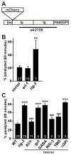
Figure 1. Inactivation of rig-3 causes hypersensitivity to aldicarb
(A) A schematic of the RIG-3 protein is shown, indicating the signal sequence (ss), Ig, FNIII, and GPI- anchoring domains, and the site utilized for mCherry tagging. The domains deleted in rig-3(ok2156) mutants are indicated by the bar. (B) Aldicarb-induced paralysis is compared following RNAi treatments with rig-3 and two negative controls, empty vector (vector) and eri-1. The number of replicate experiments for each RNAi treatment (>20 animals/replicate) is indicated. (C) Aldicarb-induced paralysis is shown for the indicated genotypes. RIG-3 transgenes are as follows: ACh neurons (unc-17 promoter), gut (vha-6 promoter), GABA (unc-25 promoter), RIG-3 (mCherry-tagged rig-3 genomic construct), TMD (membrane-anchored RIG-3 expressed in ACh neurons), ΔGPI (constitutively secreted RIG-3 expressed in ACh neurons). The number of trials (~20 animals/trial) is indicated for each genotype. Values that differ significantly from wild type (***, p < 0.001) and from rig-3 mutants (###, p < 0.001) are indicated. Error bars indicate SEM.
Figure 2
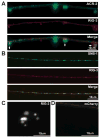
Figure 2. RIG-3 is expressed in ACh neurons
(A) The mCherry-tagged rig-3 genomic construct is expressed in cholinergic motor neurons, which are identified by expression of the acr-2::gfp reporter. Arrows indicate the cholinergic neurons that express RIG-3. (B) Distribution of mCherry::RIG-3 (expressed with the rig-3 promoter) and GFP::SNB-1 (expressed in DA neurons with the unc-129 promoter) are compared in dorsal cord axons. (C) mCherry::RIG-3 fluorescence in coelomocytes is shown. (D) Soluble mCherry (expressed in cholinergic neurons) did not label coelomocytes.
Figure 3
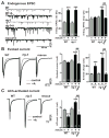
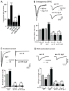
Figure 3. Aldicarb treated rig-3 mutants show an increased post-synaptic responses
Endogenous EPSCs (A), stimulus-evoked EPSCs (B), and ACh-evoked currents (C) were recorded from body wall muscle of adult worms of the indicated genotypes, with (grey) and without (black) a 60 minute aldicarb treatment. Representative traces of endogenous EPSCs, averaged traces of stimulus-evoked responses and ACh-evoked responses, and summary data for all three are shown. Rescue (resc) refers to transgenic animals expressing RIG-3 in cholinergic neurons (with the unc-17 promoter). The number of animals analyzed is indicated for each genotype. Values that differ significantly from untreated wild type (***, p <0.001) and from untreated rig-3 mutants (###, p <0.001) are indicated. Error bars indicate SEM.
Figure 4
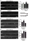
Figure 4. Aldicarb increases ACR-16 synaptic abundance in rig-3 mutants
(A) Representative images of dorsal cord ACR-16::GFP fluorescence in aldicarb treated animals is shown for the indicated genotypes. Summary data (Right) for dorsal ACR-16 puncta fluorescence is shown for control and aldicarb treated animals of the indicated genotypes. ACh rescue refers to rig-3 mutants containing a transgene expressing RIG-3 in all cholinergic neurons (using the unc-17 promoter). (B) A schematic illustrating the morphology of a cholinergic DA motor neuron (right) and representative images of RIG-3 fluorescence in the dorsal and ventral cord processes of DA neurons are shown (left). mCherry-tagged RIG-3 was expressed in DA neurons (using the unc-129 promoter). (C-D) Representative images and summary data are shown for dorsal (C) and ventral (D) ACR-16 puncta fluorescence in the indicated genotypes. DA rescue refers to rig-3 mutants containing a transgene expressing RIG-3 in DA neurons (using the unc-129 promoter). The number of animals analyzed is indicated for each genotype. A GFP-tagged ACR-16 construct was expressed in body muscles (with the myo-3 promoter). ACR-16 puncta in the dorsal and ventral cords correspond to post-synaptic receptors at dorsal and ventral NMJs, respectively. Values that differ significantly from aldicarb treated wild type controls are indicated (**, p < 0.01; ***, p < 0.001). Error bars indicate SEM.
Figure 5
Figure 5. ACR-16 is required for aldicarb-induced potentiation of post-synaptic responses in rig-3 mutants
(A) Summary data is shown for aldicarb-induced paralysis in the indicated genotypes. Transgenic animals over-expressing ACR-16 (ACR-16 OE) are indicated. The number of trials (~20 animals/trial) is indicated for each genotype. (B-C) Traces and summary data for endogenous EPSCs (B), evoked EPSCs (C), and ACh-activated currents (D) are shown for control and aldicarb treated animals of the indicated genotypes. For endogenous EPSCs, representative traces are shown. For evoked EPSCs and ACh-activated EPSCs, averaged traces are shown. The number of animals analyzed is indicated for each genotype. Values that differ significantly from wild type controls are indicated (***, p < 0.001; *, p <0.05). Error bars indicate SEM.
Figure 6
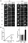
Figure 6. RIG-3 regulates ACR-16 delivery to post-synaptic elements
Representative images (A) and summary data (B) are shown for FRAP of ACR-16::GFP at dorsal cord NMJs of control and aldicarb treated animals. At time 0, ACR-16 fluorescence in a 2 μm box encompassing a single punctum was photobleached. ACR-16 fluorescence was subsequently measured at the photobleached and a neighboring control punctum. The fractional recovery of fluorescence 45 minutes after photobleaching is shown (B). The fluorescence of control unbleached puncta did not change significantly after 45 minutes of imaging (data not shown). The number of animals analyzed is indicated for each genotype. Values that differ significantly from wild type controls are indicated (**, p <0.01). Error bars indicate SEM.
Figure 7
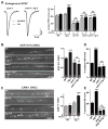
Figure 7. CAM-1 Wnt receptors are required for RIG-3’s effects on ACR-16
(A) Averaged traces and summary data are shown for endogenous EPSCs of control and aldicarb treated animals. The number of animals analyzed is indicated for each genotype. (B-C) Representative images and summary data are shown for dorsal cord ACR-16::GFP (B) and ventral cord CAM-1::GFP fluorescence (C) in control and aldicarb treated animals. The number of animals analyzed is indicated for each genotype. GFP-tagged ACR-16 and CAM-1 were expressed in body muscles (using the myo-3 promoter). (D-E) Summary data is shown for aldicarb-induced paralysis in the indicated genotypes. The number of trials (~20 animals/trial) is indicated for each genotype. In panel E, all strains (including the WT control) contain the zdIs5 transgene, which expresses GFP in the touch neurons. Values that differ significantly from wild type controls are indicated (***, p < 0.001; *, p <0.05). Error bars indicate SEM.
Figure 8
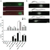
Figure 8. RIG-3 antagonizes the effects of Wnt on ALM polarity
(A) Expression of the mCherry-tagged rig-3 genomic construct is shown in ALM neurons. The ALM neurons were visualized with the zdIs5 transgene, which expresses GFP in the touch neurons (with the mec-4 promoter). (B) Representative images and schematic drawings are shown illustrating wild type, bipolar (less severe), and reversed (more severe) ALM defects. (C) Summary data for ALM polarity defects are shown for the indicated genotypes. The number of animals analyzed is indicated for each genotype. All strains contain the zdIs5 transgene, to allow visualization of ALM neurons. Values that differ significantly from wild type controls are indicated (***, p< 0.001; **, p< 0.01).

Similar articles

Cited by

References

    1. Ataman B, Ashley J, Gorczyca M, Ramachandran P, Fouquet W, Sigrist SJ, Budnik V. Rapid activity-dependent modifications in synaptic structure and function require bidirectional Wnt signaling. Neuron. 2008;57:705–718. - PMC - PubMed
    1. Barrow AD, Trowsdale J. The extended human leukocyte receptor complex: diverse ways of modulating immune responses. Immunol Rev. 2008;224:98–123. - PubMed
    1. Biederer T, Sara Y, Mozhayeva M, Atasoy D, Liu X, Kavalali ET, Sudhof TC. SynCAM, a synaptic adhesion molecule that drives synapse assembly. Science. 2002;297:1525–1531. - PubMed
    1. Brenner S. The genetics of Caenorhabditis elegans. Genetics. 1974;77:71–94. - PMC - PubMed
    1. Budnik V, Salinas PC. Wnt signaling during synaptic development and plasticity. Curr Opin Neurobiol. 2011;21:151–159. - PMC - PubMed

Publication types

MeSH terms

LinkOut - more resources