. 2011 Sep;85(18):9599-613.
doi: 10.1128/JVI.00845-11. Epub 2011 Jul 6.

Herpes simplex virus 1 protein kinase Us3 and major tegument protein UL47 reciprocally regulate their subcellular localization in infected cells

Affiliations

Herpes simplex virus 1 protein kinase Us3 and major tegument protein UL47 reciprocally regulate their subcellular localization in infected cells

Akihisa Kato et al. J Virol. 2011 Sep.

Abstract

Us3 is a serine-threonine protein kinase encoded by herpes simplex virus 1 (HSV-1). We have identified UL47, a major virion protein, as a novel physiological substrate of Us3. In vitro kinase assays and systematic analysis of mutations at putative Us3 phosphorylation sites near the nuclear localization signal of UL47 showed that serine at residue 77 (Ser-77) was required for Us3 phosphorylation of UL47. Replacement of UL47 Ser-77 by alanine produced aberrant accumulation of UL47 at the nuclear rim and impaired the nuclear localization of UL47 in a significant fraction of infected cells. The same defect in UL47 localization was produced by an amino acid substitution in Us3 that inactivated its protein kinase activity. In contrast, a phosphomimetic mutation at UL47 Ser-77 restored wild-type nuclear localization. The UL47 S77A mutation also reduced viral replication in the mouse cornea and the development of herpes stromal keratitis in mice. In addition, UL47 formed a stable complex with Us3 in infected cells, and nuclear localization of Us3 was significantly impaired in the absence of UL47. These results suggested that Us3 phosphorylation of UL47 Ser-77 promoted the nuclear localization of UL47 in cell cultures and played a critical role in viral replication and pathogenesis in vivo. Furthermore, UL47 appeared to be required for efficient nuclear localization of Us3 in infected cells. Therefore, Us3 protein kinase and its substrate UL47 demonstrated a unique regulatory feature in that they reciprocally regulated their subcellular localization in infected cells.


Figures
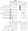
Fig. 1.
Fig. 1.
Schematic diagram of the genomes of wild-type YK304 and YK311 viruses and the relevant domains of the recombinant viruses used in this study. Line 1, linear representation of the YK304 genome carrying a bacmid (BAC) in the intergenic region between UL3 and UL4. Lines 2 and 14, domains encoding the UL46 to UL48 and Us2 to Us5 open reading frames. Lines 3 and 15, domains of the UL47 and Us3 genes. Lines 4 to 12, schematic diagrams of the recombinant viruses carrying the BAC that were used in this study. Line 13, linear representation of the genome of YK311, in which the BAC sequence was excised from the YK304 genome. Lines 16 to 19, schematic diagrams of the recombinant viruses from which the BAC sequence was excised that were used in this study.
Fig. 2.
Fig. 2.
Growth curves of recombinant viruses. Vero cells were infected at an MOI of 5 with wild-type HSV-1(F) (A, D, and E), YK524 (mRFP1-UL47) (A, B, and C), YK527 (mRFP1-UL47/Us3K220M) (B), YK528 (mRFP1-UL47/Us3K220M-repair) (B), YK532 (mRFP1-UL47S77A) (C), YK533 (mRFP1-UL47S77A-repair) (C), YK534 (mRFP1-UL47S77D) (C), YK547 (UL47S77A/ΔBAC) (D), YK548 (UL47S77A-repair/ΔBAC) (D), YK545 (ΔUL47/ΔBAC) (E), or YK546 (ΔUL47-repair/ΔBAC) (E). Total virus from the cell culture supernatants and the infected cells was harvested at the indicated times and was assayed on Vero cells.
Fig. 3.
Fig. 3.
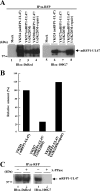
(A) Immunoblots (IB) of electrophoretically separated mRFP1-UL47 immunoprecipitates (IP) from Vero cells that were either mock infected (lanes 1 and 5) or infected with YK524 (mRFP1-UL47) (lanes 2 and 6), YK527 (mRFP1-UL47/Us3K220M) (lanes 3 and 7), or YK528 (mRFP1-UL47/Us3K220M-repair) (lanes 4 and 8) at an MOI of 3, harvested at 24 h postinfection, immunoprecipitated with an anti-RFP (α-RFP) antibody, and analyzed by immunoblotting with an anti-DsRed antibody (left) or an anti-phospho-PKA substrate antibody (100G7) (right). The position of a molecular mass marker is given on the left. (B) Quantitation of phosphorylated mRFP1-UL47, determined from the anti-phospho-PKA substrate antibody (100G7) immunoblot in panel A, relative to the amount of total mRFP1-UL47, determined from the anti-DsRed antibody immunoblot in panel A. The data were normalized to the relative amount in YK524-infected cells. (C) RFP1 immunoprecipitates of YK524-infected cells prepared as for panel A, lane 2, were either mock treated (lanes 1) or treated with λ-PPase (lanes 2) and were then analyzed by immunoblotting with an anti-DsRed antibody (left) or an anti-phospho-PKA substrate antibody (right). A molecular mass marker is indicated on the left.
Fig. 4.
Fig. 4.
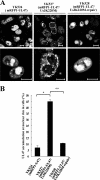
(A) Digital confocal microscope images showing the localization of mRFP1-UL47 proteins in infected Vero cells. Vero cells were infected with either YK524 (mRFP1-UL47), YK527 (mRFP1-UL47/Us3K220M), or YK528 (mRFP1-UL47/Us3K220M-repair) at an MOI of 1 and were examined by confocal microscopy at 18 h postinfection. Bars, 10 μm. (B) Quantitation of infected cells exhibiting aberrant accumulation of mRFP1-UL47 at the nuclear rim. Infected Vero cells were examined by confocal microscopy as described for panel A, and the percentages of cells showing aberrant accumulation of mRFP1-UL47 at the nuclear rim were determined for 200-cell samples, each infected with one of the recombinant viruses. Data are means and standard errors from three independent experiments. Asterisks indicate significant differences (∗, P = 3.5 × 10⁻⁵; ∗∗, P = 7.8 × 10⁻⁶) by a two-tailed Student t test.
Fig. 5.
Fig. 5.
(A) Schematic diagram of UL47. Line 1, structure of the UL47 open reading frame. The shaded areas represent arginine-rich motifs in the amino-terminal domain of the protein. Line 2, amino-terminal domain of UL47 containing residues 15 to 120, which was used in these studies to generate the MBP-UL47-P1 fusion protein. Lines 3 to 5, amino acid sequences of UL47 residues 48 to 91 in MBP-UL47-P1 and its S77A and S88A mutants. Two sites with the consensus sequence for phosphorylation by Us3 are underlined, and the UL47 NLS is boxed. (B) Purified MBP-UL47-P1 (lanes 1 and 2) and MBP-LacZ (lanes 3 and 4) were incubated in a kinase buffer containing [γ-32P]ATP and either purified GST-Us3 (lanes 1 and 3) or GST-Us3K220M (lanes 2 and 4) for 30 min, separated on a denaturing gel, and stained with CBB. A molecular mass marker is indicated on the left. (C) Autoradiograph of the gel shown in panel B. (D) Purified MBP-UL47-P1 was incubated in a kinase buffer containing [γ-32P]ATP and purified GST-Us3 for 30 min and was then either mock treated (lane 1) or treated with λ-PPase (lane 2), separated on a denaturing gel, and stained with CBB. A molecular mass marker is indicated on the left. (E) Autoradiograph of the gel shown in panel D. (F) Purified MBP-UL47-P1 (lanes 1 and 2), MBP-UL47-P1 S77A (lanes 3 and 4), and MBP-UL47-P1 S88A (lanes 5 and 6) were incubated in a kinase buffer containing [γ-32P]ATP and either purified GST-Us3 (lanes 1, 3, and 5) or GST-Us3K220M (lanes 2, 4, and 6) for 30 min, separated on a denaturing gel, and stained with CBB. A molecular mass marker is indicated on the left. (G) Autoradiograph of the gel shown in panel F.
Fig. 6.
Fig. 6.
Digital confocal microscope images showing the localization of mRFP1-UL47 proteins in infected Vero cells. Vero cells were infected with either YK524 (mRFP1-UL47), YK532 (mRFP1-UL47S77A), YK533 (mRFP1-UL47S77A-repair), or YK534 (mRFP1-UL47S77D) at an MOI of 1, and the cells were examined by confocal microscopy at 18 h postinfection. Bars, 10 μm. (B) Quantitation of infected cells exhibiting aberrant accumulation of mRFP1-UL47 and its mutants at the nuclear rim. Infected Vero cells were examined by confocal microscopy as described for panel A, and the percentages of cells showing aberrant accumulation of mRFP1-UL47 at the nuclear rim were determined for 200-cell samples, each infected with one of the recombinant viruses. Data are means and standard errors from three independent experiments. Asterisks indicate significant differences (∗, P = 1.8 × 10⁻⁴; ∗∗, P = 3.7 × 10⁻⁴; ∗∗∗, P = 4.4 × 10⁻⁵) by a two-tailed Student t test.
Fig. 7.
Fig. 7.
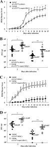
Effects of the UL47 S77A substitution and the UL47-null mutation on HSK and virus growth in the tear films in mice. Twenty (A and B) or 10 (C and D) 5-week-old female ICR mice were infected with 1 × 10⁶ PFU YK547 (UL47S77A/ΔBAC) or YK548 (UL47S77A-repair/ΔBAC) (A and B) or with 1 × 10⁶ PFU YK545 (ΔUL47/ΔBAC) or YK546 (ΔUL47-repair/ΔBAC) (C and D) per eye by corneal scarification. (A and C) Mice were scored for HSK every other day for 14 days. Each data point is the mean and standard error for the observations. Asterisks indicate P values of 6.2 × 10⁻⁴ (A) and 3.1 × 10⁻³ (C) by a two-tailed Student t test. (B and D) Viral titers in the tear films of infected mice at 2 and 5 days postinfection were determined by standard plaque assays. Each data point represents the titer in the tear film of one mouse. The average titer for each group is marked by a horizontal bar and is given below the bar. Asterisks indicate significant differences by a two-tailed Student t test. (B) ∗, P = 2.8 × 10⁻⁴; ∗∗, P = 2.5 × 10⁻². (D) ∗, P = 4.7 × 10⁻³; ∗∗, P = 1.7 × 10⁻².
Fig. 8.
Fig. 8.
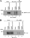
Interaction of Us3 with UL47. (A) Vero cells that were either mock infected (lanes 1 and 3) or infected with YK524 (mRFP1-UL47) (lanes 2, 4, and 5) at an MOI of 3 for 24 h were harvested, immunoprecipitated (IP) with anti-Us3 (α-Us3) (lanes 3 and 4) or with normal rabbit serum (lane 5), and analyzed by immunoblotting (IB) with an anti-DsRed antibody. One percent of the amount of Vero whole-cell extract (WCE) used in the reaction mixture for lane 3 was loaded in lane 1, and 1% of the WCE used for lanes 4 and 5 was loaded in lane 2. A molecular mass marker is indicated on the left. (B) Vero cells that were either mock infected (lanes 1 and 4) or infected with YK524 (lanes 2 and 5) or YK615 (mCherry-UL37) (lanes 3 and 6) at an MOI of 3 for 24 h were harvested, immunoprecipitated with anti-RFP, and analyzed by immunoblotting with an anti-Us3 antibody. One percent of the amount of Vero WCE used in the reaction mixtures for lanes 4, 5, and 6 was loaded in lanes 1, 2 and 3, respectively.
Fig. 9.
Fig. 9.
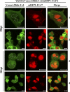
Digital confocal microscope images showing the localization of the mRFP1-UL47 and VenusA206K-Us3 proteins in infected Vero cells. Vero cells were infected with YK523 (VenusA206K-Us3/mRFP1-UL47) at an MOI of 1, and cells were examined by confocal microscopy at 18 and 24 h postinfection (hpi). Nuclear Us3 in the VenusA206K-Us3 and merge columns is circled. Bars, 10 μm.
Fig. 10.
Fig. 10.
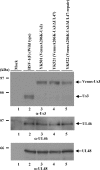
Effect of the UL47-null mutation on the expression of the neighboring UL46 and UL48 genes. Vero cells infected with either wild-type HSV-1(F), YK501 (VenusA206K-Us3), YK521 (VenusA206K-Us3/ΔUL47), or YK522 (VenusA206K-Us3/ΔUL47-repair) at an MOI of 1 for 24 h were harvested and were analyzed by immunoblotting with anti-Us3 (α-Us3), anti-UL46, and anti-UL48 antibodies. The positions of molecular mass markers are shown on the left.
Fig. 11.
Fig. 11.
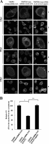
(A) Digital confocal microscope images showing the localization of VenusA206K-Us3 proteins in infected Vero cells. Vero cells were infected with either YK501 (VenusA206K-Us3), YK521 (VenusA206K-Us3/ΔUL47), or YK522 (VenusA206K-Us3/ΔUL47-repair) at an MOI of 1, and the infected cells were examined by confocal microscopy at 12, 18, and 24 h postinfection (hpi). Nuclear Us3 is circled in rows 3 and 5. Bars, 10 μm. (B) Quantitation of the fluorescence intensity (FI) of VenusA206K-Us3 in the nuclei relative to that in the cytoplasm of infected Vero cells at 24 h postinfection. Infected Vero cells were examined by confocal microscopy as described for panel A. Images of 10 infected cells were randomly selected for each virus infection. Three circular areas of the same size were randomly selected in the cytoplasm and in the nucleus of each of the 10 infected cells, and the FI of each circular area was analyzed. For each virus infection, the relative FI was calculated as the mean FI of the 30 circular areas in the nuclei divided by the mean FI of the 30 circular areas in the cytoplasm. The data are means and standard errors from three independent experiments. Asterisks indicate significant differences (*, P = 1.6 × 10⁻⁸; **, P = 3.5 × 10⁻⁹) by a two-tailed Student t test. The mean relative FI for YK501-infected cells was normalized to 100%.

Similar articles

Cited by

References

    1. Asai R., Kato A., Kawaguchi Y. 2009. Epstein-Barr virus protein kinase BGLF4 interacts with viral transactivator BZLF1 and regulates its transactivation activity. J. Gen. Virol. 90:1575–1581 - PubMed
    1. Benetti L., Roizman B. 2004. Herpes simplex virus protein kinase US3 activates and functionally overlaps protein kinase A to block apoptosis. Proc. Natl. Acad. Sci. U. S. A. 101:9411–9416 - PMC - PubMed
    1. Campbell R. E., et al. 2002. A monomeric red fluorescent protein. Proc. Natl. Acad. Sci. U. S. A. 99:7877–7882 - PMC - PubMed
    1. Chuluunbaatar U., et al. 2010. Constitutive mTORC1 activation by a herpesvirus Akt surrogate stimulates mRNA translation and viral replication. Genes Dev. 24:2627–2639 - PMC - PubMed
    1. Dobrikova E., Shveygert M., Walters R., Gromeier M. 2010. Herpes simplex virus proteins ICP27 and UL47 associate with polyadenylate-binding protein and control its subcellular distribution. J. Virol. 84:270–279 - PMC - PubMed

Publication types

MeSH terms

Substances

LinkOut - more resources