. 2012 May;227(5):1883-90.
doi: 10.1002/jcp.22916.

Afadin controls p120-catenin-ZO-1 interactions leading to endothelial barrier enhancement by oxidized phospholipids

Affiliations

Afadin controls p120-catenin-ZO-1 interactions leading to endothelial barrier enhancement by oxidized phospholipids

Anna A Birukova et al. J Cell Physiol. 2012 May.

Abstract

Afadin is a novel regulator of epithelial cell junctions assembly. However, its role in the formation of endothelial cell junctions and the regulation of vascular permeability remains obscure. We previously described protective effects of oxidized 1-palmitoyl-2-arachidonoyl-sn-glycero-3-phosphorylcholine (OxPAPC) in the in vitro and in vivo models of lung endothelial barrier dysfunction and acute lung injury, which were mediated by Rac GTPase. This study examined a role of afadin in the OxPAPC-induced enhancement of interactions between adherens junctions and tight junctions as a novel mechanism of endothelial cell (EC) barrier preservation. OxPAPC induced Rap1-dependent afadin accumulation at the cell periphery and Rap1-dependent afadin interaction with adherens junction and tight junction proteins p120-catenin and ZO-1, respectively. Afadin knockdown using siRNA or ectopic expression of afadin mutant lacking Rap1 GTPase binding domain suppressed OxPAPC-induced EC barrier enhancement and abolished barrier protective effects of OxPAPC against thrombin-induced EC permeability. Afadin knockdown also abolished protective effects of OxPAPC against ventilator-induced lung injury in vivo. These results demonstrate for the first time a critical role of afadin in the regulation of vascular barrier function in vitro and in vivo via coordination of adherens junction-tight junction interactions.


Figures

Figure 1
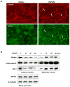
Figure 1. Effect of OxPAPC on cytoskeletal remodeling, and Rap1-dependence of OxPAPC-induced afadin translocation
A - Endothelial cells grown on glass coverslips were stimulated with OxPAPC (10 μg/ml) for 30 min followed by double immunofluorescence staining with Texas Red phalloidin to detect actin filaments and afadin antibody. Inset: control staining of cells without afadin antibody. Arrows depict peripheral actin rims (upper panel) and areas of afadin accumulation (lower panel). B - HPAEC were stimulated with OxPAPC (10 μg/ml) or vehicle for various periods of time. Membrane and cytosolic fractions were isolated as described in Methods section. The content of afadin, p120-catenin, and ZO-1 was determined by western blot analysis of cytosolic and membrane fractions with specific antibodies. Equal protein content in whole cell lysates was confirmed by detection of afadin and β-tubulin in control and OxPAPC-treated samples. Shown are representative results of three independent experiments.
Figure 2
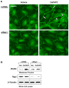
Figure 2. Effect of Rap1 knockdown on OxPAPC-induced afadin translocation
HPAEC were transfected with non-specific RNA or Rap1-specific siRNA duplexes followed by treatment with OxPAPC (10 μg/ml) or vehicle for 30 min. A - Endothelial cells grown on glass coverslips were subjected to immunofluorescence staining for afadin. Areas of afadin accumulation are marked by arrows. B - The membrane fraction was isolated, and afadin content was determined by western blotting with specific antibodies. Rap1 depletion was confirmed by western blot analysis of whole cell lysates from control (non-specific RNA) and siRap1-treated EC. Equal protein content in whole cell lysates was confirmed by detection of β-tubulin in control and OxPAPC-treated samples. Shown are representative results of three independent experiments.
Figure 3
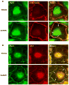
Figure 3. Effect of OxPAPC on adherens junction and tight junction remodeling
Endothelial cells grown on glass coverslips were stimulated with OxPAPC (10 μg/ml, 30 min) followed by double immunofluorescence staining for: A - afadin (green) and p120-catein (red) and B - afadin (green) and ZO-1 (red). Merged images depict areas of afadin/ZO-1 co-localization which appear in yellow and marked by arrows. Shown are representative results of three independent experiments.
Figure 4
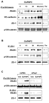
Figure 4. OxPAPC induces afadin interaction with adherens junction and tight junction proteins
A and B- Pulmonary EC were stimulated with OxPAPC (10 μg/ml), thrombin (0.1 U/ml, 10 min), or vehicle for various time periods followed by immunoprecipitation of: A - p120-catenin and B - or ZO-1. Afadin, VE-cadherin, p-120 catenin, and ZO-1 content in the immunoprecipitates was detected using specific antibodies. Equal protein loading was confirmed by membrane reprobing with p120-catenin (A) or ZO-1 (B) antibodies. Results are representative of four independent experiments. C - HPAEC transfected with Rap1-specific siRNA or non-specific RNA were treated with OxPAPC (10 μg/ml) or vehicle for 30 min followed by immunoprecipitation of p-120 catenin or ZO-1. Afadin content in the immunoprecipitates was determined using specific antibodies. Results are representative of three independent experiments.
Figure 5
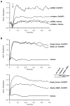
Figure 5. Involvement of afadin in the OxPAPC-induced EC barrier enhancement
A - Pulmonary EC were transfected with afadin-specific siRNA or non-specific RNA. EC were stimulated with OxPAPC (10 μg/ml) or vehicle at the time indicated by arrow, and TER changes were monitored over 5 hours. Results are representative of five independent experiments. B - EC monolayers were subjected to transfection with full length GFP-tagged afadin (upper panel), or afadin-ΔRBD mutant bearing GFP tag (lower panel). Cells transfected with empty vector served as controls. Inset - expression of recombinant wild type afadin and afadin-ΔRBD mutant in pulmonary EC detected by western blot with GFP antibody. After 48 hrs of transfection, cells were stimulated with OxPAPC. Changes in endothelial permeability were monitored by measurements of TER. Results are representative of four independent experiments. C - HPAEC were transfected with afadin-specific siRNA or non-specific RNA duplexes. EC were pretreated with OxPAPC (10 μg/ml, 30 min) or vehicle prior to thrombin (0.1 U/ml) challenge, and TER was monitored over the time. Permeability increase caused by thrombin stimulation for 10 min (538+/−73 Ohm in comparison to 1164+/−269 Ohm for non-stimulated cells) of EC transfected with non-specific RNA was taken as 100%. Results are representative of six independent experiments.
Figure 6
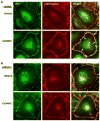
Figure 6. Afadin depletion attenuates OxPAPC-mediated adherens junction and tight junction remodeling
Pulmonary EC were transfected with: A - non-specific RNA, or B - afadin-specific siRNA. Cells were stimulated with OxPAPC (10 μg/ml, 30 min) followed by double immunofluorescence staining for ZO-1 (green) and p120-catein (red). Merged images depict areas of OxPACP-induced protein co-localization which appear in yellow and marked by arrows. This effect was attenuated in afadin-depleted EC. Shown are representative results of three independent experiments.
Figure 7
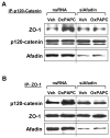
Figure 7. Afadin knockdown attenuates OxPAPC-induced interactions between ZO-1 and p120-catenin
Cells were transfected with non-specific RNA or afadin-specific siRNA followed by OxPAPC stimulation (10 μg/ml, 30 min). Co-immunoprecipitation assays using antibodies to A - p120-catenin or B - ZO-1 were performed, and afadin, p120-catenin, or ZO-1 content in the immunoprecipitates was detected using specific antibodies. Equal protein loading was confirmed by membrane reprobing with antibodies to p120-catenin (A) or ZO-1 (B). Lower panels depict siRNA-based depletion of endogenous afadin. Show are results representative of three independent experiments.
Figure 8
Figure 8. Afadin depletion in vivo abolishes protective effects by OxPAPC in the murine model of ventilator induced lung injury
Mice were transfected with non-specific or afadin-specific siRNA (72 hr) followed by high tidal volume mechanical ventilation (HTV; 30 ml/kg, 4 hr) with or without OxPAPC treatment (1.5 mg/kg, i/v). A - cell count, and B - protein concentration in BAL samples were analyzed. For each bar graph, cell counts or protein content determined in BAL from HTV-ventilated, nsRNA-treated animals were taken as 100%; n=4–6 per condition; *p<0.05. Inset depicts afadin depletion in lung samples assessed by western blot. Probing of the membrane with β-tubulin antibody served as a loading control.

Similar articles

Cited by

References

    1. Arthur WT, Quilliam LA, Cooper JA. Rap1 promotes cell spreading by localizing Rac guanine nucleotide exchange factors. The Journal of cell biology. 2004;167(1):111–122. - PMC - PubMed
    1. Birukov KG, Bochkov VN, Birukova AA, Kawkitinarong K, Rios A, Leitner A, Verin AD, Bokoch GM, Leitinger N, Garcia JG. Epoxycyclopentenone-containing oxidized phospholipids restore endothelial barrier function via Cdc42 and Rac. Circ Res. 2004;95(9):892–901. - PubMed
    1. Birukova AA, Alekseeva E, Cokic I, Turner CE, Birukov KG. Cross talk between paxillin and Rac is critical for mediation of barrier-protective effects by oxidized phospholipids. Am J Physiol Lung Cell Mol Physiol. 2008;295(4):L593–602. - PMC - PubMed
    1. Birukova AA, Chatchavalvanich S, Oskolkova O, Bochkov VN, Birukov KG. Signaling pathways involved in OxPAPC-induced pulmonary endothelial barrier protection. Microvasc Res. 2007a;73(3):173–181. - PMC - PubMed
    1. Birukova AA, Fu P, Chatchavalvanich S, Burdette D, Oskolkova O, Bochkov VN, Birukov KG. Polar head groups are important for barrier protective effects of oxidized phospholipids on pulmonary endothelium. Am J Physiol Lung Cell Mol Physiol. 2007b;292(4):L924–935. - PubMed

Publication types

MeSH terms

LinkOut - more resources