. 2011 Jul 3;13(8):914-923.
doi: 10.1038/ncb2281.

A SNX3-dependent retromer pathway mediates retrograde transport of the Wnt sorting receptor Wntless and is required for Wnt secretion

Affiliations

A SNX3-dependent retromer pathway mediates retrograde transport of the Wnt sorting receptor Wntless and is required for Wnt secretion

Martin Harterink et al. Nat Cell Biol. .

Abstract

Wnt proteins are lipid-modified glycoproteins that play a central role in development, adult tissue homeostasis and disease. Secretion of Wnt proteins is mediated by the Wnt-binding protein Wntless (Wls), which transports Wnt from the Golgi network to the cell surface for release. It has recently been shown that recycling of Wls through a retromer-dependent endosome-to-Golgi trafficking pathway is required for efficient Wnt secretion, but the mechanism of this retrograde transport pathway is poorly understood. Here, we report that Wls recycling is mediated through a retromer pathway that is independent of the retromer sorting nexins SNX1-SNX2 and SNX5-SNX6. We have found that the unrelated sorting nexin, SNX3, has an evolutionarily conserved function in Wls recycling and Wnt secretion and show that SNX3 interacts directly with the cargo-selective subcomplex of the retromer to sort Wls into a morphologically distinct retrieval pathway. These results demonstrate that SNX3 is part of an alternative retromer pathway that functionally separates the retrograde transport of Wls from other retromer cargo.


Figures

Fig. 1
Fig. 1
SNX-3 is required for EGL-20 (Wnt) signaling and MIG-14 (Wls) recycling in C. elegans. (A) The final positions of the QL.paa and QL.pap cells relative to the invariant positions of the seam cells V1 to V6 (n>100). Both snx-1(tm847) and snx-6(tm3790) are viable as single or double mutants and could be propagated as homozygous strains, excluding a contribution of maternally provided protein in our assays. (B) Expression of the EGL-20 target gene mab-5 in the QL descendants QL.a and QL.p. Cell nuclei are shown by DAPI staining. Scale bar is 10 μm. (C) Staining of EGL-20::proteinA with rabbit anti-goat-Cy5 in wild type, vps-35(hu68) and snx-3(tm1595). Expression is visible within the egl-20 expressing cells (closed line) and as a punctate posterior to anterior gradient (dotted line). In all images, anterior is to the left and dorsal is up. Scale bar is 10 μm. (D) Confocal images of MIG-14::GFP (huSi2) at identical exposure settings in wild type and in snx-1(tm847); snx-6(tm3790), vps-26(tm1523) and snx-3(tm1595). Scale bar is 10 μm. (E) Western blot quantification of MIG-14::GFP (huSi2) protein levels. (F) Confocal images of MIG-14::GFP (huIs71) (green) and LMP-1::mCherry (red) in wild type, vps-35(hu68) and snx-3(tm1595). Arrowheads indicate examples of co-localization between MIG-14::GFP and LMP-1::mCherry. Scale bar is 10 μm.
Fig. 2
Fig. 2
DSnx3 is required for Wg secretion and Wls recycling in the Drosophila wing imaginal disc. (A, B, C) Immunostaining of Wg, Wls and Senseless in wild type wing disc. (D, E, G, H, I, J, K) Expression of Dsnx6 or Dsnx3 RNAi transgenes was induced in the posterior compartment of the wing disc (marked by mCD8-GFP in green) using a hhGal4 driver (see Fig. S3A, B for quantification of knock down efficiency). (D, E) Immunostaining of Senseless (red). Arrowheads indicate loss of senseless expression in the Dsnx3 RNAi expressing posterior compartment. (F) Dsnx3 RNAi was induced in the posterior compartment using hhGal4 or in clones using an actinGal4 driver. Arrowheads indicate notches and loss of sensory bristles. (G, H) Immunostaining of total Wg (red). Arrowheads indicate Wg accumulation in the Dsnx3 RNAi expressing posterior compartment. (I) Immunostaining of extracellular Wg (red). Arrowheads indicate loss of extracellular Wg staining. (J, K) Immunostaining of Wls (red). Arrowheads indicate loss of Wls in wg expressing cells in the Dsnx3 RNAi expressing posterior compartment. Scale bars, 50 μm.
Fig. 3
Fig. 3
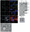
Co-localization and physical interaction of SNX3 with the cargo-selective sub-complex of the retromer. (A) SNX3 partially co-localizes with VPS26-positive early endosomes. HeLa cells lentivirally transduced to express GFP-SNX3 (green) were fixed and stained for VPS26, SNX1, EEA1 or LAMP1 (red). Co-localization between GFP-SNX3 and VPS26, SNX1, EEA1, LAMP1, Rab5 and Rab7 was quantified as 0.43 ± 0.05, 0.55 ± 0.04, 0.38 ± 0.02 and 0.07 ± 0.04, 0.61 ± 0.02, and 0.34 ± 0.02, respectively (Pearson’s coefficient, mean ± SD, n=3 with 30 cells per condition, for Rab5 and Rab7, n=20 cells). Scale bar, 11 μm. (B) At the ultrastructural level, SNX3 and VPS26 localize to common vesicular endosomal profiles. GFP-SNX3 is labeled with 10 nm gold and mCherry-VPS26 with 6 nm gold. The image is representative of that observed from the analysis of 5 other endosomal vacuoles. Scale bar, 100 nm. (C) SNX3 interacts with the cargo-selective sub-complex of the retromer. Cell extracts derived from HeLa cells lentivirally transduced with GFP, GFP-SNX3 or both GFP-SNX1 and GFP-SNX5 (GFP-SNX1/5), were subjected to GFP-nanotrap. The classical retromer SNX-BARs form heterodimeric complexes leading to the presence of both endogenous SNX1 and SNX5 in the GFP-SNX1/5 IPs. (D) 3xFLAG-VPS26-VPS29-VPS35-His6 complex (His-VPS) was isolated from BL21 E. coli onto TALON resin and incubated with 2 μM of recombinant SNX3, SNX1 or SNX5 for 2 hours at 4°C. Supernatant (S) and TALON containing resin (P) were isolated. SNX3 directly associates with His-VPS as do SNX1 and SNX5 although this is less well pronounced (longer exposures are shown). Control: boiled His-VPS resin.
Fig. 4
Fig. 4
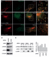
SNX3 co-localizes with Wls and facilitates membrane association of the cargo-selective sub-complex of the retromer. (A) Co-localization between SNX3-GFP (green) and Wls-mCherry (red) in HeLa cells was quantified as 0.25 ± 0.02% (Pearson’s coefficient; mean ± SEM, n=2 with 23 and 11 cells). Arrowheads show examples of co-localization. Scale bar, 10 μm. (B, C) Co-localization between Wls-mCherry and endogenous VPS26 (green) in HeLa cells treated with control or SNX3 siRNA was quantified as 0.19 ± 0.02% and 0.08 ± 0.02%, respectively (Pearson’s coefficient; mean ± SEM, n=4 with 7-10 cells each). Arrowheads show examples of co-localization. (D) HeLa cells were transfected with control or SNX3 siRNA and assayed for endogenous Wls, VPS26, SNX3 and tubulin protein levels. (E) HeLa cells treated with control, SNX3 or RAB7 siRNA were separated into a supernatant (S) fraction containing cytosol and a pellet fraction (P) containing membranes and were stained for endogenous VPS26, SNX1 and LAMP1. The amount of VPS26 in the supernatant and pellet fractions was quantified using densitometry and is shown as percentage of the total. Data are presented as mean ± SEM and represent three independent experiments. There was no significant change in SNX1 membrane association upon SNX3 knock down (17.8 ± 3.8% versus 22 ± 8.1%). Knock down of RAB7 was included as a positive control.
Fig. 5
Fig. 5
Wls is contained within SNX3 positive vesicular carriers but is absent from SNX1 retromer decorated tubular carriers. (A) RPE-1 cells were transiently co-transfected with pEGFP-SNX1 (green) and Wls-mCherry (red) and cells were subsequently imaged live after a 16 hour incubation period. Frames depicting the formation and scission of a GFP-SNX1 tubule from a vesicle positive for both SNX1 and Wls are shown (arrows indicate the dual expressing vesicle, while arrowheads indicate the carrier post scission) (see Supplementary Information, Movie 1). Scale bars represent 6 μm. Of the 100 SNX1 decorated tubulating endosomes that were analyzed, 22 were positive for Wls; 18/22 tubules emanating from these endosomes were negative for Wls, while 4/22 were weakly positive. Quantification of Wls-mCherry and GFP-SNX1 levels in an endosome and corresponding tubule is shown in Fig. S4D. (B) Further examples of SNX1 retromer tubules negative for Wls. (i) An example of a SNX1 retromer positive endosome and tubule both of which are negative for Wls. (ii and iii) Further examples of SNX1-labelled endosomes positive for Wls, but with tubules negative for Wls. Scale bars represent 6 μm. (C) RPE-1 cells were transiently co-transfected with pEGFP-SNX3 (green) and Wls-mCherry and cells were subsequently imaged live after a 16 hour incubation period. Frames depicting the formation and scission of GFP-SNX3 labeled buds from vesicles positive for both SNX3 and Wls are shown. Note the 1 second delay between acquisitions for a given image pair. Arrows and arrowheads show two examples of buds positive for both Wls and SNX3 (see Supplementary Information, Movie 2). Scale bars represent 6 μm.

Comment in

Similar articles

Cited by

References

    1. Carlton J, et al. Sorting nexin-1 mediates tubular endosome-to-TGN transport through coincidence sensing of high-curvature membranes and 3-phosphoinositides. Curr Biol. 2004;14:1791–1800. - PubMed
    1. Carlton JG, et al. Sorting nexin-2 is associated with tubular elements of the early endosome, but is not essential for retromer-mediated endosome-to-TGN transport. J Cell Sci. 2005;118:4527–4539. - PMC - PubMed
    1. Wassmer T, et al. A loss-of-function screen reveals SNX5 and SNX6 as potential components of the mammalian retromer. J Cell Sci. 2007;120:45–54. - PubMed
    1. Wassmer T, et al. The retromer coat complex coordinates endosomal sorting and dynein-mediated transport, with carrier recognition by the trans-Golgi network. Dev Cell. 2009;17:110–122. - PMC - PubMed
    1. Seaman MN. Recycle your receptors with retromer. Trends Cell Biol. 2005;15:68–75. - PubMed

Publication types

MeSH terms