. 2011 Jul;138(14):3021-31.
doi: 10.1242/dev.059980.

Apical deficiency triggers JNK-dependent apoptosis in the embryonic epidermis of Drosophila

Affiliations

Apical deficiency triggers JNK-dependent apoptosis in the embryonic epidermis of Drosophila

Golnar Kolahgar et al. Development. 2011 Jul.

Abstract

Epithelial homeostasis and the avoidance of diseases such as cancer require the elimination of defective cells by apoptosis. Here, we investigate how loss of apical determinants triggers apoptosis in the embryonic epidermis of Drosophila. Transcriptional profiling and in situ hybridisation show that JNK signalling is upregulated in mutants lacking Crumbs or other apical determinants. This leads to transcriptional activation of the pro-apoptotic gene reaper and to apoptosis. Suppression of JNK signalling by overexpression of Puckered, a feedback inhibitor of the pathway, prevents reaper upregulation and apoptosis. Moreover, removal of endogenous Puckered leads to ectopic reaper expression. Importantly, disruption of the basolateral domain in the embryonic epidermis does not trigger JNK signalling or apoptosis. We suggest that apical, not basolateral, integrity could be intrinsically required for the survival of epithelial cells. In apically deficient embryos, JNK signalling is activated throughout the epidermis. Yet, in the dorsal region, reaper expression is not activated and cells survive. One characteristic of these surviving cells is that they retain discernible adherens junctions despite the apical deficit. We suggest that junctional integrity could restrain the pro-apoptotic influence of JNK signalling.


Figures
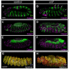
Fig. 1.
Fig. 1.
The ventral epidermis undergoes apoptosis in crb mutant Drosophila embryos. (A,B) Anti-Caspase 3 immunoreactivity (magenta) at stage 11 in control (A, crb heterozygous) and crb homozygous (B) embryos. At this stage, activated Caspase 3 is already clearly elevated in the head region (bracket) and a moderate segmental increase can be seen in the trunk (arrows). Anti-Engrailed (En, green) was used as a reference along the anteroposterior axis. (C-F) At stage 13, anti-Caspase 3 staining has further increased in the trunk region. These embryos were triple stained for activated Caspase 3, Engrailed and GFP expressed under the control of pannier-Gal4. Activated Caspase 3 and Engrailed are shown in C (control, crb heterozygous) and D (crb homozygous), whereas activated Caspase 3 and GFP are shown in E (crb heterozygous) and F (crb homozygous). The domain of pannier expression is largely devoid of Caspase 3-positive cells in crb mutants. The dashed line marks the dorsal edge of the epidermis. (G,H) Stage 14 control (G, crb heterozygous) and crb homozygous (H) embryos expressing Apoliner under the control of the ubiquitous tubulin-Gal4 driver. With this sensor, live cells appear yellow. A band of live cells remains on the dorsal side, whereas no live cells remain ventrally in the crb mutant. (A-F) Projections of a confocal stack; (G,H) Generated by 3D rendering. See also Fig. S1 in the supplementary material. Scale bars: 50 μm (in A for A-F; in G for G,H).
Fig. 2.
Fig. 2.
Apoptosis in the epidermis of crb mutant embryos relies on the transcriptional activation of rpr. (A-J) Expression of the pro-apoptotic genes rpr (A-D), skl (E,F), hid (G,H) and grim (I,J) in control and crb mutant Drosophila embryos at stage 11 (A,B,E-J) or 13 (C,D). Whereas hid or grim are similarly expressed in the two genotypes, rpr and skl are both upregulated in the crb mutant background. Co-staining with anti-Engrailed (A-D) provides a spatial landmark, showing that there is a general correlation between the pattern of rpr expression and that of Caspase 3 activation (see K). (K-N) Caspase 3 immunoreactivity in stage 13 crb mutant embryos lacking various combinations of pro-apoptotic genes. Engrailed (red) marks the posterior of each segment. Almost no anti-Caspase 3 staining can be detected in Def(XR38), crb (L), which removes rpr and skl, or in Def(H99), crb (N), where the contribution of hid, rpr and grim is absent. By contrast, the levels of Caspase 3 staining are similar in crb and Def(X14), crb embryos (where the contribution of hid is removed) (M). The dorsal edge of the epidermis is marked with a dashed line. (O) Model of the effect of crb loss of function on pro-apoptotic gene expression.
Fig. 3.
Fig. 3.
Transcriptional targets of JNK signalling are upregulated in the epidermis of crb mutant embryos. (A-F) Expression pattern of dpp and scarface at stage 13, as determined by RNA in situ hybridisation in control (A,D), crb single-mutant (B,E) and Def(H99) crb double-mutant (C,F) Drosophila embryos. In control embryos, JNK-dependent expression of dpp and scarface can be detected in the most dorsal epidermal cells, which abut the amnioserosa (A,D). In the absence of Crb, expression of dpp and scarface expands laterally in a segmented fashion (B,E). This is also true when apoptosis is suppressed in Def(H99) crb double mutants (C,F). See also Fig. S2 in the supplementary material.
Fig. 4.
Fig. 4.
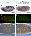
JNK activity is required for apoptosis in Drosophila crb mutant embryos. Comparison of crb embryos with crb embryos overexpressing puc under the control of the tubulin-Gal4 driver (JNK signalling prevented). (A,B) Puc overexpression prevents activation of rpr expression as assayed by RNA in situ hybridisation at stage 11 (lateral views). In Puc-overexpressing crb mutants, expression of rpr is similar to that in wild-type embryos (not shown). (C,D) Puc overexpression also prevents caspase activation (red), as shown here in ventral views of stage 13 embryos. GFP is expressed from a UAS transgene activated by tubulin-Gal4. (E,F) Puc overexpression also rescues the cuticle phenotype of crb mutants. Whereas crb mutant embryos deposit ‘crumbs’ of cuticle (refracting objects in E) at the end of embryogenesis, a continuous sheet of cuticle is apparent in rescued embryos (F). Dorsal closure is defective (as is the case in wild-type embryos made to overexpress Puc), but segmentally repeated denticle belts can be discerned. See also Fig. S3 in the supplementary material.
Fig. 5.
Fig. 5.
Endogenous Puc restrains rpr expression. (A) Expression of rpr in a stage 11 homozygous crb mutant Drosophila embryo that is also heterozygous for pucE69. The pattern is seemingly identical to that in single crb mutants (compare with Fig. 2B). (B) In a pucE69 crb double mutant embryo of the same stage, expression of rpr expands into the dorsal epidermis. Dashed line marks the boundary between the dorsal epidermis and the amnioserosa. (C) Expression of rpr in a +/pucE69, +/crb embryo remains low, as it does in the wild type. (D) Expression of rpr is specifically activated in the dorsal-most cells of homozygous pucE69 embryos. Genotypes: (A) pucE69, crb2/+, crb2; (B) pucE69, crb2; (C) pucE69, crb2/TDY; (D) pucE69.
Fig. 6.
Fig. 6.
Loss of apical, but not basolateral, identity causes JNK-dependent rpr expression. (A-C) Expression of rpr in control (A), sdt (B) and baz (C) Drosophila embryos. Upregulation is seen in both sdt and baz mutants. (D,E) Removal of zygotic lgl activity partially prevents upregulation of rpr expression in crb mutants. To ensure identical treatments, double-mutant embryos (E) were compared with single crb mutants that are heterozygous at the lgl locus (D). (F) Complete absence of a basolateral determinant (maternal zygotic lgl mutant) does not cause widespread rpr expression. (G,H) Upregulation of scarface expression in crb mutant embryos (G; the embryo shown is also heterozygous at lgl) is prevented if zygotic lgl activity is completely removed (H; lgl; crb). (I) Inhibition of JNK signalling by puc overexpression prevents scarface expression in a crb background. Ventral views of stage 11 (A-F) and stage 13 (G-I) embryos are shown, with expression determined by RNA in situ hybridisation. See also Figs S6-S8 in the supplementary material. Genotypes: (A) sdt7D22/FM7 ftzlacZ; (B) sdt7D22; (C) bazxi106; (D,G) lgl4, FRT40A/CyOKrG4>UASGFP; UAS-CD8-GFP, crb2; (E,H) lgl4, FRT40A; UAS-CD8-GFP, crb2; (F) lgl27S3 (M–Z–); (I) w; UAS Puc14C/+; tubulin-Gal4, crb2 e/UAS-CD8-GFP, crb2 e.
Fig. 7.
Fig. 7.
JNK signalling and junctional disruption activate rpr expression. (A,B) Distribution of E-cadherin (E-Cad) at stage 13 in a wild-type (A) and a crb mutant (B) Drosophila embryo. Stacks of confocal micrographs are shown. Note the residual honeycomb pattern in the dorsal epidermis of the crb mutant. (C-J) Optical transverse sections through the dorsal and ventral epidermis (as indicated) stained for (C,D,G,H) E-cadherin (green), Dlg (a basolateral marker, red) and nuclei (TOPRO-3, blue) and stained for (E,F,I,J) Stranded at second (Sas, red) and Fasciclin III (FasIII, green). (K) rpr expression is mildly upregulated in the ventral-lateral epidermis of embryos homozygous for a shg antimorphic allele (embryo shown is also heterozygous for crb). No rpr upregulation is seen in the dorsal region. (L) Expression of rpr in a stage 13 homozygous crb mutant embryo that is also heterozygous for the same shg antimorphic allele. Expression is undistinguishable from that in a single crb mutant. (M) In a shg crb double mutant embryo of the same stage, expression of rpr expands into the dorsal epidermis. White dashed line marks the boundary between the dorsal epidermis and the amnioserosa. (N) Diagram highlighting the correlation between junctional disruption and the ability of JNK signalling to activate rpr expression. See also Fig. S9 in the supplementary material. Genotypes: (A,C,E,G,I) crb2/TTG; (B,D,F,H,J) crb2; (K) shgg317; UAS-CD8-GFP, crb2/TDY; (L) shgg317/CyOKrG4>UASGFP; UAS-CD8-GFP, crb2; (M) shgg317; UAS-CD8-GFP, crb2. Scale bars: 10 μm.
Fig. 8.
Fig. 8.
Signalling upstream and downstream of JNK. (A) Apical disruption leads to JNK activation, either as a direct consequence of altered cell polarity (1, blue arrow), or indirectly, via the disruption of junctional integrity (2, green arrow). (B) Two mechanisms restrain the ability of JNK signalling to trigger apoptosis. Junctional integrity could act either at the level of JNK signalling or further downstream. As a feedback inhibitor, Puc is likely to limit the extent of JNK signalling. However, we cannot exclude the possibility that Puc has additional activities.

Similar articles

Cited by

References

    1. Abrams J. M., White K., Fessler L. I., Steller H. (1993). Programmed cell death during Drosophila embryogenesis. Development 117, 29-43 - PubMed
    1. Adachi-Yamada T., O'Connor M. B. (2002). Morphogenetic apoptosis: a mechanism for correcting discontinuities in morphogen gradients. Dev. Biol. 251, 74-90 - PubMed
    1. Affolter M., Nellen D., Nussbaumer U., Basler K. (1994). Multiple requirements for the receptor serine/threonine kinase thick veins reveal novel functions of TGF beta homologs during Drosophila embryogenesis. Development 120, 3105-3117 - PubMed
    1. Agrawal N., Kango M., Mishra A., Sinha P. (1995). Neoplastic transformation and aberrant cell-cell interactions in genetic mosaics of lethal(2)giant larvae (lgl), a tumor suppressor gene of Drosophila. Dev. Biol. 172, 218-229 - PubMed
    1. Baehrecke E. H. (2002). How death shapes life during development. Nat. Rev. Mol. Cell Biol. 3, 779-787 - PubMed

Publication types

MeSH terms

Substances