. 2011 Jul;138(14):2957-68.
doi: 10.1242/dev.063784.

Sprouty genes prevent excessive FGF signalling in multiple cell types throughout development of the cerebellum

Affiliations

Sprouty genes prevent excessive FGF signalling in multiple cell types throughout development of the cerebellum

Tian Yu et al. Development. 2011 Jul.

Abstract

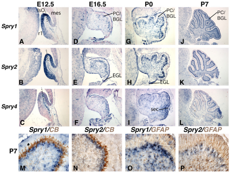
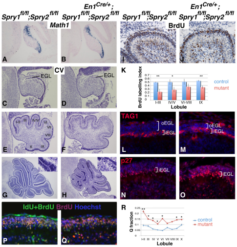
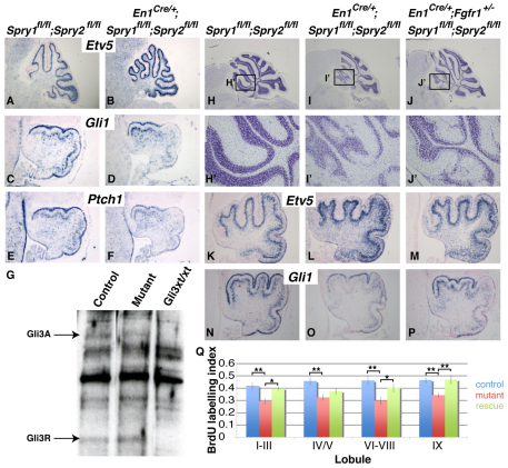
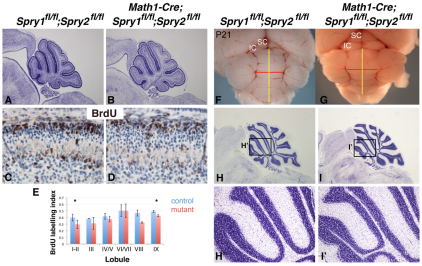
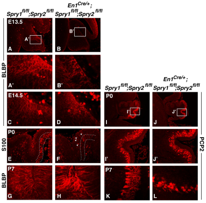
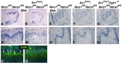
Fibroblast growth factors (FGFs) and regulators of the FGF signalling pathway are expressed in several cell types within the cerebellum throughout its development. Although much is known about the function of this pathway during the establishment of the cerebellar territory during early embryogenesis, the role of this pathway during later developmental stages is still poorly understood. Here, we investigated the function of sprouty genes (Spry1, Spry2 and Spry4), which encode feedback antagonists of FGF signalling, during cerebellar development in the mouse. Simultaneous deletion of more than one of these genes resulted in a number of defects, including mediolateral expansion of the cerebellar vermis, reduced thickness of the granule cell layer and abnormal foliation. Analysis of cerebellar development revealed that the anterior cerebellar neuroepithelium in the early embryonic cerebellum was expanded and that granule cell proliferation during late embryogenesis and early postnatal development was reduced. We show that the granule cell proliferation deficit correlated with reduced sonic hedgehog (SHH) expression and signalling. A reduction in Fgfr1 dosage during development rescued these defects, confirming that the abnormalities are due to excess FGF signalling. Our data indicate that sprouty acts both cell autonomously in granule cell precursors and non-cell autonomously to regulate granule cell number. Taken together, our data demonstrate that FGF signalling levels have to be tightly controlled throughout cerebellar development in order to maintain the normal development of multiple cell types.


Figures

Fig. 1.
Fig. 1.
Sprouty gene expression in the cerebellum during late embryogenesis and early postnatal development. (A-L) The expression of sprouty1 (Spry1; A,D,G,J), sprouty2 (Spry2; B,E,H,K) and sprouty4 (Spry4; C,F,I,L) was determined by in situ hybridisation (blue) on sagittal sections of E12.5 (A-C), E16.5 (D-F), P0 (G-I) and P7 (J-L) mouse cerebella, counterstained with Nuclear Fast Red (pink). (M-P) P7 cerebellar sections immunostained (brown) with antibodies against calbindin (CB) (M,N) or GFAP (O,P) after in situ hybridisation. Dashed lines in A-C indicate the IsO. EGL, external granular layer; mes, mesencephalon; PC/BGL, Purkinje cell/Bergmann glial cell layer; r1, rhombomere 1; sec, secondary fissure.
Fig. 2.
Fig. 2.
Sprouty loss of function results in expansion of the vermis and abnormal cytoarchitecture. (A-C) Dorsal views of control Spry1fl/fl;Spry2fl/fl (A), En1cre/+;Spry1fl/fl;Spry2fl/fl (B) and En1cre/+;Spry1fl/fl;Spry2fl/fl;Spry4fl/fl (C) whole-mount P21 mouse cerebella. The yellow line indicates the anteroposterior length of the vermis, and the red line indicates the mediolateral width. (D-F′) Cresyl Violet-stained sagittal sections of P21 control (D,D′), En1cre/+;Spry1fl/fl;Spry2fl/fl (E,E′) and En1cre/+;Spry1fl/fl;Spry2fl/fl;Spry4fl/fl (F,F′) cerebella; anterior is to the left. D′-F′ are higher magnification views of boxed areas in D-F. (G-I) GFAP immunostaining (red) on P21 sagittal sections to detect Bergmann glial fibres, counterstained with Hoechst (blue). High magnification views of the molecular layer are shown. (J-L) PCP2 immunostaining (red) on P21 sagittal sections to detect Purkinje cells. IC, inferior colliculus; SC, superior colliculus.
Fig. 3.
Fig. 3.
Sprouty genes function as negative regulators of FGF signalling at the IsO in the early embryo, whereafter they are required for the normal production of granule neurons. (A-I) Whole-mount in situ hybridisation of Etv5 (A-C), Gbx2 (D-F) and Otx2;Hoxa2 (G-I) on E9.5 control (Spry1fl/fl;Spry2fl/fl; A,D,G); En1cre/+;Spry1fl/fl;Spry2fl/fl (B,E,H) and En1cre/+;Spry1fl/fl;Spry2fl/fl;Spry4fl/fl (C,F,I) mouse embryos. Red dashed lines in D-F indicate the anterior limit of Gbx2 expression and white arrows the approximate size of anterior r1 (ar1) as marked by Gbx2. Red dashed lines in G-I mark the anterior and posterior limits of r1 as indicated by Otx2 and Hoxa2, respectively, and black arrows the approximate size of r1. The white dashed lines in H and I indicate the approximate position of the Gbx2 expression limit in the control embryo for comparison. (J) Dorsal view of a whole-mount Nestin-Cre;Spry1fl/fl;Spry2fl/fl cerebellum at P21. (K,K′) Cresyl Violet-stained sagittal sections from the brain shown in J. K′ is a higher magnification view of the boxed area in K. IC, inferior colliculus; SC, superior colliculus.
Fig. 4.
Fig. 4.
Reduced proliferation and enhanced differentiation of GCps in sprouty mutants. (A,B) The presence of Math1+ GCps as visualised in sagittal sections from E13.5 control (Spry1fl/fl;Spry2fl/fl) and En1cre/+;Spry1fl/fl;Spry2fl/fl mouse cerebella by in situ hybridisation. (C-H) Cresyl Violet (CV) staining of E16.5 (C,D), P0 (E,F) and P7 (G,H) control and mutant sagittal sections. Insets in G and H are high magnification views of the external granular layer (EGL). (I,J) Sagittal sections through P0 cerebella from control and mutant animals treated with BrdU and immunostained with an antibody against BrdU (brown), counterstained with Haematoxylin. BrdU-positive GCps and total GCps in the EGL were counted in 100×100 μm areas as indicated by the boxes and used to calculate the BrdU labelling index. (K) Comparison of BrdU labelling indices in different lobules at P0. Note the significantly reduced labelling indices in mutants (red bar) compared with controls (blue bar) (*P<0.05, **P<0.001, Student's t-test, n=4 per genotype). Error bars represent s.e.m. (L,M) TAG1 immunohistochemistry (red) to mark the inner EGL (iEGL), counterstained with Hoechst (blue) to visualise the outer EGL (oEGL). Note the alteration in relative thickness of the oEGL and iEGL (white brackets). (N,O) p27 immunohistochemistry (red) to visualise cells that are exiting the cell cycle and initiation differentiation. (P,Q) Sagittal sections of P4.5 control and mutant brains after BrdU and IdU injections as described in Materials and methods, immunostained with monoclonal antibodies against both BrdU and IdU (green), BrdU alone (red), and counterstained with Hoechst (blue). (R) Q fractions in different lobules (I-X) from control (blue) and mutant (red) cerebella. Note the increased cell cycle exit (differentiation) in mutants compared with controls (*P<0.05, **P<0.001, Student's t-test, n=3 per genotype).
Fig. 5.
Fig. 5.
Increased FGF signalling in sprouty mutant cerebella is associated with reduced SHH signalling and reduced GCp proliferation. (A-F) The expression of Etv5 at P6 (A,B), Gli1 (C,D) and Ptch1 (E,F) at E18.5 in control (Spry1fl/fl;Spry2fl/fl) and En1cre/+;Spry1fl/fl;Spry2fl/fl mutant mouse cerebella was determined by in situ hybridisation; sagittal sections are shown, anterior to the left. (G) GLI3 western blot from total cell lysates prepared from E18.5 cerebella. Control=Spry1fl/fl;Spry2fl/fl, Mutant=En1cre/+;Spry1fl/fl;Spry2fl/fl. Note the absence of GLI3 protein in homozygous Gli3xt/xt (Gli3xt/xt) mutant cerebellum (Schimmang et al., 1992). (H-J′) Sagittal sections from P21 control, En1cre/+;Spry1fl/fl;Spry2fl/fl and En1cre/+;Spry1fl/fl;Spry2fl/fl;Fgfr1+/− mutants stained with Cresyl Violet. H′,I′ and J′ are high magnification views of the boxed areas in H,I and J, respectively. (K-P) The expression of Etv5 (K-M) and Gli1 (N-P) on sagittal sections from newborn control, En1cre/+;Spry1fl/fl;Spry2fl/fl and En1cre/+;Spry1fl/fl;Spry2fl/fl;Fgfr1+/− cerebella. (Q) BrdU labelling index comparison between P0 control (blue), En1cre/+;Spry1fl/fl;Spry2fl/fl mutant (red) and En1cre/+;Spry1fl/fl;Spry2fl/fl;Fgfr1+/− rescue (green) cerebella. Note significant differences (*P<0.05, **P<0.001, one-way ANOVA, n=4 per genotype) between control and mutants in all lobules, and between mutant and rescue samples in all lobules apart from IV/V. Error bars represent s.e.m.
Fig. 6.
Fig. 6.
Sprouty gene deletion in granule cell precursors has a minor effect on cell proliferation. (A,B) Cresyl Violet staining of sagittal sections through P3 control (Spry1fl/fl;Spry2fl/fl) and Math1-Cre;Spry1fl/fl;Spry2fl/fl mouse cerebella. (C,D) BrdU immunostaining (brown) on sagittal sections counterstained with Hematoxylin (blue). (E) BrdU labelling indices at P3 show a minor reduction in proliferation in the anterior and posterior lobules of the mutant cerebella (*P<0.05, Student's t-test, n=3 per genotype). Error bars represent s.e.m. (F-I′) Whole-mount views (F,G) of and Cresyl Violet-stained sagittal sections (H-I′) through P21 control (Spry1fl/fl;Spry2fl/fl) and Math1-Cre;Spry1fl/fl;Spry2fl/fl cerebella. H′ and I′ are higher magnification views of the boxed areas in H and I, respectively. IC, inferior colliculus; SC, superior colliculus.
Fig. 7.
Fig. 7.
Sprouty gene function is required for the normal differentiation of Bergmann glia and Purkinje cells. (A-H) Sagittal sections of E13.5 (A-B′), E14.5 (C,D), P0 (E,F) and P7 (G,H) control (Spry1fl/fl;Spry2fl/fl) and En1cre/+;Spry1fl/fl;Spry2fl/fl mouse cerebella immunostained with anti-BLBP (A-D,G,H) or S100 (E,F) antibodies. A′ and B′ are high magnification views of the boxed areas in A and B, respectively. Area outlined in E and F is the PC/BG cell layer. (I-L) Sagittal sections of P0 (I-J′) and P7 (K,L) control (Spry1fl/fl;Spry2fl/fl) and En1cre/+;Spry1fl/fl;Spry2fl/fl cerebella immunostained with anti-PCP2 antiserum. I′ and J′ are high magnification views of the boxed areas in I and J, respectively.
Fig. 8.
Fig. 8.
SHH expression is reduced in sprouty-deficient cerebella. (A-B′) Shh expression was assayed on sagittal sections from E18.5 control (Spry1fl/fl;Spry2fl/fl) and mutant (En1cre/+;Spry1fl/fl;Spry2fl/fl) mouse cerebella. A′ and B′ are high magnification views of A and B, respectively. (C,D) SHH protein distribution was determined by immunohistochemistry (green) on control and En1cre/+;Spry1fl/fl;Spry2fl/fl sections at P7, with nuclei counterstained with Hoechst (blue). White arrowheads in C indicate individual PCs with high levels of cytoplasmic SHH protein and open arrowheads in D indicate PCs with reduced SHH content. (E-G′) In situ hybridisation for Shh gene expression on sections from P0 control (E,E′), En1cre/+;Spry1fl/fl;Spry2fl/fl mutant (F,F′) and En1cre/+;Spry1fl/fl;Spry2fl/fl;Fgfr1+/− rescue (G,G') cerebella. E′, F′ and G′ are high magnification views of E, F and G, respectively.

Similar articles

Cited by

References

    1. Baptista C. A., Hatten M. E., Blazeski R., Mason C. A. (1994). Cell-cell interactions influence survival and differentiation of purified Purkinje cells in vitro. Neuron 12, 243-260 - PubMed
    1. Basson M. A., Akbulut S., Watson-Johnson J., Simon R., Carroll T. J., Shakya R., Gross I., Martin G. R., Lufkin T., McMahon A. P., et al. (2005). Sprouty1 is a critical regulator of GDNF/RET-mediated kidney induction. Dev. Cell 8, 229-239 - PubMed
    1. Basson M. A., Echevarria D., Ahn C. P., Sudarov A., Joyner A. L., Mason I. J., Martinez S., Martin G. R. (2008). Specific regions within the embryonic midbrain and cerebellum require different levels of FGF signaling during development. Development 135, 889-898 - PMC - PubMed
    1. Berry M., Bradley P. (1976). The growth of the dendritic trees of Purkinje cells in irradiated agranular cerebellar cortex. Brain Res. 116, 361-387 - PubMed
    1. Bizzoca A., Virgintino D., Lorusso L., Buttiglione M., Yoshida L., Polizzi A., Tattoli M., Cagiano R., Rossi F., Kozlov S., et al. (2003). Transgenic mice expressing F3/contactin from the TAG-1 promoter exhibit developmentally regulated changes in the differentiation of cerebellar neurons. Development 130, 29-43 - PubMed

Publication types

MeSH terms