. 2011 Jun 17;30(14):2829-42.
doi: 10.1038/emboj.2011.193.

Sgf29 binds histone H3K4me2/3 and is required for SAGA complex recruitment and histone H3 acetylation

Affiliations

Sgf29 binds histone H3K4me2/3 and is required for SAGA complex recruitment and histone H3 acetylation

Chuanbing Bian et al. EMBO J. .

Abstract

The SAGA (Spt-Ada-Gcn5 acetyltransferase) complex is an important chromatin modifying complex that can both acetylate and deubiquitinate histones. Sgf29 is a novel component of the SAGA complex. Here, we report the crystal structures of the tandem Tudor domains of Saccharomyces cerevisiae and human Sgf29 and their complexes with H3K4me2 and H3K4me3 peptides, respectively, and show that Sgf29 selectively binds H3K4me2/3 marks. Our crystal structures reveal that Sgf29 harbours unique tandem Tudor domains in its C-terminus. The tandem Tudor domains in Sgf29 tightly pack against each other face-to-face with each Tudor domain harbouring a negatively charged pocket accommodating the first residue alanine and methylated K4 residue of histone H3, respectively. The H3A1 and K4me3 binding pockets and the limited binding cleft length between these two binding pockets are the structural determinants in conferring the ability of Sgf29 to selectively recognize H3K4me2/3. Our in vitro and in vivo functional assays show that Sgf29 recognizes methylated H3K4 to recruit the SAGA complex to its targets sites and mediates histone H3 acetylation, underscoring the importance of Sgf29 in gene regulation.


Conflict of interest statement

The authors declare that they have no conflict of interest.

Figures
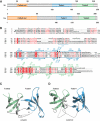
Figure 1
Figure 1
Crystal structures of human and yeast Sgf29 tandem Tudor domains. (A) Domain structures of budding yeast Sgf29 (Sc) and human SGF29 (Hs). The coiled-coil domain is coloured in orange, and the two Tudor domains are coloured in blue and green, respectively. The starting and ending residues of each domain are numbered. (B) Structure-based sequence alignment of Sgf29 homologues. The two conserved Tudor domains are framed and coloured in blue and green, respectively. The secondary structure elements of scSgf29 and hsSGF29 are indicated above and below the sequence alignment, respectively. H3K4me and H3A1 binding residues are numbered and marked by stars and dots, respectively. The sequence identity of the full-length and the tandem Tudor domains of yeast Sgf29 with the other three homologues are indicated beside the first and the third row, respectively. The alignment was created with Espript (http://espript.ibcp.fr/ESPript/ESPript/). Sc, S. cerevisiae; Sp, Schizosaccharomyces pombe; Dm, Drosophila melanogaster; Hs, Homo sapiens. β and η represent β strands and 310 helix, respectively. (C, D) Cartoon representation of the crystal structures of yeast Sgf29 and human SGF29 tandem Tudor domains, respectively. The two Tudor domains are coloured in blue and green, respectively, and the secondary structure regions in both proteins are marked.
Figure 2
Figure 2
Selective binding of H3K4me2/3 by the SGF29 tandem Tudor domains. Interactions between yeast (A) and human SGF29 (B) tandem Tudor domains and H3K4me3 peptide. The Sgf29 tandem Tudor domains are shown in cartoon representation and coloured in blue. The H3K4me3 peptide is shown as a stick model. The conserved and non-conserved hydrogen bonds are coloured in red and black, respectively. (C, D) Electrostatic surface representation of yeast (C) and human (D) Sgf29–H3K4me3 complex. Sgf29 is shown in surface representation, and histone peptide is shown in a stick model.
Figure 3
Figure 3
Architecture of different double Tudor domains. (A) Human SGF29. (B) Yeast Sgf29. (C) 53BP1 (PDB code 2G3R). (D) FXR1 (PDB code 3O8V). (E) Hybrid Tudor domain protein JMJD2A (PDB code 2GFA). (F) The extended Tudor domain of SND1 (PDB code 3OMC). The Tudor domains are coloured in rainbow spectrum on the basis of topology, except those of JMJD2A and SND1 that are coloured based on the sequence boundaries of Tudor domains. The N-terminal and C-terminal extensions are coloured in grey.
Figure 4
Figure 4
Sgf29 is important for histone H3 acetylation. (A) Global acetylation levels of H3K9, H3K14, H3K18, H3K23 and H4, and expression of scGcn5 in various yeast mutant strains: lane 1: BY4742; lane 2: ΔAda3; lane 3: ΔGcn5; lane 4: ΔSgf29. (B) Global acetylation levels of H3K9, H3K14, H3K18 and H3K23, and expression of scGcn5 in mutant strains in the yeast cells expressing Sgf29 with different deletions or point mutations. Lane 1: BY4742 (WT); lane 2: ΔSgf29, different Sgf29 plasmids were transformed into Sgf29 deletion yeast strain, respectively; lane 3: WT Sgf29; lane 4: D163A; lane 5: E165A; lane 6: Y205A; lane 7: T209A; lane 8: T210A; lane 9: Y212A; lane 10: F229A; lane 11: ΔT1; and lane 12: ΔT2 (deletion of T1 and T2 domains). Quantified values for the acetylation levels in the above western blot panel are present in a bar chart with values as percentage of WT strain. (C) Expression levels of scSgf29 and its mutants. (D) Efficiency of human GCN5 and hsSGF29 knockdown by shRNA. (E) Global acetylation levels of histone H3K9, H3K14, H3K18, H3K23 and H4. Lane 1: knockdown of GCN5; lane 2: control; lane 3: knockdown of hsSGF29. The bands are quantified to better show the differences.
Figure 5
Figure 5
Sgf29 is required for proper H3K9 acetylation and SAGA localization at Gal1 locus. (A) Purified SAGA from WT and Sgf29 deletion strains via Spt8-TAP was combined, respectively, with.0.5 μg of various SAGA substrates: no peptide, core histones, nucleosomes, unmodified histone H3 peptide (1–20), tetra-acetylated histone H3 peptide (1–20), mono-, di- and tri-methylated histone H3K4 peptide (1–20) and tri-methylated histone H3K9 peptide (6–20). Activity was measured as disintegrations per minute (DPM). (B) mRNA expression levels of WT, ΔSgf29, Sgf29ΔT1, Sgf29ΔT2, ΔSpp1 and ΔSwd1 were determined by RT–PCR in real time. GAL1 was normalized to ACT1 levels and values are expressed as fold induction changes with WT values in dextrose medium set at 1.0. (C) H3K9ac ChIP (anti-H3K9Ac) was used to evaluate the H3K9 acetylation levels of WT, ΔSgf29, Sgf29ΔT1, Sgf29ΔT2, Spp1Δ and Swd1Δ at the GAL1 UAS, 5′ and 3′ end. (D) SAGA ChIP (anti-Ada2) was used to evaluate the SAGA levels of WT, ΔSgf29, Sgf29ΔT1, Sgf29ΔT2, Spp1Δ and Swd1Δ at the GAL1 UAS, 5′ and 3′ end.
Figure 6
Figure 6
Both genome-wide and SAGA-specific gene array analysis indicate that Sgf29 is required for H3K9acetylation and for proper SAGA localization. (A) Whole-genome median gene analysis of H3K9 acetylation localization in WT strain (solid grey line), sgf29Δ strain (solid black line), SAGA-specific gene analysis of H3K9 acetylation localization in WT strain (dotted grey lines) and sgf29Δ strain (dotted black lines). (B) Identical analysis as in (A), but represented as mutant/WT for sgf29Δ in the H3K9Ac for the whole-genome (All) and SAGA-specific genes (SAGA). (C) Whole-genome median gene analysis of SAGA localization (via Spt8Flag) in WT strain (solid grey line) and sgf29Δ strain (solid black line) and SAGA-specific gene analysis of SAGA localization in WT strain (dotted grey lines) and sgf29Δ strain (dotted black lines). (D) Identical analysis as in (C), but represented as mutant/WT for sgf29Δ in the Spt8 ChIP for the whole-genome (All) and SAGA-specific genes (SAGA). All traces are averages of two (in the case of SAGA) and three (histone ChIPs) independent experiments, shown as IP/IN. The grey box represents the ORF, which for this analysis is divided into 40 equally sized bins. The data are represented on a log2 scale (y axis).

Similar articles

Cited by

References

    1. Adams-Cioaba MA, Guo Y, Bian C, Amaya MF, Lam R, Wasney GA, Vedadi M, Xu C, Min J (2010) Structural studies of the tandem Tudor domains of fragile X mental retardation related proteins FXR1 and FXR2. PLoS One 5: e13559. - PMC - PubMed
    1. Adams-Cioaba MA, Min J (2009) Structure and function of histone methylation binding proteins. Biochem Cell Biol 87: 93–105 - PubMed
    1. Baker SP, Grant PA (2007) The SAGA continues: expanding the cellular role of a transcriptional co-activator complex. Oncogene 26: 5329–5340 - PMC - PubMed
    1. Botuyan MV, Lee J, Ward IM, Kim JE, Thompson JR, Chen J, Mer G (2006) Structural basis for the methylation state-specific recognition of histone H4-K20 by 53BP1 and Crb2 in DNA repair. Cell 127: 1361–1373 - PMC - PubMed
    1. Boyer LA, Latek RR, Peterson CL (2004) The SANT domain: a unique histone-tail-binding module? Nat Rev Mol Cell Biol 5: 158–163 - PubMed

Publication types

MeSH terms