. 2011 Aug 26;286(34):29861-71.
doi: 10.1074/jbc.M111.241521. Epub 2011 Jun 16.

Evidence of a role for soluble N-ethylmaleimide-sensitive factor attachment protein receptor (SNARE) machinery in HIV-1 assembly and release

Affiliations

Evidence of a role for soluble N-ethylmaleimide-sensitive factor attachment protein receptor (SNARE) machinery in HIV-1 assembly and release

Anjali Joshi et al. J Biol Chem. .

Abstract

Retrovirus assembly is a complex process that requires the orchestrated participation of viral components and host-cell factors. The concerted movement of different viral proteins to specific sites in the plasma membrane allows for virus particle assembly and ultimately budding and maturation of infectious virions. The soluble N-ethylmaleimide-sensitive factor attachment protein receptor (SNARE) proteins constitute the minimal machinery that catalyzes the fusion of intracellular vesicles with the plasma membrane, thus regulating protein trafficking. Using siRNA and dominant negative approaches we demonstrate here that generalized disruption of the host SNARE machinery results in a significant reduction in human immunodeficiency virus type 1 (HIV-1) and equine infectious anemia virus particle production. Further analysis of the mechanism involved revealed a defect at the level of HIV-1 Gag localization to the plasma membrane. Our findings demonstrate for the first time a role of SNARE proteins in HIV-1 assembly and release, likely by affecting cellular trafficking pathways required for Gag transport and association with the plasma membrane.


Figures

FIGURE 1.
FIGURE 1.
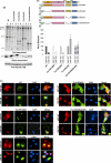
Generalized disruption of the SNARE machinery inhibits retrovirus release. A, top, HeLa cells were transfected with control, NSF, or SNAP-23 siRNA. 24 h later, cells were infected with HIV-1 particles pseudotyped with vesicular stomatitis virus G. 24 h after infection, cells were labeled with [35S]Met/Cys for 3 h. Virus was pelleted by ultracentrifugation, and cell and virus lysates were immunoprecipitated with HIV-Ig and subjected to SDS-PAGE and fluorography. Positions of the Gag precursor protein Pr55Gag (Pr55) and mature CA (p24) are indicated. Relative virus release efficiency (virus release) was calculated as described under “Materials and Methods.” Bottom, efficiency of NSF and SNAP-23 siRNA-mediated depletion is shown. HeLa cells were transfected with HA-tagged NSF or SNAP-23 along with the control, NSF-, or SNAP-23-specific siRNA. Cell lysates were analyzed by anti-HA Western blotting (WB). B–D, WT HIV-1 (B) or EIAV (C), or MLV expression vectors (D) were cotransfected with control vector or vectors expressing HA-tagged NSF-WT or NSF-DN at a DNA ratio of 4:1. Virus release was determined as described above; anti-EIAV horse antiserum was used for the EIAV Gag IP, and goat anti-MLV serum was used for MLV Gag IP. In B, the effect of NSF overexpression on HIV-1 Gag processing, as measured by the ratio of Pr55Gag (Pr55) to p24 (CA) is also shown. Data represent mean ± S.D. (error bars), n = 5 (A and B) or 3 (C and D).
FIGURE 2.
FIGURE 2.
NSF-DN does not inhibit release or localization of NC-deficient Gag. A, domain organization of WT HIV-1 Gag, showing MA, CA, SP1, NC, SP2, and p6 domains, or the Fyn10-tagged derivatives used in this study. The 10 amino acid residues at the N terminus were derived from the PM-associated Fyn protein (light green), which bears one myristylation (M) and two palmitylation (Palm) signals. B, HeLa cells cotransfected with pNL4-3 clones expressing WT HIV-1 Gag or the Fyn10-tagged derivates and NSF-WT (lanes 1) or NSF-DN (lanes 2) at a DNA ratio of 4:1. 24 h after transfection, cells were radiolabeled; cell and viral lysates were prepared and processed as described in the Fig. 1 legend. Relative virus release data are graphed from three experiments ± S.D. (error bars). In each case, virus release efficiency was normalized to the levels obtained in the presence of NSF-WT expression and was set at a relative value of 100%. C, HeLa cells cotransfected with pNL4-3 clones expressing WT Gag or the Fyn10-tagged derivatives and HA-tagged NSF-WT or NSF-DN at a DNA ratio of 1.6:1. Cells were fixed 24 h after transfection, stained with anti-p24 (green) and anti-HA antibodies (red), and analyzed by fluorescence microscopy. Nuclei are shown in blue (DAPI).
FIGURE 3.
FIGURE 3.
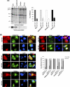
NSF-DN inhibits the production of K29E/K31E MA-mutant and late domain-defective HIV-1. A, HeLa cells were cotransfected with WT pNL4-3 or derivatives expressing the K29E/K31E MA mutant or PTAP late domain mutant and vectors expressing NSF-WT (lane 1) or NSF-DN (lane 2) at a DNA ratio of 4:1. Cells were metabolically radiolabeled, and virus and cell lysates were prepared and analyzed as described under “Materials and Methods ” and in the Fig. 1 legend. Data represent means ± S.D. (error bars), n = 3. B, HeLa cells were cotransfected with the indicated pNL4-3 derivatives and vectors expressing HA-tagged WT or DN NSF at a DNA ratio of 1.6:1. Cells were fixed 24 h after transfection, stained with anti-p24 and anti-HA antibodies, and analyzed by fluorescence microscopy. Graph represents quantitation of diffuse or punctate Gag staining pattern in NSF-WT- or NSF-DN-expressing cells, n = 30. Cells with more than 75% diffuse or punctate Gag staining were scored as diffuse versus punctate, respectively.
FIGURE 4.
FIGURE 4.
NSF-DN induces the cytosolic localization of CD63 and CD81. A and B, HeLa cells were transfected with vectors expressing HA-tagged NSF-WT or NSF-DN. 24 h after transfection, cells were fixed, stained with anti-HA antibody along with anti-CD63 (A) or anti-CD81 antibody (B) and analyzed by fluorescence microscopy. C, HeLa cells were transfected with control pcDNA3.1, NSF-WT, or NSF-DN expression vectors. Cells were harvested approximately 24 h after transfection, and membrane and cytosolic fractions were isolated using the Subcellular Protein Fractionation kit (Pierce) and analyzed for CD63 expression using Western blotting. M, membrane fraction; C, cytosolic fraction; L, protein ladder. One representative of two independent experiments is shown.
FIGURE 5.
FIGURE 5.
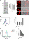
Expression of NSF-DN leads to defects in HIV-1 Env glycoprotein processing and virion infectivity. A, HeLa cells were cotransfected with WT pNL4-3 and vectors expressing NSF-WT or NSF-DN as described previously (DNA ratio 4:1). 24 h after transfection cells were radiolabeled and processed as described in the Fig. 1 legend. Data represent mean ± S.D. (error bars), n = 3. This panel represents a portion of Fig. 1B. B, HeLa cells were transfected as described in A. Virus supernatants were collected 24 h after transfection, normalized for reverse transcriptase activity, and used to infect the TZM-bl indicator cell line. Luciferase activity was measured 24 h after infection. Data are mean ± S.D. of triplicate observations from three independent experiments. C, HeLa cells were transfected with the WT HIV-1 clone along with control or HA-tagged NSF-WT or NSF-DN expression constructs. 24 h later the indicated wells were treated with 2 μg/ml BFA for 1 h. Cells were then incubated on ice for 5 min followed by staining with HIV-1 anti-gp120 antibody and anti-human Alexa Fluor 594 antibody on ice. Cells were then fixed, stained with anti-HA antibody, and analyzed by fluorescence microscopy. Results are representative of 20–35 cells scored for each treatment.
FIGURE 6.
FIGURE 6.
The defect in HIV-1 particle production induced by DN-NSF is distinct from the effects of BFA and CytoD. A, HeLa cells were transfected with WT pNL4-3. 24 h after transfection cells were treated with 1 μg/ml BFA or 5 μg/ml CytoD for 5 h. Cells were then metabolically radiolabeled with [35S]Met/Cys in media containing BFA or CytoD for an additional 3 h. Cell and virus lysates were prepared and immunoprecipitated with HIV-Ig as described under “Materials and Methods ” and in the Fig. 1 legend. Data represent mean ± S.D. (error bars), n = 3. B, HeLa cells were labeled with 10 μm CFSE and then transfected with control plasmid, NSF-WT, or NSF-DN plasmids or treated with 1 μg/ml BFA or 5 μg/ml CytoD as in A. Cells were analyzed for CFSE uptake by flow cytometry immediately following CFSE labeling or for CFSE dilution 24 h after labeling/transfection. Data represent mean ± S.D. of triplicate observations. One representative of two independent experiments is shown. C, HeLa cells transfected with vectors expressing NSF-WT or NSF-DN or were treated with BFA or CytoD as in B. 24 h after transfection cells were labeled with the membrane dye DiI and analyzed by fluorescence microscopy. D, TZM cells were transfected with control, NSF-WT, or NSF-DN expression constructs. 24 h later the indicated wells were treated with 1 μg/ml BFA for 5 h. Cells were then stained with CD4-FITC antibody and analyzed by flow cytometry. Data are mean ± S.D. from triplicate wells. One representative of two independent experiments is shown.
FIGURE 7.
FIGURE 7.
Hypothetical model for the action of NSF-DN on retroviral particle production. HIV-1 Gag (brown circle) is shown to traffic to the PM via a direct route or via late endosomes or transport vesicles. Env glycoproteins (blue lollipops) traffic to the PM via the ER-Golgi secretory pathway. Gag is targeted to cholesterol-enriched membrane raft microdomains (blue bar) and PI(4,5)P2 (yellow star) at the PM. NSF-DN is proposed to inhibit the function of t- and v-SNAREs and intra-Golgi membrane traffic.

Similar articles

Cited by

References

    1. Swanstrom R., Wills J. W. (1997) in Retroviruses (Coffin J. M., Hughes S. H., Varmus H. E. eds) pp. 263–334, Cold Spring Harbor Laboratory, Cold Spring Harbor, NY - PubMed
    1. Vogt V. M. (1997) in Retroviruses (Coffin J. M., Hughes S. H., Varmus H. E. eds) pp. 27–70, Cold Spring Harbor Laboratory, Cold Spring Harbor, NY
    1. Adamson C. S., Freed E. O. (2007) Adv. Pharmacol. 55, 347–387 - PubMed
    1. Bieniasz P. D. (2006) Virology 344, 55–63 - PubMed
    1. Freed E. O. (1998) Virology 251, 1–15 - PubMed

Publication types

MeSH terms

Substances

LinkOut - more resources