. 2012 Apr;227(4):1553-60.
doi: 10.1002/jcp.22886.

The RhoA-Rok-myosin II pathway is involved in extracellular matrix-mediated regulation of prolactin signaling in mammary epithelial cells

Affiliations
Free PMC article

The RhoA-Rok-myosin II pathway is involved in extracellular matrix-mediated regulation of prolactin signaling in mammary epithelial cells

Jyun-Yi Du et al. J Cell Physiol. 2012 Apr.
Free PMC article

Abstract

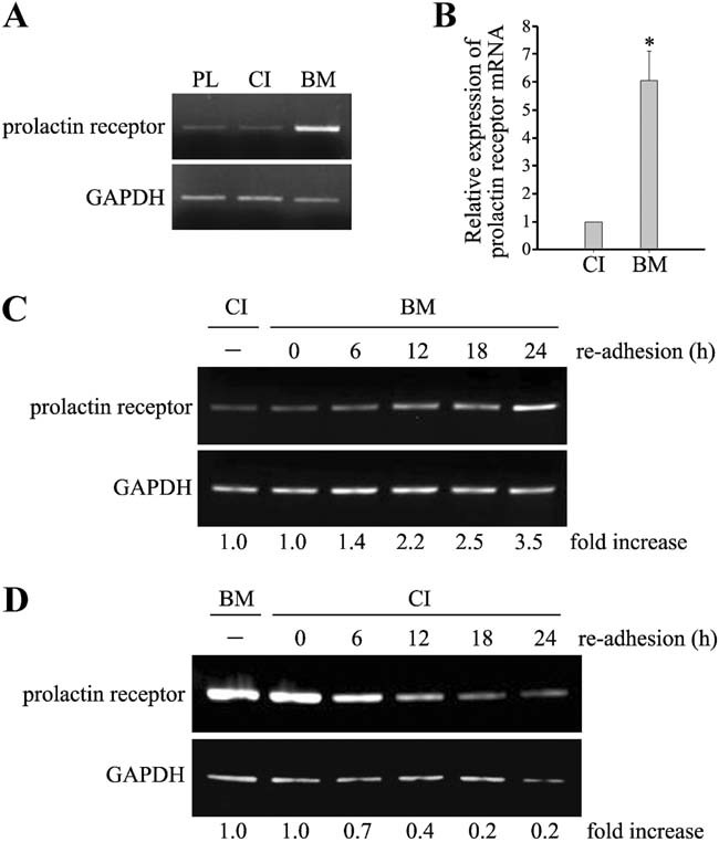
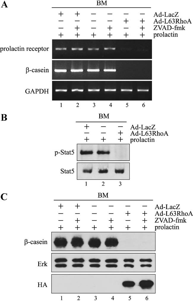
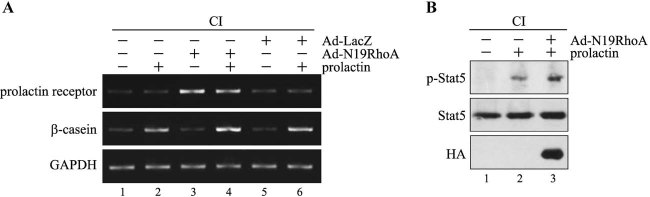
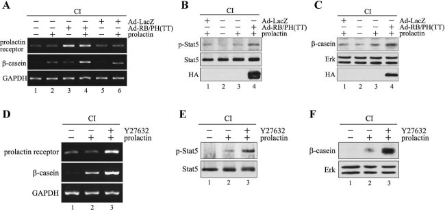
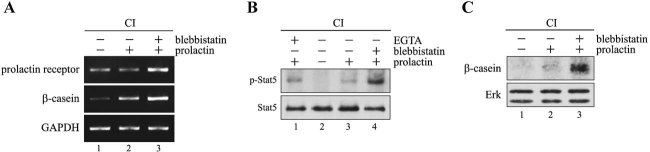
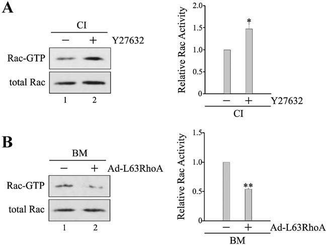
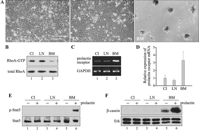
In mammary epithelial cells (MECs), prolactin-induced signaling and gene expression requires integrin-mediated cell adhesion to basement membrane (BM). In the absence of proper cell-BM interactions, for example, culturing cells on collagen-coated plastic dishes, signal propagation is substantially impaired. Here we demonstrate that the RhoA-Rok-myosin II pathway accounts for the ineffectiveness of prolactin signaling in MECs cultured on collagen I. Under these culture conditions, the RhoA pathway is activated, leading to downregulation of prolactin receptor expression and reduced prolactin signaling. Enforced activation of RhoA in MECs cultured on BM suppresses prolactin receptor levels, and prevents prolactin-induced Stat5 tyrosine phosphorylation and β-casein expression. Overexpression of dominant negative RhoA in MECs cultured on collagen I, or inhibiting Rok activity, increases prolactin receptor expression, and enhances prolactin signaling. In addition, inhibition of myosin II ATPase activity by blebbistatin also exerts a beneficial effect on prolactin receptor expression and prolactin signaling, suggesting that tension exerted by the collagen substratum, in collaboration with the RhoA-Rok-myosin II pathway, contributes to the failure of prolactin signaling. Furthermore, MECs cultured on laminin-coated plastic have similar morphology and response to prolactin as those cultured on collagen I. They display high levels of RhoA activity and are inefficient in prolactin signaling, stressing the importance of matrix stiffness in signal transduction. Our results reveal that RhoA has a central role in determining the fate decisions of MECs in response to cell-matrix interactions.


Figures

Fig. 1
Fig. 1
MECs cultured on plastic and collagen I exhibit higher RhoA activity than those cultured on BM. MECs were cultured on plastic (PL), collagen I (CI) or BM. Cell lysates were incubated with GST-Rhotekin Rho binding domain bound to glutathione-agarose beads to precipitate GTP-bound Rho. Total lysates and precipitates were then analyzed by immunoblotting using antibody to RhoA.
Fig. 2
Fig. 2
ECM regulates the expression of prolactin receptor. A: MECs were cultured on plastic (PL), collagen I (CI) or BM for 4 days. Total RNA was reverse transcribed and PCR-amplified with primers for prolactin receptor and GAPDH. B: MECs were cultured on collagen I or BM for 5 days. Total RNA was reverse transcribed and subjected to real-time PCR using primers for prolactin receptor and MAPK1. Relative expression of prolactin receptor normalized to MAPK1 is expressed as fold increase with respect to cells cultured on collagen I. *P < 0.05. C: MECs were either cultured on collagen I for 4 days, or cultured on collagen I for 3 days, then trypsinized, and replated on BM for 0–24 h. D: MECs were either cultured on BM for 4 days, or cultured on BM for 3 days, then trypsinized and replated on collagen I for 0–24 h. Total RNA was reverse transcribed and PCR-amplified with primers for prolactin receptor and GAPDH. Relative expression of prolactin receptor normalized to GAPDH is expressed as fold increase.
Fig. 3
Fig. 3
Constitutively active RhoA inhibits prolactin signaling in MECs cultured on BM. MECs cultured on BM were trypsinized, infected with adenovirus carrying HA-tagged constitutively active RhoA (Ad-L63RhoA) or LacZ (Ad-LacZ), and replated on BM for 24 h. A: Cells were then stimulated with 3 µg/ml prolactin in the absence or presence 100 µM ZVAD-fmk for 24 h. Total RNA was extracted, reverse transcribed, and PCR-amplified with primers for prolactin receptor, β-casein, and GAPDH. B: Cells were serum-starved for 8 h, and then stimulated with prolactin for 15 min. Cell lysates were analyzed by immunoblotting with antibodies to phospho-Stat5 (p-Stat5) and Stat5. C: Cells were stimulated with prolactin in the absence or presence ZVAD-fmk for 36 h. Total cell lysates were analyzed by immunoblotting with antibodies to β-casein, Erk, and HA. Levels of Erk were used as loading control.
Fig. 4
Fig. 4
Expression of dominant negative RhoA improves prolactin signaling in MECs cultured on collagen I. MECs cultured on collagen I (CI) were infected with adenovirus carrying HA-tagged dominant negative RhoA (Ad-N19RhoA) or LacZ (Ad-LacZ) in situ for 24 h. A: Cells were then stimulated without or with 3 µg/ml prolactin for 24 h. Total RNA was extracted, reverse transcribed, and PCR-amplified with primers for prolactin receptor, β-casein, and GAPDH. Due to the low levels of β-casein expression in these cells, the number of cycles for PCR-amplification of β-casein cDNA was increased as described in Materials and Methods section. (B) Cells were serum-starved for 8 h, and then stimulated with prolactin for 15 min. Cell lysates were analyzed by immunoblotting with antibodies to phospho-Stat5 (p-Stat5), Stat5, and HA.
Fig. 5
Fig. 5
Expression of dominant negative Rok or application of Rok inhibitor Y27632 enhances prolactin signaling in MECs cultured on collagen I. A–C: MECs cultured on collagen I (CI) were infected with adenovirus carrying HA-tagged dominant negative Rok (Ad-RB/PH (TT)) or LacZ (Ad-LacZ) in situ for 24 h. D–F: MECs cultured on collagen I were pretreated with 10 µM Y27632. A,D: Cells were then stimulated without or with 3 µg/ml prolactin for 24 h. Total RNA was isolated, reverse transcribed, and PCR-amplified with primers for prolactin receptor, β-casein, and GAPDH. B,E: Cells were stimulated without or with prolactin for 15 min. Cell lysates were analyzed by immunoblotting with antibodies to phospho-Stat5 (p-Stat5), Stat5 or HA. C,F: Cells were stimulated without or with prolactin for 36 h. Total cell lysates were analyzed by immunoblotting with antibodies to β-casein, Erk, or HA.
Fig. 6
Fig. 6
Blebbistatin augments prolactin signaling in MECs cultured on collagen I. A,C: MECs cultured on collagen I (CI) were pretreated without or with 25 µM blebbistatin for 1 h, and then stimulated with 3 µg/ml prolactin for 24 or 36 h. Total RNA was isolated after 24-h stimulation, reverse transcribed, and PCR-amplified with primers for prolactin receptor, β-casein, and GAPDH (A). Total cell lysates were prepared after 36-h stimulation, and analyzed by immunoblotting with antibodies to β-casein and Erk (C). B: MECs were untreated, pretreated with 25 µM blebbistatin for 12 h, or pretreated with 5 mM EGTA for 1 h, and then stimulated with prolactin for 15 min. Cell lysates were analyzed by immunoblotting with antibodies to phospho-Stat5 (p-Stat5) and Stat5.
Fig. 7
Fig. 7
Application of Y27632 into MECs cultured on collagen I increases Rac activity, whereas expression of constitutively active RhoA in MECs cultured on BM decreases Rac activity. A: MECs cultured on collagen I (CI) were treated with 15 µM Y27632 for 12 h. B: MECs cultured on BM were trypsinized, mock-infected, or infected with adenovirus carrying constitutively active RhoA (Ad-L63RhoA), and replated on BM for 24 h. Cell lysates were incubated with GST-PAK1 p21-binding domain bound to glutathione-agarose beads to precipitate GTP-bound Rac. Total lysates and precipitates were then analyzed by immunoblotting using antibody to Rac. Immunoblots from three independent experiments were analyzed by densitometry. Relative Rac activity is indicated by the amount of Rac-GTP normalized to that of total Rac, and values are expressed as fold stimulation with respect to untreated cells or mock-infected cells. *P < 0.05; **P < 0.01.
Fig. 8
Fig. 8
MECs cultured on 2D collagen I and 2D laminin are less effective in prolactin signaling than those cultured on 3D BM. MECs were cultured on 2D collagen I (CI), 2D laminin (LN), or 3D BM for 4 days. A: Phase contrast micrographs of the cells. B: Cell lysates were incubated with GST-Rhotekin Rho binding domain bound to glutathione-agarose beads to precipitate GTP-bound Rho. Total lysates and precipitates were then analyzed by immunoblotting using antibody to RhoA. C: Total RNA was reverse transcribed and PCR-amplified with primers for prolactin receptor and GAPDH. D: Total RNA was reverse transcribed and subjected to real-time PCR using primers for prolactin receptor and MAPK1. Relative expression of prolactin receptor normalized to MAPK1 is expressed as fold increase with respect to cells cultured on collagen I (n = 3). E: Cells were serum-starved for 8 h, and then stimulated without or with 3 µg/ml prolactin for 15 min. Cell lysates were analyzed by immunoblotting with antibodies to phospho-Stat5 (p-Stat5) and Stat5. F: Cells were stimulated without or with prolactin for 36 h. Total cell lysates were analyzed by immunoblotting with antibodies to β-casein and Erk.

Similar articles

Cited by

References

    1. Akhtar N, Streuli CH. Rac1 links integrin-mediated adhesion to the control of lactational differentiation in mammary epithelia. J Cell Biol. 2006;173:781–793. - PMC - PubMed
    1. Akhtar N, Marlow R, Lambert E, Schatzmann F, Lowe ET, Cheung J, Katz E, Li W, Wu C, Dedhar S, Naylor MJ, Streuli CH. Molecular dissection of integrin signalling proteins in the control of mammary epithelial development and differentiation. Development. 2009;136:1019–1027. - PMC - PubMed
    1. Alcaraz J, Xu R, Mori H, Nelson CM, Mroue R, Spencer VA, Brownfield D, Radisky DC, Bustamante C, Bissell MJ. Laminin and biomimetic extracellular elasticity enhance functional differentiation in mammary epithelia. Embo J. 2008;27:2829–2838. - PMC - PubMed
    1. Arthur WT, Petch LA, Burridge K. Integrin engagement suppresses RhoA activity via a c-Src-dependent mechanism. Curr Biol. 2000;10:719–722. - PubMed
    1. Bustos RI, Forget MA, Settleman JE, Hansen SH. Coordination of Rho and Rac GTPase function via p190B RhoGAP. Curr Biol. 2008;18:1606–1611. - PMC - PubMed

Publication types

MeSH terms

LinkOut - more resources