. 2012 Apr;227(4):1592-603.
doi: 10.1002/jcp.22876.

Essential role of maternal UCHL1 and UCHL3 in fertilization and preimplantation embryo development

Affiliations

Essential role of maternal UCHL1 and UCHL3 in fertilization and preimplantation embryo development

Namdori R Mtango et al. J Cell Physiol. 2012 Apr.

Abstract

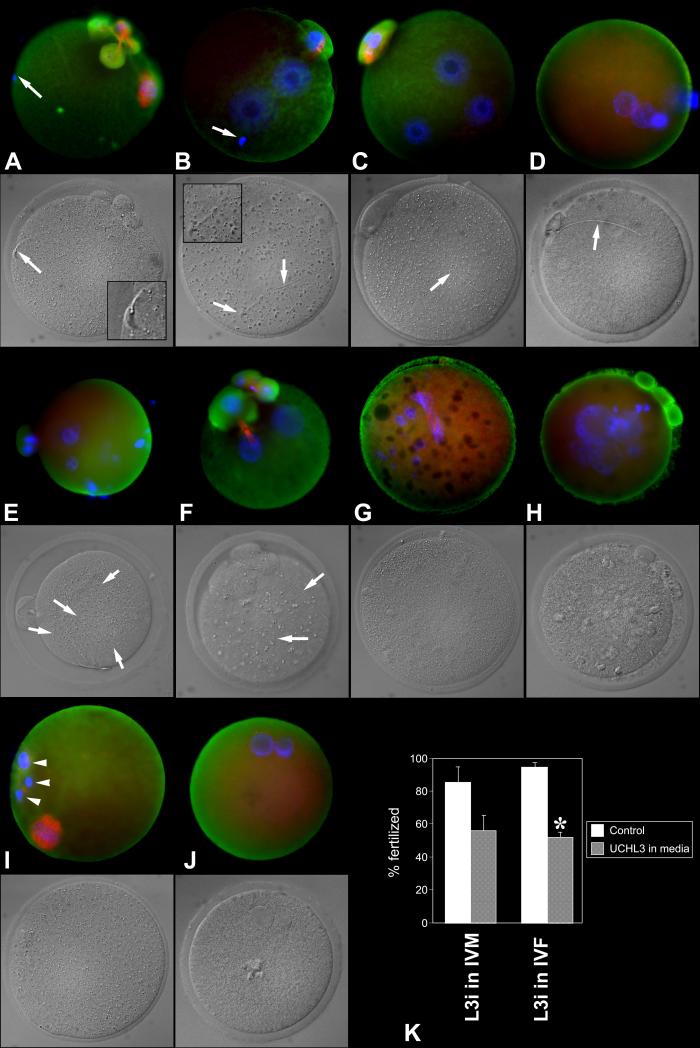
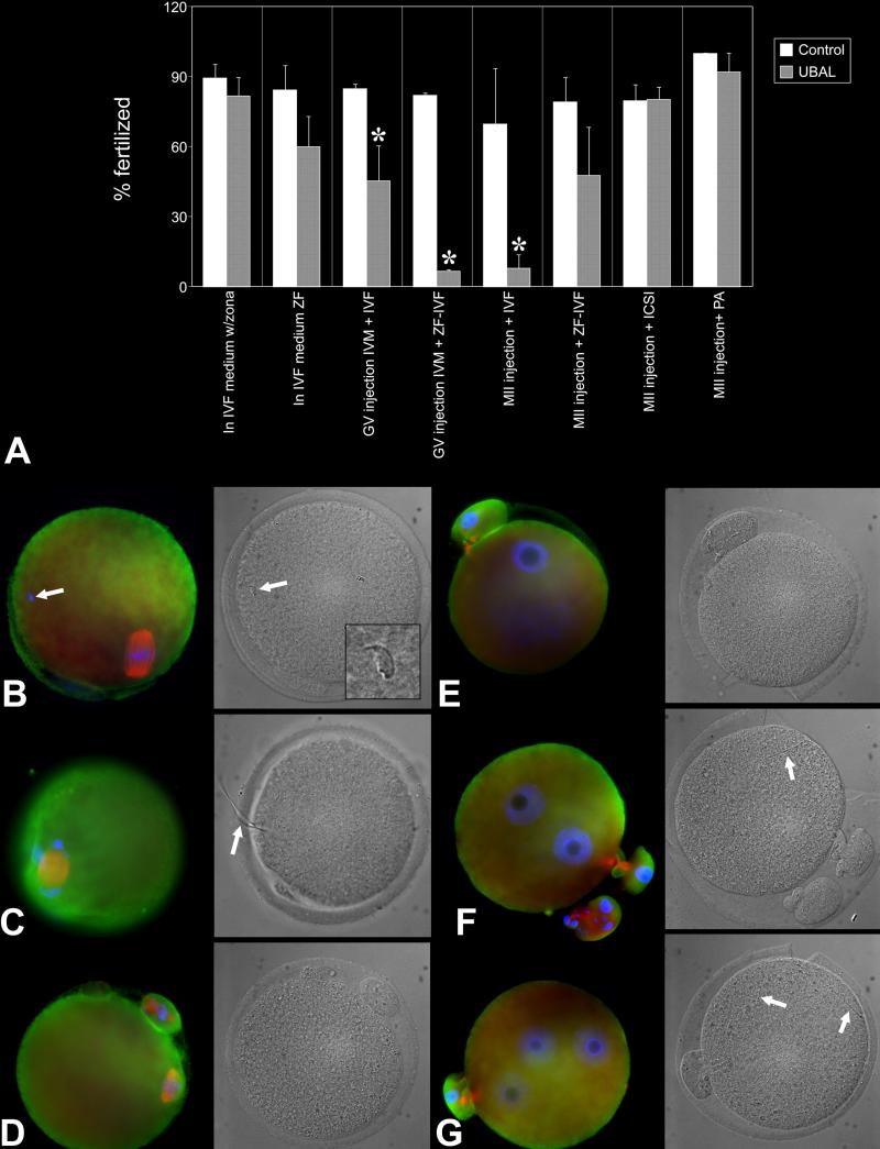
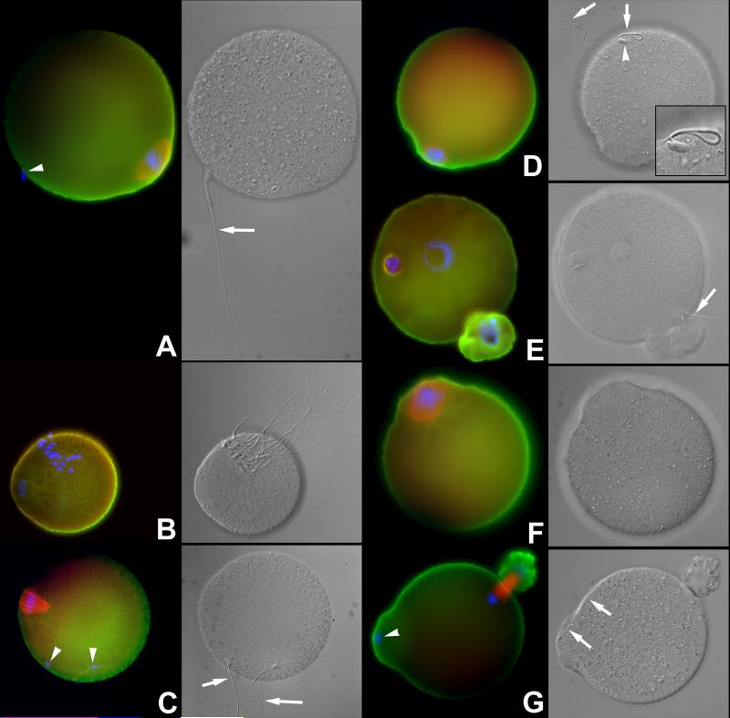
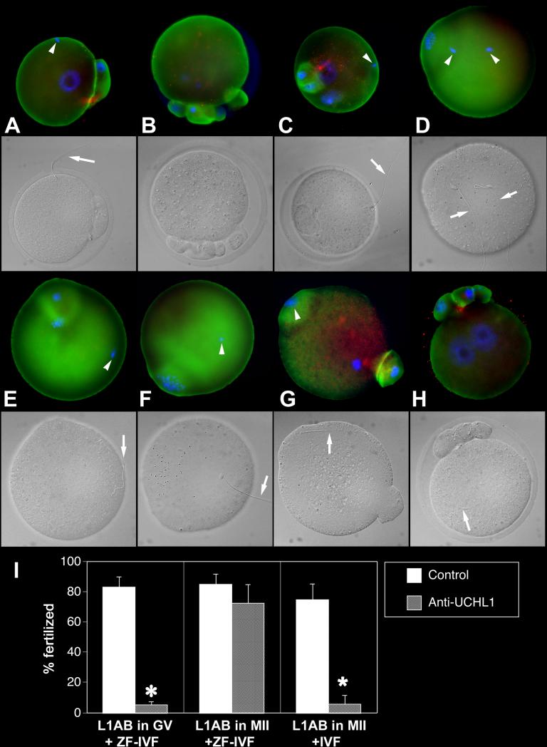
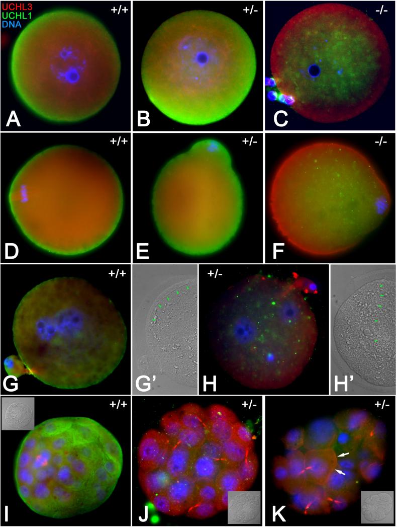
Post-translational protein modification by ubiquitination, a signal for lysosomal or proteasomal proteolysis, can be regulated and reversed by deubiquitinating enzymes (DUBs). This study examined the roles of UCHL1 and UCHL3, two members of ubiquitin C-terminal hydrolase (UCH) family of DUBs, in murine fertilization and preimplantation development. Before fertilization, these proteins were associated with the oocyte cortex (UCHL1) and meiotic spindle (UCHL3). Intracytoplasmic injection of the general UCH-family inhibitor ubiquitin-aldehyde (UBAL) or antibodies against UCHL3 into mature metaphase II oocytes blocked fertilization by reducing sperm penetration of the zona pellucida and incorporation into the ooplasm, suggesting a role for cortical UCHL1 in sperm incorporation. Both UBAL and antibodies against UCHL1 injected at the onset of oocyte maturation (germinal vesicle stage) reduced the fertilizing ability of oocytes. The subfertile Uchl1(gad-/-) mutant mice showed an intriguing pattern of switched UCH localization, with UCHL3 replacing UCHL1 in the oocyte cortex. While fertilization defects were not observed, the embryos from homozygous Uchl1(gad-/-) mutant females failed to undergo morula compaction and did not form blastocysts in vivo, indicating a maternal effect related to UCHL1 deficiency. We conclude that the activity of oocyte UCHs contributes to fertilization and embryogenesis by regulating the physiology of the oocyte and blastomere cortex.


Figures

Fig. 1
Fig. 1
Fertilization anomalies in the oocyte matured under control conditions (no inhibitor) and fertilized in the presence of UCHL3-inhibitor. A. Spermatozoon (arrow) penetrated ZP but failed to fuse with the oolemma and/or become incorporated. Oocyte displays abnormal spindle and a fragmented PB1. B. Sperm incorporation failed (arrow) but the ovum was activated, forming two female PN. C. Monospermic zygote with two female PN. D. Monospermic zygote with supernumerary, abnormally small pronuclei. E. Polyspermic egg showing multiple sperm tails in ooplasm. F. Dispermic ovum with supernumerary pronuclei and PBs. G. Fertilization failure with disrupted spindle and multiple female pronuclei/karyomeres. H. Fertilization failure with multiple parthenogenetic female pronuclei/karyomeres. I. Fertilization failure with a fragmented first polar body (arrowheads; incomplete PB2 extrusion). J. Spontaneous parthenogenote showing two abnormally small pronuclei. K. Diagram summarizing fertilization rates of ova exposed to UCHL3-inhibitor during in vitro maturation (two left columns; n=176 ova, two replicates) or fertilization (two right columns; n=486 ova, three replicates). Asterisk denotes a statistically significant difference between control and treatment, at P < 0.05.
Fig. 2
Fig. 2
Intracytoplasmic injection of ubiquitin-aldehyde (UBAL) prevents murine fertilization but does not cause sperm accumulation in the perivitelline space. A. Diagram summarizing the effect of UBAL on fertilization rate under various scenarios including addition of UBAL in IVF medium (n=639 ova, four replicates), injection of UBAL in GV or MII oocytes followed by IVF (n= 164 ova and 301 ova, respectively; two replicates for GV, four for MII oocytes), injection of UBAL in GV or MII oocytes followed zona-free IVF (ZF-IVF; n=169 ova, two replicates for GV; n=660 ova, three replicates for MII), injection of UBAL in MII oocytes followed ICSI (n=733 ova, five replicates) or injection of UBAL in MII oocytes followed parthenogenetic activation (PA; n=412 ova, three replicates). Asterisk denotes a statistically significant difference between control and treatment, at P < 0.05.B. Metaphase II-stage, UBAL preinjected ovum. Only one spermatozoon is visible in perivitelline space (arrow). C. An UBAL-preinjected ovum in which the sperm head became incorporated in the oocyte, but the sperm flagellum (arrow) incorporation has not been completed. D-G. Control oocytes that remained unfertilized at metaphase-II (D), failed to fertilize but underwent spontaneous parthenogenetic activation (E; one PN is visible), were fertilized by a single spermatozoon (F, arrow) or fertilized by two spermatozoa (G, two sperm tails are identified in ooplasm by arrows).
Fig. 3
Fig. 3
A. Fertilization /sperm incorporation anomalies in the oocytes pre-injected with UBAL at the GV or MII-stage of maturation, and fertilized zona-free. Arrows point to sperm tails, arrowheads to sperm nuclei. A Failed sperm-oolemma fusion and sperm incorporation documented by the presence of oolemma-bound intact spermatozoon and lack of oocyte activation (metaphase-II plate still present). B. Rare case in which multiple oolemma-bound spermatozoa were observed in an oocyte preinjected with UBAL. C Sperm incorporation and oocyte activation failure with two oolemma-bound spermatozoa. D. Sperm incorporation and oocyte activation failure, with a single oolemma-bound spermatozoon. E. Incomplete sperm tail incorporation in the presence of a male and female pronucleus and PB2 extrusion. The male pronucleus is abnormal. F. Complete fertilization failure in an UBAL-preinjected ovum. G. Control, vehicle-injected ovum showing normal incorporation/fertilization cone with concomitant incorporation of the sperm head and tail. Labeling of UCHL1 is green, UCHL3 red and DNA blue. Corresponding DIC images are shown in grayscale.
Fig. 4
Fig. 4
Fertilization failures of oocytes pre-injected with anti-UCHL1-antibody (L1AB) at the MII-stage of maturation. Labeling of UCHL1 is green, UCHL3 red and DNA blue. Arrows point to sperm tails, arrowheads to sperm nuclei. Corresponding DIC images are shown in grayscale. A. Injection of L1AB followed by IVF resulted in a sperm incorporation failure. B. An oocyte pre-injected with L1AB at MII shows abnormal PB1 extrusion and maternal chromosome partition. C. A complete sperm incorporation block is observed in the presence of oocyte activation and female PN-development (sperm-oolemma fusion occurred normally) after the L1AB injection. D-F. An ovum injected with L1AB at MII-stage, showing sperm incorporation and oocyte activation failure. G. Non-immune serum injection at MII and zona-free-IVF; normal sperm incorporation features a fertilization cone with concomitant incorporation of the sperm head and tail. H. Ovum pre-injected with non-immune serum and fertilized with intact zona, shows two normal pronuclei; PB1 is already fragmented H. Diagram summarizing fertilization rates of oocytes pre-injected with L1AB at GV-stage and fertilized zona-free (ZF-IVF), oocytes pre-injected with L1AB at MII stage and fertilized zona free (total of four replicates using 761 ova), and oocytes pre-injected with L1Ab at MII stage and fertilized with an intact zona (IVF; four replicates, 544 ova). Asterisk denotes a statistically significant difference between control and treatment, at P < 0.05.
Fig. 5
Fig. 5
Oocyte maturation, fertilization and embryo development in Uchl1gad mice. A-F Homozygous Uchl1 gad−/− oocytes (−/−), heterozygous Uchl1gad−/− oocytes (+/−) and wild type Uchl1gad+/+ oocytes (+/+) were obtained from the properly phenotyped daughters of Uchl1gad+/− female mice mated with Uchl1gad+/− males. Red labeling denotes UCHL3 while UCHL1 is shown in green. A-C. The GV-stage oocytes; note the replacement of cortical UCHL1 with UCHL3 in Uchl1gad−/− ovum. D-F. Metaphase-II oocyte; UCHL3 translocation to oocyte cortex becomes even more obvious in Uchl1gad−/− oocyte. G-K. Wild type Uchl1gad+/+ zygotes/embryos (G, G’, I) were obtained from Uchl1gad+/+ females mated with Uchl1gad+/+ males. Heterozygous Uchl1gad+/− zygotes and later stage embryos (H, H’, J and K) were obtained from Uchl1gad−/− females mated with Uchl1gad+/+ males; G, H. Neither the wild type nor the Uchl1gad+/− oocyte showed polyspermy or fertilization failure. Normal fertilization is revealed by the presence of two pronuclei (blue) and one sperm tail (arrows in panels G’ and H’, showing corresponding DIC images). I-K. Day 4 embryos; wild type, Uchl1gad+/+ embryo has a normal blastocyst appearance with a dividing blastomere within its inner cell mass. The Uchl1gad+/− embryos failed to reach blastocyst stage and became arrested at pre-compaction morula stage. Note the accumulation of UCHL3 in blastomere cortex in Uchl1gad+/− morula (arrows, panel K). Insets show corresponding DIC images.

Similar articles

Cited by

References

    1. Assou S, Cerecedo D, Tondeur S, Pantesco V, Hovatta O, Klein B, Hamamah S, De Vos J. A gene expression signature shared by human mature oocytes and embryonic stem cells. BMC Genomics. 2009;10:10. - PMC - PubMed
    1. Boudreaux DA, Maiti TK, Davies CW, Das C. Ubiquitin vinyl methyl ester binding orients the misaligned active site of the ubiquitin hydrolase UCHL1 into productive conformation. Proc Natl Acad Sci U S A. 2010;107(20):9117–9122. - PMC - PubMed
    1. Chakravarty S, Bansal P, Sutovsky P, Gupta SK. Role of proteasomal activity in the induction of acrosomal exocytosis in human spermatozoa. Reprod Biomed Online. 2008;16(3):391–400. - PubMed
    1. Doelling JH, Phillips AR, Soyler-Ogretim G, Wise J, Chandler J, Callis J, Otegui MS, Vierstra RD. The ubiquitin-specific protease subfamily UBP3/UBP4 is essential for pollen development and transmission in Arabidopsis. Plant Physiol. 2007;145(3):801–813. - PMC - PubMed
    1. Ellederova Z, Halada P, Man P, Kubelka M, Motlik J, Kovarova H. Protein patterns of pig oocytes during in vitro maturation. Biol Reprod. 2004;71(5):1533–1539. - PubMed

Publication types

MeSH terms

LinkOut - more resources