. 2011;6(6):e20616.
doi: 10.1371/journal.pone.0020616. Epub 2011 Jun 1.

Biosynthesis of promatrix metalloproteinase-9/chondroitin sulphate proteoglycan heteromer involves a Rottlerin-sensitive pathway

Affiliations

Biosynthesis of promatrix metalloproteinase-9/chondroitin sulphate proteoglycan heteromer involves a Rottlerin-sensitive pathway

Nabin Malla et al. PLoS One. 2011.

Abstract

Background: Previously we have shown that a fraction of the matrix metalloproteinase-9 (MMP-9) synthesized by the macrophage cell line THP-1 was bound to a chondroitin sulphate proteoglycan (CSPG) core protein as a reduction sensitive heteromer. Several biochemical properties of the enzyme were changed when it was bound to the CSPG.

Methodology/principal findings: By use of affinity chromatography, zymography, and radioactive labelling, various macrophage stimulators were tested for their effect on the synthesis of the proMMP-9/CSPG heteromer and its components by THP-1 cells. Of the stimulators, only PMA largely increased the biosynthesis of the heteromer. As PMA is an activator of PKC, we determined which PKC isoenzymes were expressed by performing RT-PCR and Western Blotting. Subsequently specific inhibitors were used to investigate their involvement in the biosynthesis of the heteromer. Of the inhibitors, only Rottlerin repressed the biosynthesis of proMMP-9/CSPG and its two components. Much lower concentrations of Rottlerin were needed to reduce the amount of CSPG than what was needed to repress the synthesis of the heteromer and MMP-9. Furthermore, Rottlerin caused a minor reduction in the activation of the PKC isoenzymes δ, ε, θ and υ (PKD3) in both control and PMA exposed cells.

Conclusions/significance: The biosynthesis of the proMMP-9/CSPG heteromer and proMMP-9 in THP-1 cells involves a Rottlerin-sensitive pathway that is different from the Rottlerin sensitive pathway involved in the CSPG biosynthesis. MMP-9 and CSPGs are known to be involved in various physiological and pathological processes. Formation of complexes may influence both the specificity and localization of the enzyme. Therefore, knowledge about biosynthetic pathways and factors involved in the formation of the MMP-9/CSPG heteromer may contribute to insight in the heteromers biological function as well as pointing to future targets for therapeutic agents.


Conflict of interest statement

Competing Interests: The authors have declared that no competing interests exist.

Figures

Figure 1
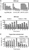
Figure 1. Cell viability in the presence of PMA, ConA, MCSF and Rottlerin.
Cell viability was detected by the MTS assay (A, B) and by counting viable cells in a Bürker chamber (C) using the Trypan Blue exclusion test. In (A) 6×10⁴ cells in serum free medium were seeded per well in 96 well plates in the absence or presence of PMA, ConA or MCSF at the indicated concentrations, and incubated for 72 h. In (B) and (C) the cells were incubated with various concentrations of Rottlerin (as indicated in the insert in c) either in the absence or presence of 10−7 M PMA. In (B) 6×10⁴ cells were seeded per well in 96 well plates and in (C) 4×10⁵ cells were added to each well in 12 well plates (0.6 ml/well) and incubated in serum free medium for the indicated time period. The results presented are from a typical experiment where N = 4 in (A), 3 in (B) and 2 in (C); *p<0.05 compared to control.
Figure 2
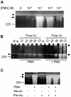
Figure 2. Synthesis of proMMP-9/CSPG in the absence and presence of PMA.
THP-1 cells were incubated in the absence or presence of PMA in serum free medium. Harvested medium was thereafter applied to Q-Sepharose chromatography and the presence of proMMP-9/CSPG was detected with gelatin zymography as described in Materials and Methods. In (A), cells were incubated for 72 h in the presence of various concentrations of PMA as indicated. In (B), cells were incubated for various time periods (as indicated) in the absence or presence of 10−7 M PMA. The samples (containing CSPG and proMMP-9/CSPG complex) from cells not exposed to PMA were five times more concentrated than the samples from the PMA treated cells when applied to the gel. In (C), cells were either incubated for 72 h in the absence (−) or presence (+) of 10−7 M PMA at serum free conditions, or pre-incubated for 3 h in the absence (−) or presence (+) of 10−7 M PMA and/or 10% fetal calf serum. After the pre-incubation, cells were washed three times in PBS and thereafter incubated in serum free medium for 72 h. Arrowhead shows the border between the separating and stacking gel, and arrows show the position of the proMMP-9/CSPG complexes. Purified proMMP-9 was used as a standard and the position of the 225 kDa homodimer form is shown at the left. The gels are representative for several similar experiments.
Figure 3
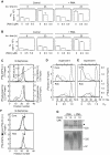
Figure 3. Synthesis of proMMP-9 and CSPG in the absence and presence of PMA.
AC: Typical gelatin zymographies of 20 times diluted conditioned medium from THP-1 cells. A: Cells were incubated for 72 h in the presence of various concentrations of PMA as indicated. B: Cells were incubated for various time periods (as indicated) in the absence or presence of 10−7 M and 10−5 M PMA. C: Cells were either incubated for 72 h in the absence (−) or presence (+) of 10−7 M PMA at serum free conditions, or pre-incubated for 3 h in the absence (−) or presence (+) of 10−7 M PMA and/or 10% fetal calf serum. After the pre-incubation, cells were washed three times in PBS and thereafter incubated in serum free medium for 72 h. A–C: The position of proMMP-9 homodimer (225 kDa), monomer (92 kDa) and proMMP-2 (72 kDa) standards is indicated. D and E: Conditioned medium from THP-1 cells incubated with [35S]sulphate was passed over a G-50 Sepharose column in order to separate labelled macromolecules from free [35S]sulphate. The amount of labelled macromolecules was determined by counting the entire pass through fraction in a liquid scintillation spectrometer. D: Cells were incubated in the absence or presence of various concentrations of PMA for 72 h as indicated and in (E) the cells were incubated for various time periods in the absence or presence of 10−7 M PMA. (D: upper and lower panel) Results (mean ± s.d) were normalized against the control (without PMA). Lower panel, results were in addition normalized against the number of viable cells. E: Results (mean ± s.d) are presented as cpm (upper panel) and cpm/viable cells (lower panel). The results in (D) and (E) are from a typical experiment with four parallels in (D) and three parallels in (E), where *p<0.05 compared to control without PMA.
Figure 4
Figure 4. PKC and PKD isoenzymes expressed in THP-1 cells.
RT-PCR was used to detect the PKC and PKD isoenzyme mRNA present in cells incubated in the absence (−) or presence (+) of 10-7 M PMA. To ensure that equal amounts of cDNA has been used in samples from control and PMA stimulated cells, rRNA was used as a standard. The probes used are described in Materials and Methods. Ladder is a marker with DNA fragments of known size (in bp).
Figure 5
Figure 5. Effect of PKC inhibitors on THP-1 cells synthesis of proMMP-9/CSPG heteromer and proMMP-9.
Shown is a typical experiment where cells in the absence (−) and presence (+) of PMA (10−7 M) were incubated with various concentrations of the PKC inhibitors Gö6976 and Gö6983 (A, B) and Rottlerin (C, D). In the presence of Gö6976 and Gö6983 (A, B), cells were incubated for 72 h, while in the presence of Rottlerin (C, D) the cells were incubated for 12 h in serum free medium. To detect the effect of the PKC inhibitors on the synthesis of the proMMP-9/CSPG heteromer (A, C), the harvested media was applied to Q-Sepharose chromatography as described in Materials and Methods prior to gelatin zymography. To determine the effect of the inhibitors on the synthesised proMMP-9 (B, D), the harvested medium was diluted 20 times and then applied to gelatin zymography. Arrowhead shows the border between the separating and stacking gel and the position of purified proMMP-9 monomer (92 kDa) and proMMP-9 homodimer (225 kDa) used as a standard (Std) is shown.
Figure 6
Figure 6. Effect of Rottlerin on biosynthesis and molecular size of CSPG and the CS-chains.
A, B: Cells in the absence (control) or presence of 10−7 M PMA were incubated with increasing concentrations of Rottlerin for 8 h and 23 h in serum free medium containing either [35S]sulphate (A) or [3H]glucosamine (B). A typical experiment is shown, where the results (total cpm not adjusted to the amount of living cells) are presented as mean ± s.d. (n = 2). CF: Cells were incubated for 24 h in serum free medium containing either [35S]sulphate or [3H]glucosamine. C: [35S]sulphate and [3H]glucosamine labelled macromolecules were applied to Q-Sepharose chromatography and the bound CSPG were eluted with a 0.15–1.5 M NaCl gradient (----) as shown in the upper graph. ○, absence of Rottlerin; Δ, presence of 1 µM Rottlerin. D, E: Eluted CSPG from the Q-Sepharose column was either untreated (solid line) or treated with cABC (---) or 0.5 M NaOH (…….) prior to application on a Superose 6 gel chromatography column as described in Material and Methods. Cont., control without Rottlerin; Rotl., presence of 1 µM Rottlerin and Fr.No., fraction number. F: [35S]CSPG (•) and free [35S]CS-chains (○) from control and Rottlerin-treated cells were subjected to Q-Sepharose chromatography as in (C). Arrow shows the elution position of shark cartilage CS. G: [35S]Sulphate labelled CSPG (isolated from control, PMA (10−7 M) and Rottlerin treated cells) was subjected to SDS-PAGE (upper panel: 4% stacking gel and 7.5% separating gel; lower panel: 4–12% gradient gel) followed by autoradiography (see Materials and Methods). An equal amount of radioactivity (based on scintillation counting) was loaded to each well in order to be able to compare the bands. Arrowhead shows the border between the separating and stacking gel and the position of molecular size markers are shown. Small arrow shows the bottom of the application well.
Figure 7
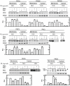
Figure 7. Effect of Rottlerin on cytosolic and plasma membrane bound PKC isoenzymes.
Isolated cytosol and plasma membranes from untreated (−) or PMA and Rottlerin treated (+) THP-1 cells were subjected to Western blotting using PKC specific antibodies. The amount of total protein loaded (PL) to each well is shown. As an additional loading control, ERK2 was used. The diagram under each blot shows the relative amounts (mean ±s.d.) of the PKC isoenzymes in cytosol and membranes, where the results were normalised against the untreated cytosol and membrane controls. All results are based on equal protein loading and in all cases n = 2 except for PKC δ and υ (PKD3) where n = 3. The arrowhead shows the active PKD3 at 100 kDa, while the two bands with reduced molecular size may be truncated variants of PKD3. The position of the molecular mass markers at 100 and 80 kDa are shown at the left. In order to show the increased level of PKC ε in the cytosol from PMA treated cells (353 µg/well) in the presence of Rottlerin compared to the absence of this compound, a largely increased developmental exposure time of the membrane in the presence of the Luminol substrate was needed.
Figure 8
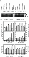
Figure 8. Effects of various compounds on the synthesis of proMMP-9/CSPG and CSPG.
THP-1 cells were incubated in the absence or presence of the compounds shown in serum free medium for 72 h. In (A), the harvested medium was applied to Q-Sepharose chromatography and the presence of proMMP-9/CSPG was detected with gelatin zymography as described in Materials and Methods. Arrowhead shows the border between the separating and stacking gel and arrow shows the position of the 300 kDa proMMP-9/CSPG heteromer. The position of the pro-MMP-9 homodimer (225 kDa) is shown. B: Cells were incubated with [35S]sulphate. The harvested serum free medium (open bars) and the lysed cell preparations (grey bars) were passed over a G-50 Sepharose column in order to separate labelled macromolecules from free [35S]sulphate. The entire pass through fraction that contains the labelled macromolecules was then counted in a liquid scintillation spectrometer. All results (mean ± s.d) were normalized against the controls, i.e. the synthesis in absence of added compounds. In lower panel with ConA and MCSF, the results were in addition normalized against the number of viable cells. The results in (B) are from a typical experiment with four parallels.

Similar articles

Cited by

References

    1. Hascall VC, Calabro A, Midura RJ, Yanagishita M. Isolation and characterization of proteoglycans. Methods Enzymol. 1994;230:390–417. - PubMed
    1. Kjellen L, Lindahl U. Proteoglycans: structures and interactions [published erratum appears in Annu Rev Biochem 1992;61:following viii]. Annu Rev Biochem. 1991;60:443–475. - PubMed
    1. Edwards IJ, Xu H, Obunike JC, Goldberg IJ, Wagner WD. Differentiated macrophages synthesize a heparan sulphate proteoglycan and an oversulphated chondroitin sulphate proteoglycan that bind lipoprotein lipase. Arterioscler Thromb Vasc Biol. 1995;15:400–409. - PubMed
    1. Kolset SO, Gallagher JT. Proteoglycans in haemopoietic cells. Biochim Biophys Acta. 1990;1032:191–211. - PubMed
    1. Kolset SO, Prydz K, Pejler G. Intracellular proteoglycans. Biochem J. 2004;379:217–227. - PMC - PubMed

Publication types

MeSH terms