Review
. 2011;87(6):287-327.
doi: 10.2183/pjab.87.287.

Calpain chronicle--an enzyme family under multidisciplinary characterization

Affiliations
Review

Calpain chronicle--an enzyme family under multidisciplinary characterization

Hiroyuki Sorimachi et al. Proc Jpn Acad Ser B Phys Biol Sci. 2011.

Abstract

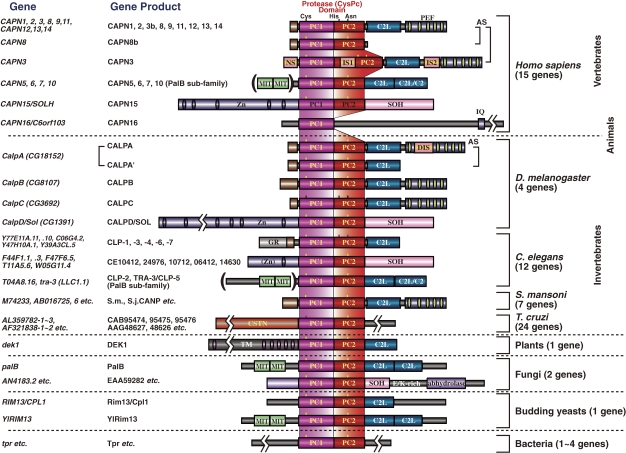
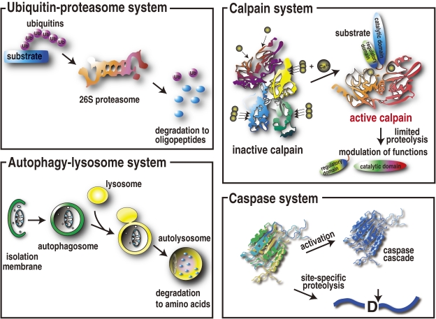
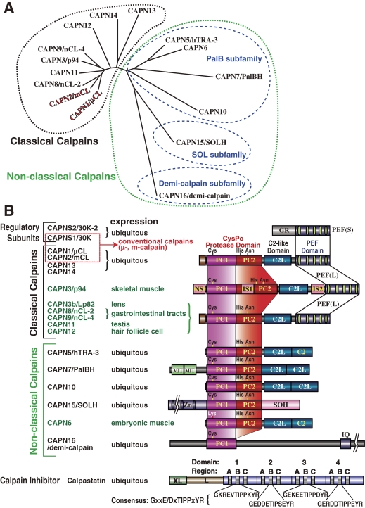
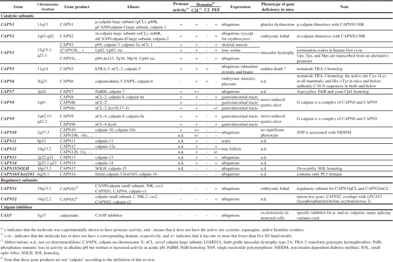
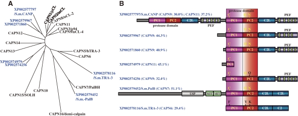
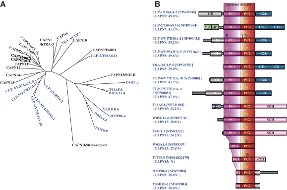
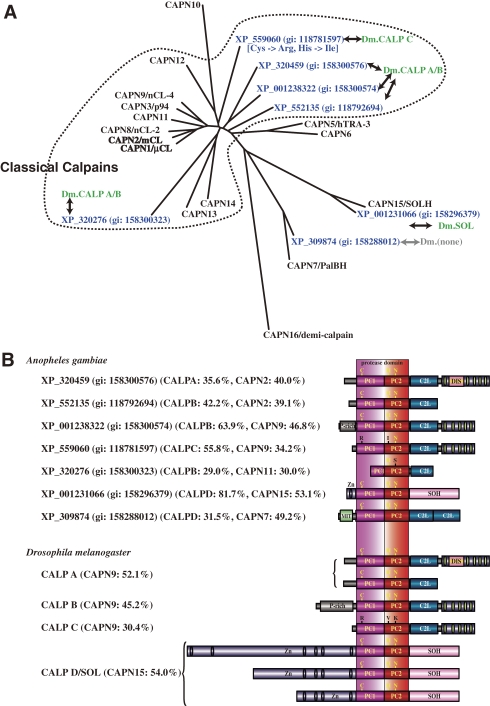
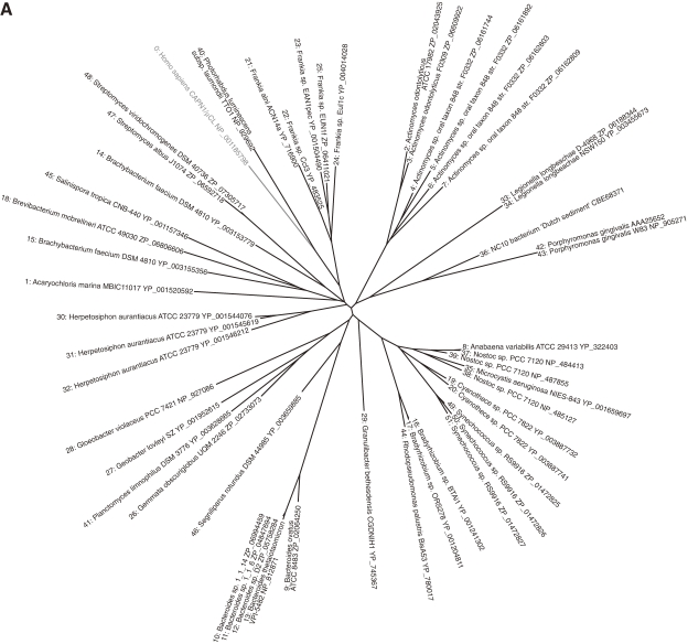
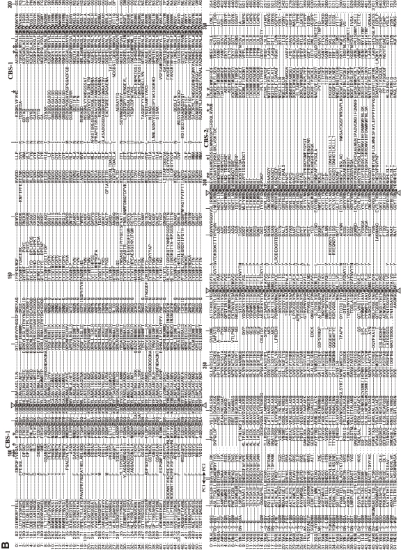
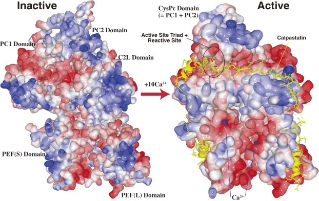
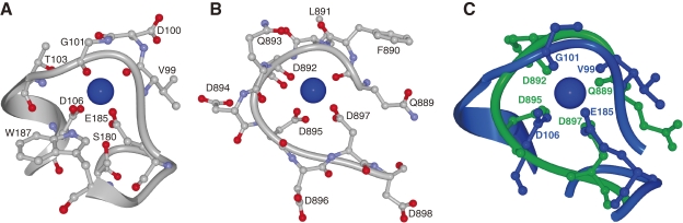
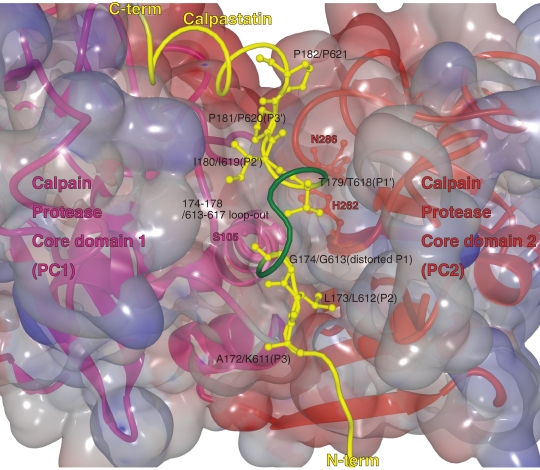
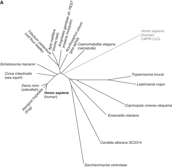
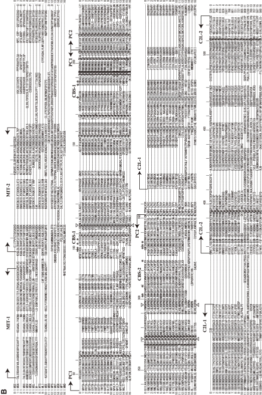
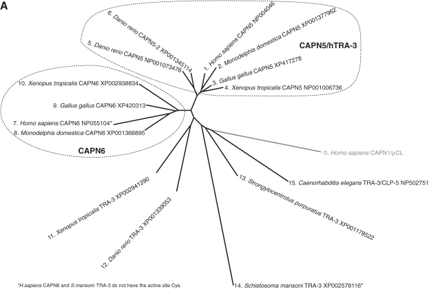
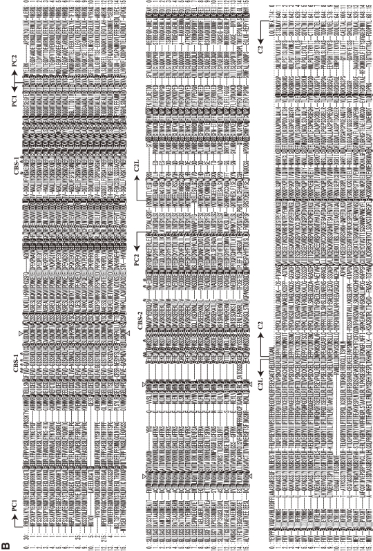
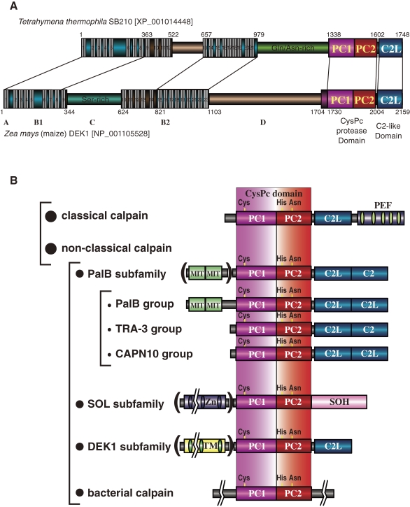
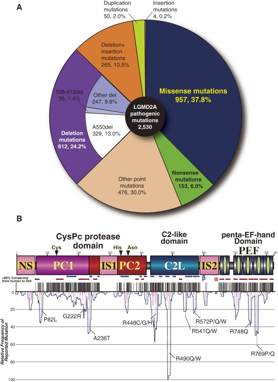
Calpain is an intracellular Ca2+-dependent cysteine protease (EC 3.4.22.17; Clan CA, family C02) discovered in 1964. It was also called CANP (Ca2+-activated neutral protease) as well as CASF, CDP, KAF, etc. until 1990. Calpains are found in almost all eukaryotes and a few bacteria, but not in archaebacteria. Calpains have a limited proteolytic activity, and function to transform or modulate their substrates' structures and activities; they are therefore called, "modulator proteases." In the human genome, 15 genes--CAPN1, CAPN2, etc.--encode a calpain-like protease domain. Their products are calpain homologs with divergent structures and various combinations of functional domains, including Ca2+-binding and microtubule-interaction domains. Genetic studies have linked calpain deficiencies to a variety of defects in many different organisms, including lethality, muscular dystrophies, gastropathy, and diabetes. This review of the study of calpains focuses especially on recent findings about their structure-function relationships. These discoveries have been greatly aided by the development of 3D structural studies and genetic models.


Figures

Figure 1.
Figure 1.
Schematic structures of calpain superfamily members. Calpain homologs have been identified in almost all eukaryotes and in some bacteria. Historically, there are several nomenclatures for domains. To avoid confusion in this review, the domains are called by the abbreviation corresponding to the structure, i.e., PC1 (protease core domain 1), PC2 (protease core domain 2), C2L (C2 domain-like), PEF (penta-EF-hand) etc. Symbols: N, N-terminal region; PEF(L) and PEF(S), PEF domains of large catalytic and small regulatory subunits, respectively; GR, glycine-rich hydrophobic domain; AS, alternative splicing products; NS/IS1/IS2, CAPN3-characteristic sequences; C2, C2 domain; MIT, microtubule-interacting-and-transport motif; Zn, Zn-finger-containing domain; SOH, SOL-homology domain; DIS, CALPA-specific insertion sequence; TM, transmembrane domain; CSTN, calpastatin-like domain; IQ, a motif interactive with calmodulin.
Figure 2.
Figure 2.
Major intracellular proteolytic systems. The ubiquitin–proteasome system degrades and eliminates specific substrate proteins in an ubiquitin-tagging system consisting of more than 1,000 ubiquitin ligases. The autophagy–lysosome system primarily degrades non-specific cell components, including proteins and micro-organisms, by compartmentalization by isolation membranes. The caspase system functions mainly for apoptosis. In contrast, calpains primarily use proteolytic processing, rather than degradation, to modulate or modify their substrates’ activity, specificity, longevity, localization, and structure.
Figure 3.
Figure 3.
Phylogenetic tree and schematic structures of human calpain-related molecules. A. Phylogenetic tree of human calpains. Method: The tree was drawn by the neighbor-joining/bootstrap method after aligning all the sequences using MAFFT v6.240 (at http://align.genome.jp/mafft/, strategy: E-INS-i). Atypical calpains are more divergent than classical calpains. The PalB subfamily consists of the strict PalB group, the TRA-3 group, and the CAPN10 group. B. Schematic structures: Black and green letters indicate ubiquitous and tissue/organ-specific calpains, respectively. See also Fig. 4. Symbols: L and XL, N-terminal and extended N-terminal regions of calpastatin. See Fig. 1 legend for other symbols.
Figure 4.
Figure 4.
Human calpain-related genes. See also Fig. 3.
Figure 5.
Figure 5.
Phylogenetic tree and schematic structures of S. mansoni calpains. A. Phylogenetic tree of S. mansoni and human calpains. See Fig. 3A for methods. B. Schematic structures. The human calpain most similar to each S. mansoni calpain is indicated in parentheses, with the aa sequence identity. The idnentity is given for each of the tandem classical calpain structures in XP_002577797. USP: ubiquitin-specific peptidase. See Fig. 1 legend for other symbols.
Figure 6.
Figure 6.
Phylogenetic tree and schematic structures of C. elegans calpains. A. Phylogenetic tree of C. elegans and human calpains. See Fig. 3A for methods. B. Schematic structures. The human calpain most similar to each C. elegans calpain is indicated in parentheses, with the aa sequence identity. CI and CII stand for conserved regions I and II with unknown function, respectively. See Fig. 1 legend for other symbols.
Figure 7.
Figure 7.
Phylogenetic tree and schematic structures of insect calpains. A. Phylogenetic tree of A. gambiae and human calpains. See Fig. 3A for methods. D. melanogaster (DM) homologs are shown in green letters. B. Schematic structures. The human calpain most similar to each insect calpain is indicated in parentheses, along with the aa sequence identity. The D. melanogaster calpain most similar to A. gambiae calpain is also indicated. P-rich: Pro-rich region. DIS: Drosophila-type insertion sequence. See Fig. 1 legend for other symbols.
Figure 8.
Figure 8.
Phylogenetic tree and sequence alignment of bacterial calpains. A. Phylogenetic tree of bacterial calpains and human CAPN1/μCL. See Fig. 3A for methods. B. Sequence alignment of the bacterial calpain protease domains and human CAPN1/μCL. Only the sequences of the protease domains are compared, with the start and end aa residue numbers indicated, as other sequences and lengths are somewhat divergent. Reversed fonts and gray shadow indicate residues conserved in all sequences or more than half, respectively. Triangles: active site residues. Asterisks: residues involved in Ca2+-binding of the protease domain of human CAPN1/μCL. Sequences: 0: H. sapiens NP_001185798; 1: Acaryochloris marina MBIC11017 YP_001520592; 2: Actinomyces odontolyticus ATCC 17982 ZP_02043925; 3: Actinomyces odontolyticus F0309 ZP_06609922; 4: Actinomyces sp. oral taxon 848 str. F0332 ZP_06161744; 5: Actinomyces sp. oral taxon 848 str. F0332 ZP_06161892; 6: Actinomyces sp. oral taxon 848 str. F0332 ZP_06162803; 7: Actinomyces sp. oral taxon 848 str. F0332 ZP_06162809; 8: Anabaena variabilis ATCC 29413 YP_322403; 9: Bacteroides ovatus ATCC 8483 ZP_02064250; 10: Bacteroides sp. 1_1_14 ZP_06994459; 11: Bacteroides sp. 1_1_6 ZP_04847694; 12: Bacteroides sp. D2 ZP_05758284; 13: Bacteroides thetaiotaomicron VPI-5482 NP_812871; 14: Brachybacterium faecium DSM 4810 YP_003153779; 15: Brachybacterium faecium DSM 4810 YP_003155356; 16: Bradyrhizobium sp. BTAi1 YP_001241302; 17: Bradyrhizobium sp. ORS278 YP_001204811; 18: Brevibacterium mcbrellneri ATCC 49030 ZP_06806606; 19: Cyanothece sp. PCC 7822 YP_003887732; 20: Cyanothece sp. PCC 7822 YP_003887741; 21: Frankia alni ACN14a YP_716900; 22: Frankia sp. CcI3 YP_483525; 23: Frankia sp. EAN1pec YP_001504490; 24: Frankia sp. EuI1c YP_004014028; 25: Frankia sp. EUN1f ZP_06411021; 26: Gemmata obscuriglobus UQM 2246 ZP_02733073; 27: Geobacter lovleyi SZ YP_001952615; 28: Gloeobacter violaceus PCC 7421 NP_927086; 29: Granulibacter bethesdensis CGDNIH1 YP_745367; 30: Herpetosiphon aurantiacus ATCC 23779 YP_001544076; 31: Herpetosiphon aurantiacus ATCC 23779 YP_001545619; 32: Herpetosiphon aurantiacus ATCC 23779 YP_001546212; 33: Legionella longbeachae D-4968 ZP_06188344; 34: Legionella longbeachae NSW150 YP_003455673; 35: Microcystis aeruginosa NIES-843 YP_001659697; 36: NC10 bacterium ‘Dutch sediment’ CBE68371; 37: Nostoc sp. PCC 7120 NP_484413; 38: Nostoc sp. PCC 7120 NP_485127; 39: Nostoc sp. PCC 7120 NP_487855; 40: Photorhabdus luminescens subsp. laumondii TTO1 NP_929692; 41: Planctomyces limnophilus DSM 3776 YP_003628665; 42: Porphyromonas gingivalis AAA25652; 43: Porphyromonas gingivalis W83 NP_905271; 44: Rhodopseudomonas palustris BisA53 YP_780017; 45: Salinispora tropica CNB-440 YP_001157346; 46: Segniliparus rotundus DSM 44985 YP_003659885; 47: Streptomyces albus J1074 ZP_06592718; 48: Streptomyces viridochromogenes DSM 40736 ZP_07305717; 49: Synechococcus sp. RS9916 ZP_01472825; 50: Synechococcus sp. RS9916 ZP_01472826; 51: Synechococcus sp. RS9916 ZP_01472827.
Figure 8.
Figure 8.
Phylogenetic tree and sequence alignment of bacterial calpains. A. Phylogenetic tree of bacterial calpains and human CAPN1/μCL. See Fig. 3A for methods. B. Sequence alignment of the bacterial calpain protease domains and human CAPN1/μCL. Only the sequences of the protease domains are compared, with the start and end aa residue numbers indicated, as other sequences and lengths are somewhat divergent. Reversed fonts and gray shadow indicate residues conserved in all sequences or more than half, respectively. Triangles: active site residues. Asterisks: residues involved in Ca2+-binding of the protease domain of human CAPN1/μCL. Sequences: 0: H. sapiens NP_001185798; 1: Acaryochloris marina MBIC11017 YP_001520592; 2: Actinomyces odontolyticus ATCC 17982 ZP_02043925; 3: Actinomyces odontolyticus F0309 ZP_06609922; 4: Actinomyces sp. oral taxon 848 str. F0332 ZP_06161744; 5: Actinomyces sp. oral taxon 848 str. F0332 ZP_06161892; 6: Actinomyces sp. oral taxon 848 str. F0332 ZP_06162803; 7: Actinomyces sp. oral taxon 848 str. F0332 ZP_06162809; 8: Anabaena variabilis ATCC 29413 YP_322403; 9: Bacteroides ovatus ATCC 8483 ZP_02064250; 10: Bacteroides sp. 1_1_14 ZP_06994459; 11: Bacteroides sp. 1_1_6 ZP_04847694; 12: Bacteroides sp. D2 ZP_05758284; 13: Bacteroides thetaiotaomicron VPI-5482 NP_812871; 14: Brachybacterium faecium DSM 4810 YP_003153779; 15: Brachybacterium faecium DSM 4810 YP_003155356; 16: Bradyrhizobium sp. BTAi1 YP_001241302; 17: Bradyrhizobium sp. ORS278 YP_001204811; 18: Brevibacterium mcbrellneri ATCC 49030 ZP_06806606; 19: Cyanothece sp. PCC 7822 YP_003887732; 20: Cyanothece sp. PCC 7822 YP_003887741; 21: Frankia alni ACN14a YP_716900; 22: Frankia sp. CcI3 YP_483525; 23: Frankia sp. EAN1pec YP_001504490; 24: Frankia sp. EuI1c YP_004014028; 25: Frankia sp. EUN1f ZP_06411021; 26: Gemmata obscuriglobus UQM 2246 ZP_02733073; 27: Geobacter lovleyi SZ YP_001952615; 28: Gloeobacter violaceus PCC 7421 NP_927086; 29: Granulibacter bethesdensis CGDNIH1 YP_745367; 30: Herpetosiphon aurantiacus ATCC 23779 YP_001544076; 31: Herpetosiphon aurantiacus ATCC 23779 YP_001545619; 32: Herpetosiphon aurantiacus ATCC 23779 YP_001546212; 33: Legionella longbeachae D-4968 ZP_06188344; 34: Legionella longbeachae NSW150 YP_003455673; 35: Microcystis aeruginosa NIES-843 YP_001659697; 36: NC10 bacterium ‘Dutch sediment’ CBE68371; 37: Nostoc sp. PCC 7120 NP_484413; 38: Nostoc sp. PCC 7120 NP_485127; 39: Nostoc sp. PCC 7120 NP_487855; 40: Photorhabdus luminescens subsp. laumondii TTO1 NP_929692; 41: Planctomyces limnophilus DSM 3776 YP_003628665; 42: Porphyromonas gingivalis AAA25652; 43: Porphyromonas gingivalis W83 NP_905271; 44: Rhodopseudomonas palustris BisA53 YP_780017; 45: Salinispora tropica CNB-440 YP_001157346; 46: Segniliparus rotundus DSM 44985 YP_003659885; 47: Streptomyces albus J1074 ZP_06592718; 48: Streptomyces viridochromogenes DSM 40736 ZP_07305717; 49: Synechococcus sp. RS9916 ZP_01472825; 50: Synechococcus sp. RS9916 ZP_01472826; 51: Synechococcus sp. RS9916 ZP_01472827.
Figure 9.
Figure 9.
Schematic of the 3D structure of inactive and active m-calpain. Surface-type schematic 3D structures of inactive (Ca2+ free) and active (Ca2+- and calpastatin-bound) forms of m-calpain using PDB data, 1KFX109) and 3DF0.111) The oligopeptides represented by the yellow ribbon + ball-and-stick indicate portions of calpastatin bound to active m-calpain. The dotted lines indicate portions that were too mobile for the 3D structure to be determined. The active protease domain (CysPc) is formed by the fusion of core domains PC1 and PC2 upon the binding of one Ca2+ to each of the core domains. The active site is circled in black. Blue balls represent Ca2+ (not all are visible).
Figure 10.
Figure 10.
A comparison of 3D structures of the Ca2+-binding sites of the CAPN1/μCL protease domain IIa and SLO1 (Ca-bowl). Ca2+-binding sites in (A) the CAPN1/μCL protease domain (2R9F) and (B) the high-conductance voltage- and Ca2+-activated K+ channel (BK or SLO1 channel) (3MT5). (C) The structures were superimposed for comparison.126)
Figure 11.
Figure 11.
Calpastatin region B binds and inhibits calpain. Enlarged view of the interaction between calpastatin (yellow) and the catalytic cleft in the protease core domain PC1 (pink)–PC2 (red) of m-calpain (the 3D structure is from 3DF0). Calpastatin binds in the substrate orientation indicated by positions P3 to P3′. At P1, calpastatin distorts from the substrate path and projects residues 174–178 or 613–617, which form a kink between the P2 and P1′ anchor sites.
Figure 12.
Figure 12.
Phylogenetic tree and sequence alignment of PalB calpains. A. Phylogenetic tree of strict PalB calpains and human CAPN1/μCL. See Fig. 3A for methods. B. Sequence alignment of PalBs and human CAPN1/μCL (the start and end aa residue numbers are indicated). Triangles: active site residues. Asterisks: residues involved in Ca2+-binding of the protease domain of CAPN1/μCL. Arrows: domain boundaries. Deletion (−) and multiple (+) residue positions are indicated. Residue numbers of human CAPN1/μCL are given above the sequences. Residues conserved in all the sequences, all except human CAPN1/μCL, and in more than half the sequences are indicated by black reversed fonts, gray reversed fonts, and gray shadow, respectively. Sequences: 0: H. sapiens (human) CAPN1/μCL (NP_005177); 1: H. sapiens CAPN7/PalBH (NP_055111); 2: X. tropicalis (frog) (NP_998853); 3: D. rerio (zebrafish) (NP_001128580); 4: Ciona intestinalis (sea squirt) (XP_002121253); 5: Tribolium castaneum (red flour beetle) (XP_967682); 6: Apis mellifera (honey bee) (XP_001121978); 7: Acyrthosiphon pisum (pea aphid) (XP_001945029); 8: A. gambiae str. PEST (malaria mosquito) (XP_309874); 9: S. mansoni (XP_002579452); 10: C. elegans (NP_497964); 11: T. brucei TREU927 (XP_828540); 12: L. major (CAJ06528); 13: Coprinopsis cinerea okayama7#130 (common ink cap) (EAU91907); 14: E. nidulans FGSC-A4 (XP_657860); 15: C. albicans SC5314 (EAL03290); 16: S. cerevisiae (Q03792).
Figure 12.
Figure 12.
Phylogenetic tree and sequence alignment of PalB calpains. A. Phylogenetic tree of strict PalB calpains and human CAPN1/μCL. See Fig. 3A for methods. B. Sequence alignment of PalBs and human CAPN1/μCL (the start and end aa residue numbers are indicated). Triangles: active site residues. Asterisks: residues involved in Ca2+-binding of the protease domain of CAPN1/μCL. Arrows: domain boundaries. Deletion (−) and multiple (+) residue positions are indicated. Residue numbers of human CAPN1/μCL are given above the sequences. Residues conserved in all the sequences, all except human CAPN1/μCL, and in more than half the sequences are indicated by black reversed fonts, gray reversed fonts, and gray shadow, respectively. Sequences: 0: H. sapiens (human) CAPN1/μCL (NP_005177); 1: H. sapiens CAPN7/PalBH (NP_055111); 2: X. tropicalis (frog) (NP_998853); 3: D. rerio (zebrafish) (NP_001128580); 4: Ciona intestinalis (sea squirt) (XP_002121253); 5: Tribolium castaneum (red flour beetle) (XP_967682); 6: Apis mellifera (honey bee) (XP_001121978); 7: Acyrthosiphon pisum (pea aphid) (XP_001945029); 8: A. gambiae str. PEST (malaria mosquito) (XP_309874); 9: S. mansoni (XP_002579452); 10: C. elegans (NP_497964); 11: T. brucei TREU927 (XP_828540); 12: L. major (CAJ06528); 13: Coprinopsis cinerea okayama7#130 (common ink cap) (EAU91907); 14: E. nidulans FGSC-A4 (XP_657860); 15: C. albicans SC5314 (EAL03290); 16: S. cerevisiae (Q03792).
Figure 13.
Figure 13.
Phylogenetic tree and sequence alignment of TRA-3 calpains. A. Phylogenetic tree of TRA-3 calpains and human CAPN1/μCL. See Fig. 3A for methods. B. Sequence alignment of TRA-3 and human CAPN1/μCL (the start and end aa residue numbers are indicated). Sequences: 0: H. sapiens CAPN1/μCL NP005177; 1: H. sapiens CAPN5 NP004046; 2: M. domestica CAPN5 XP001377962; 3: G. gallus CAPN5 XP417278; 4: X. tropicalis CAPN5 NP001006736; 5: D. rerio CAPN5 NP001073476; 6: D. rerio CAPN5-2 XP001345114; 7: H. sapiens CAPN6 NP055104; 8: M. domestica CAPN6 XP001366895; 9: G. gallus CAPN6 XP420313; 10: X. tropicalis CAPN6 XP002938834; 11: X. tropicalis TRA-3 XP002941290; 12: D. rerio TRA-3 XP001339053; 13: Strongylocentrotus purpuratus CAPN5 XP001178522; 14: S. mansoni XP002578116; 15: C. elegans TRA-3/CLP-5 NP502751. For other explanation, see Fig. 12B.
Figure 13.
Figure 13.
Phylogenetic tree and sequence alignment of TRA-3 calpains. A. Phylogenetic tree of TRA-3 calpains and human CAPN1/μCL. See Fig. 3A for methods. B. Sequence alignment of TRA-3 and human CAPN1/μCL (the start and end aa residue numbers are indicated). Sequences: 0: H. sapiens CAPN1/μCL NP005177; 1: H. sapiens CAPN5 NP004046; 2: M. domestica CAPN5 XP001377962; 3: G. gallus CAPN5 XP417278; 4: X. tropicalis CAPN5 NP001006736; 5: D. rerio CAPN5 NP001073476; 6: D. rerio CAPN5-2 XP001345114; 7: H. sapiens CAPN6 NP055104; 8: M. domestica CAPN6 XP001366895; 9: G. gallus CAPN6 XP420313; 10: X. tropicalis CAPN6 XP002938834; 11: X. tropicalis TRA-3 XP002941290; 12: D. rerio TRA-3 XP001339053; 13: Strongylocentrotus purpuratus CAPN5 XP001178522; 14: S. mansoni XP002578116; 15: C. elegans TRA-3/CLP-5 NP502751. For other explanation, see Fig. 12B.
Figure 14.
Figure 14.
Schematic structures of T. thermophila and Z. mays DEK1s. A. The number of TM spanners is taken from prediction programs and may be corrected in the future. Domain names for Z. mays DEK1 are taken from ref. 62). B. Structural classification and consensus domain structures of calpain super family members. See Fig. 1 legend for symbols.
Figure 15.
Figure 15.
Pathogenic CAPN3 mutations in calpainopathy patients. A. Summary of calpainopathy mutation types. B. Calpainopathy missense mutation positions and frequencies (relative to the maximum value). Black vertical lines indicate aa residues that are conserved more than 80% of vertebrates as shown in Fig. 16B. α and β indicate α-helix and β-strand secondary structures of the corresponding m-calpain 3D structure. See Fig. 1 legend for other symbols.
Figure 16.
Figure 16.
Phylogenetic tree and sequence alignment of CAPN3 proteins. A. Phylogenetic tree of vertebrate CAPN3. See Fig. 3A for methods. Explicit CAPN3 homologs are found only in vertebrates. B. Sequence alignment of CAPN3s. Fishes have two CAPN3 homologs (type-1 and -2); the latter does not have explicit IS1 or IS2 regions. Sequences: 1: H. sapiens, NP_000061; 2: M. domestic, ENSMODP00000022330; 3: G. gallus, NP_001004405; 4: A. carolinensis, ENSACAP00000015362; 5: X. tropicalis, ENSXETP00000026925; 6: Takifugu rubripes (type-1), ENSTRUP00000046251; 7: Hippoglossus hippoglossus (type-1), ACY78226; 8: Salmo salar (type-1), NP_001158880; 9: D. rerio (type-1), XP_001337065; 10: T. rubripes (type-2), ENSTRUP00000016935; 11: D. rerio (type-2), ENSDARP00000094244. For other explanation, see Fig. 12B.
Figure 17.
Figure 17.
Pal-PacC pathway and corresponding molecules in yeast and human. Schematics of signaling pathways involving PalB of fungi. In fungi, a membrane protein (PalH) senses ambient pH and transduces the alkaline signal to PalI. An arrestin homolog, PalF, further transduces the signal to PalA, which forms a complex with AnPef1. At the endosomal membrane, PalA forms the PalB active complex to proteolyze PacC at the C-terminus, thus activating this key transcription factor. Activated PacC regulates gene expression under alkaline conditions. This pathway is highly conserved in yeast and possibly conserved in human; corresponding molecules: PalH (fungi)-Rim21 (yeast)-? (human), PalI-Rim9-?, PalF-Rim8-arrestin, PalA-Rim20-Alix/AIP1, PalC-Ygr122w-?, AnPef1-Pef1/Ygr058w-ALG-2, AnSnf7-Snf7/Vps32-CHMP4s, PalB-Rim13/Cpl1-CAPN7/PalBH, and PacC-Rim101-C2H2 Zn-finger proteins? See text for details.

Similar articles

Cited by

References

    1. Guroff G. (1964) A neutral calcium-activated proteinase from the soluble fraction of rat brain. J. Biol. Chem. 239, 149–155 - PubMed
    1. Lazarovici P. (2000) Obituary: Dr. Gordon Guroff (1933–1999). J. Mol. Neurosci. 14, 1–2 - PubMed
    1. Huston R.B., Krebs E.G. (1968) Activation of skeletal muscle phosphorylase kinase by Ca2+. II. Identification of the kinase activating factor as a proteolytic enzyme. Biochemistry 7, 2116–2122 - PubMed
    1. Meyer W.L., Fischer E.H., Krebs E.G. (1964) Activation of skeletal muscle phosphorylase kinase by Ca2+. Biochemistry 3, 1033–1039 - PubMed
    1. Busch W.A., Stromer M.H., Goll D.E., Suzuki A. (1972) Ca2+-specific removal of Z lines from rabbit skeletal muscle. J. Cell Biol. 52, 367–381 - PMC - PubMed

Publication types