. 2011 Jun 14;20(6):775-87.
doi: 10.1016/j.devcel.2011.04.018.

Overlapping roles and collective requirement for the coreceptors GAS1, CDO, and BOC in SHH pathway function

Affiliations

Overlapping roles and collective requirement for the coreceptors GAS1, CDO, and BOC in SHH pathway function

Benjamin L Allen et al. Dev Cell. .

Abstract

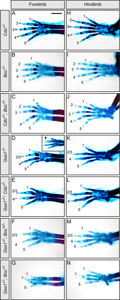
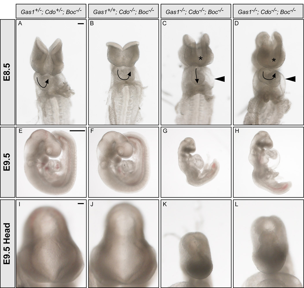
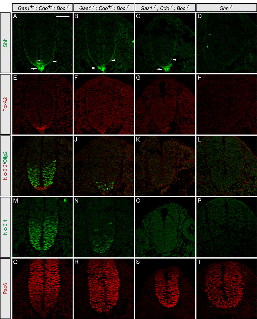
Secreted Hedgehog (HH) ligands signal through the canonical receptor Patched (PTCH1). However, recent studies implicate three additional HH-binding, cell-surface proteins, GAS1, CDO, and BOC, as putative coreceptors for HH ligands. A central question is to what degree these coreceptors function similarly and what their collective requirement in HH signal transduction is. Here we provide evidence that GAS1, CDO, and BOC play overlapping and essential roles during HH-mediated ventral neural patterning of the mammalian neural tube. Specifically, we demonstrate two important roles for these molecules: an early role in cell fate specification of multiple neural progenitors and a later role in motor neuron progenitor maintenance. Most strikingly, genetic loss-of-function experiments indicate an obligatory requirement for GAS1, CDO, and BOC in HH pathway activity in multiple tissues.


Figures

Figure 1
Figure 1. Gas1, Cdo and Boc equally promote Shh-dependent specification of ventral neural progenitors
HH stage 21–22 chick neural tubes electroporated with pCIG (A–D), Gas1-pCIG (E–H), Cdo-pCIG (I–L), Boc-pCIG (M–P), or SmoM2-pCIG (Q–T) were sectioned at the forelimb level and stained with antibodies raised against Nkx6.1 (red; A, E, I, M, Q) and Nkx2.2 (red; C, G, K, O, S). GFP expressing cells (green; B, F, J, N, R and D, H, L, P, T) indicate electroporated cells on one side, while the un-electroporated half of the neural tube serves as an internal negative control. Arrows denote ectopic expression of Nkx6.1 (E, I, M, Q) and Nkx2.2 (G, K, O, S). Arrowheads indicate ectopic Nkx6.1 and Nkx2.2 expression throughout the dorsal neural tube in embryos expressing SmoM2 (Q–T), whereas ectopic induction of these markers following Gas1 (E–H), Cdo (I–L) and Boc (M–P) electroporation was restricted to the ventral neural tube. Scale bar: A, 50µm.
Figure 2
Figure 2. Defective ventral neural patterning in E10.5Cdo−/−; Boc−/− and Gas1−/−; Boc−/− mouse embryos
Immunofluorescent analysis of E10.5 forelimb level sections detects expression of Shh (green; A–H), FoxA2 (red; I–P), Nxk2.2 and Olig2 (red and green, respectively; Q–X) in wt (A, I, Q), Boc−/− (B, J, R), Cdo−/− (C, K, S), Cdo+/−; Boc−/− (D, L, T), Cdo−/−; Boc−/− (E, M, U), Gli2−/− (F, N, V), Gas1−/− (G, O, W), and Gas1−/−; Boc−/− (H, P, X) embryos. Arrowheads denote FoxA2 and Nkx2.2 double positive floorplate cells in Cdo−/− embryos (S), but not Boc−/− embryos (R). Despite the complete loss of FoxA2+ FP cells in both Cdo−/−; Boc−/−, and Gli2−/− embryos (M and N, respectively), there is a selective loss of Olig2+ but not Nkx2.2+ cells in Cdo−/−; Boc−/−embryos (U). In contrast, Gli2−/− embryos lack Nkx2.2+ cells, but maintain Olig2 expression (V). Gas1−/−; Boc−/− embryos display complete loss of FoxA2 (P), Nkx2.2 and Olig2 (X) at E10.5. Scale bar: A, 50µm. See Figure S1 for a more detailed analysis of neural patterning in Boc−/−embryos. Refer to Figure S2 for Gas1, Cdo and Boc protein distribution in E10.5 mouse neural tubes.
Figure 3
Figure 3. Motor neuron progenitors are specified, but not maintained in embryos with reduced Shh signaling
Antibody detection of Shh (green; A–C, J–K), FoxA2 (red; D–F, L–M), Nkx2.2 and Olig2 (red and green, respectively; G–I, N–O) in forelimb level sections of E9.5 Boc−/− (A, D, G), Cdo−/−; Boc−/− (B, E, H), Gli2−/− (C, F, I), Gas1−/−; Cdo−/− (J, L, N), and Gas1−/−; Boc−/−(K, M, O) embryos. At E9.5 Cdo−/−; Boc−/− embryos contain similar numbers of Olig2+ progenitors to Boc−/− or Gli2−/− embryos (G and I, respectively) in marked contrast to E10.5 embryos. Note the variable presence of a few FoxA2+ and Nkx2.2+ cells in Cdo−/−; Boc−/−, Gas1−/−; Cdo−/− and Gas1−/−; Boc−/− embryos at the forelimb level (E, H, L–O). Neither cell type appears to be specified in Gli2−/− embryos (F, I). Immunofluorescent detection of Shh (green; P–R), Nkx2.2 and Olig2 (red and green, respectively; S–U) in forelimb level sections of E12.5 wt (P, S, V), Gas1−/− (Q, T, W), and Gli2−/− (R, U, X) embryos. Nuclei are identified with DAPI (G–I). Insets (A–C) indicate notochord expression of Shh. Note that in Gas1−/− embryos (E), only a few Olig2+ cells are present, while the number of Nkx2.2+ cells is comparable to wt (D). Olig2 + cells are reduced and fail to cluster normally in Gli2−/− embryos (F). Scale bar: A,P, 50µm.
Figure 4
Figure 4. Delayed cyclopamine administration selectively affects motor neuron maintenance in HH stage 22 chick embryos
Cyclopamine (E–H, M–P) or ethanol (A–D, I–L) were administered to HH stage 17–18 chick embryos for 24 hours, followed by immunofluorescent detection of nuclei (DAPI; A, E, I, M), Nkx6.1 (green; B, D, F, H), Pax3 (red; C, D, G, H), Nkx2.2 (red; J, L, N, P), and Olig2 (green; K, L, O, P). Note the normal maintenance of Nkx6.1, Pax3, and Nkx2.2 in cyclopamine-treated embryos (F–H, N–P). In contrast, there is a significant reduction in the number of Olig2+ cells following cyclopamine administration (N–P). The number of Olig2 cells varies from moderate (O, P) to severe (inset, N). Comparison of Nkx2.2 and Olig2 cell numbers in EtOH-treated embryos and moderately affected cyclopamine-treated embryos is quantitated in Q. Error bars represent the mean +/− SD calculated from analysis of sections from three different embryos. P-values calculated by two-tailed Student’s t-test are listed. NS, not significant. Scale bar: A, 50µm. Refer to Figure S3 for a similar analysis with a second Hh pathway antagonist, SANT-1.
Figure 5
Figure 5. Analysis of Cdo−/−; Boc−/− and Gas1−/−; Boc−/− limb development
Forelimbs (A–G) and hindlimbs (H–N) of E18.5 embryos stained with alcian blue and alizarin red to visualize cartilage and bone, respectively, in the limb skeleton. Numbers denote specific digits (1 most anterior, and 5 most posterior). Cdo−/− (A, H), Boc−/− (B, I) and Cdo−/−; Boc−/− (C, J) embryos display normal digit patterning. In contrast, Gas1−/− embryos display fusion and loss of digits 2/3 (D, K). A similar phenotype is seen in Gas1−/−; Cdo−/− (E, L) and Gas1−/−; Boc+/− (F, M). In contrast, Gas1−/−; Boc−/− embryos display a significantly more severe digit patterning defect; only digits 1 and 5 are identifiable in both the forelimb (G) and hindlimb (N), and a third digit (labeled as “?”), possibly a fusion of digits 3 and 4, is at the anterior-posterior intersect. Scale bar: A, 1mm. Gas1, Cdo and Boc expression in the developing limb is provided in Figure S4, along with analysis of Shh, Ptch1, and Gli1 transcript levels in E10.5 Gas1−/−; Boc−/− forelimb buds.
Figure 6
Figure 6. Cyclopia, holoprosencephaly and heart looping defects in E8.5 and E9.5 Gas1−/−; Cdo−/−; Boc−/− embryosGas1+/−; Cdo+/−; Boc−/− (A, E, I), Gas1+/+; Cdo−/−; Boc−/− (B, F, J) and two Gas1−/−; Cdo−/−; Boc−/−
(C–D, G–H, K–L) embryos are shown. En face images of E8.5 (10–12 somite) embryos (A–D). Arrows indicate the direction of heart looping, while arrowheads denote pericardial edema that is present in Gas1−/−; Cdo−/−; Boc−/− embryos (C, D). 50% of Gas1−/−; Cdo−/−; Boc−/− embryos display a linear heart tube (N = 4/8 embryos). Asterisks indicate abnormal forebrain development that is a hallmark of the failure to specify ventral midline cell fates. Examination of embryos of the same genotype at E9.5 (20–25 somites; E–H) demonstrates a failure to complete the turning process in Gas1−/−; Cdo−/−; Boc−/− embryos (G, H). Higher magnification views of the heads of E9.5 embryos (I–L) reveal holoprosencephaly in Gas1−/−; Cdo−/−; Boc−/− embryos (K, L). Scale bars: A, 100um; E, 500µm; I, 100µm.
Figure 7
Figure 7. Simultaneous genetic removal of Gas1, Cdo and Boc results in complete loss of Shh-dependent ventral cell specification
Antibody detection of Shh (green; A–D), FoxA2 (red; E–H), Nkx2.2 and Olig2 (red and green, respectively; I–L), Nkx6.1 (green; M–P), and Pax6 (red; Q–T) in Gas1+/−; Cdo+/−; Boc−/− (A, E, I, M, Q), Gas1−/−; Cdo+/−; Boc−/− (B, F, J, N, R), Gas1−/−; Cdo/-; Boc−/− (C, G, K, O, S), and Shh−/− (D, H, L, P, T) E9.5 embryos. Arrows (A–C) indicate Shh expression in the notochord. Arrowheads (A–C) denote secreted Shh protein. Shh expression in the floorplate is detected only in Gas1+/−; Cdo+/−; Boc−/− embryos (asterisk in A). Scale bar: A, 50µm.

Similar articles

Cited by

References

    1. Airaksinen MS, Holm L, Hatinen T. Evolution of the GDNF family ligands and receptors. Brain Behav Evol. 2006;68:181–190. - PubMed
    1. Allen BL, Tenzen T, McMahon AP. The Hedgehog-binding proteins Gas1 and Cdo cooperate to positively regulate Shh signaling during mouse development. Genes Dev. 2007;21:1244–1257. - PMC - PubMed
    1. Beachy PA, Hymowitz SG, Lazarus RA, Leahy DJ, Siebold C. Interactions between Hedgehog proteins and their binding partners come into view. Genes Dev. 2010;24:2001–2012. - PMC - PubMed
    1. Briscoe J, Ericson J. Specification of neuronal fates in the ventral neural tube. Curr Opin Neurobiol. 2001;11:43–49. - PubMed
    1. Cabrera JR, Sanchez-Pulido L, Rojas AM, Valencia A, Manes S, Naranjo JR, Mellstrom B. Gas1 is related to the glial cell-derived neurotrophic factor family receptors alpha and regulates Ret signaling. J Biol Chem. 2006;281:14330–14339. - PubMed

Publication types

MeSH terms

LinkOut - more resources