. 2011 Aug 5;286(31):27506-14.
doi: 10.1074/jbc.M111.238501. Epub 2011 Jun 9.

Protein kinase R as mediator of the effects of interferon (IFN) gamma and tumor necrosis factor (TNF) alpha on normal and dysplastic hematopoiesis

Affiliations

Protein kinase R as mediator of the effects of interferon (IFN) gamma and tumor necrosis factor (TNF) alpha on normal and dysplastic hematopoiesis

Bhumika Sharma et al. J Biol Chem. .

Abstract

IFNγ and TNFα are potent inhibitors of hematopoiesis and have been implicated in the pathophysiology of bone marrow failure and myelodysplastic syndromes (MDS). We examined the role of protein kinase R (PKR) in the generation of the inhibitory effects of these myelosuppressive cytokines on hematopoiesis. Our data demonstrate that PKR is rapidly phosphorylated/activated in response to engagement of IFNγ or TNFα receptors in normal human hematopoietic progenitors. Such engagement of PKR is important for the suppressive effects of these cytokines on normal hematopoiesis. Pharmacological targeting of PKR using a specific inhibitor or siRNA-mediated PKR knockdown results in partial reversal of the suppressive effects of IFNγ and TNFα on normal human CD34+-derived myeloid (colony-forming unit-granulocyte-monocytic) and erythroid (burst-forming unit-erythroid) progenitors. Importantly, inhibition of PKR activity or expression increases hematopoietic colony formation from human MDS progenitors, suggesting that drugs that target PKR may provide a novel approach for the treatment of MDS and marrow failure syndromes. Altogether, our data establish that beyond its key role in the induction of IFN-antiviral responses, PKR plays important roles in signaling for IFNγ and other myelosuppressive cytokine receptors as a common mediator of signals for hematopoietic suppression.


Figures
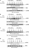
FIGURE 1.
FIGURE 1.
TNFα and IFNγ induce phosphorylation/activation of PKR in leukemia cells. U937 cells stably expressing CTRL shRNA or PKR shRNA were treated with human TNFα (A and C) or IFNγ (B and D) for the indicated times. Total lysates were resolved by SDS-PAGE and immunoblotted with the indicated antibodies. CTRL, control.
FIGURE 2.
FIGURE 2.
TNFα- or IFNγ-dependent activation of p38 MAPK is PKR-independent phosphorylation. U937 cells stably expressing control (CTRL) shRNA or PKR shRNA were treated with human TNFα (A) or IFNγ (B) for the indicated times. Total lysates were resolved by SDS-PAGE and immunoblotted with the indicated antibodies.
FIGURE 3.
FIGURE 3.
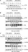
PKR engagement is required for activation of ERK1/2 or JNK in response to TNFα or IFNγ. A, U937 cells stably expressing control (CTRL) shRNA or PKR shRNA were treated with human IFNγ for the indicated times, and cell lysates were resolved by SDS-PAGE and immunoblotted with anti-phospho-Thr-202/Tyr-204-Erk or anti-Erk, as indicated. B, U937 cells stably expressing control shRNA or PKR shRNA were treated with human IFNγ for the indicated times, and cell lysates were resolved by SDS-PAGE and immunoblotted with antibodies against phosphor-JNK or GAPDH as indicated (top two panels). Equal amounts of cell lysates from the same experiment were resolved separately by SDS-PAGE and immunoblotted with antibodies against JNK or GAPDH, as indicated (bottom two panels). C, U937 cells stably expressing CTRL shRNA or PKR shRNA were treated with TNFα for the indicated times, and cell lysates were resolved by SDS-PAGE and immunoblotted with antibodies against phosphor-JNK or GAPDH as indicated (top two panels). Equal amounts of cell lysates from the same experiment were resolved separately by SDS-PAGE and immunoblotted with antibodies against JNK or GAPDH, as indicated (bottom two panels).
FIGURE 4.
FIGURE 4.
Requirement for PKR kinase mediates the antiproliferative effects of TNFα and IFNγ on the growth of U937 cells. A and B, U937 cells stably expressing control shRNA or PKR shRNA were incubated for 7 days in the presence or absence of the indicated concentrations of human TNFα (A) or human IFNγ (B), and cell viability was assessed by 3-(4,5-dimethylthiazol-2-yl)-2,5-diphenyltetrazolium bromide assays. The data are expressed as percentages of control of untreated cells and represent the means ± S.E. of five experiments. C, U937 cells stably expressing control shRNA or PKR shRNA were incubated in the presence or absence of IFNγ as indicated and leukemic progenitor (CFU-L) colonies were scored after 7 days in methylcellulose cultures. The data are expressed as percentages of control colony formation from untreated cells and represent the means ± S.E. of five experiments. Paired t test analysis showed p = 0.0014 for the effects of IFNγ on PKR shRNA versus control shRNA expressing cells. CTRL, control.
FIGURE 5.
FIGURE 5.
Cytokine-mediated engagement of PKR in expanded human progenitors. CFU-E progenitor cells were treated with human TNFα (A) or human IFNγ (B) for the indicated times. Total lysates were separated by SDS-PAGE and immunoblotted with an antibody against phosphorylated PKR (Thr-446). The same blots were subsequently stripped and reprobed with an antibody against PKR.
FIGURE 6.
FIGURE 6.
Requirement of PKR activity for activation of MAPK pathways in primitive hematopoietic precursors. A, expanded human CFU-E cells were nucleofected with control siRNA or PKR siRNA, as described under “Materials and Methods.” Expression of PKR mRNA was assessed by real time RT-PCR and is shown as a percentage of the levels of control siRNA-treated cells. The data shown represent the means ± S.E. of two experiments. B, CFU-E cells were nucleofected with control siRNA or PKR siRNA. Equal amounts of protein were separated by SDS-PAGE and then immunoblotted with antibodies against PKR or GAPDH. C–F, CFU-E cells were nucleofected with either control siRNA or PKR siRNA for 24 h and were then treated with human TNFα (C and D) or IFNγ (E and F) for the indicated times. Equal amounts of protein were separated by SDS-PAGE and then immunoblotted with the indicated antibodies. CTRL, control.
FIGURE 7.
FIGURE 7.
Effects of pharmacological inhibition of PKR activity on eIF2a and Erk phosphorylation in normal hematopoietic progenitors. Expanded human CFU-E cells were pretreated with inactive inhibitor or PKR inhibitor (1 μm) for 1 h and were then treated with human IFNγ (A and C) or TNFα (B and D) for the indicated times. Equal amounts of protein were separated by SDS-PAGE and then immunoblotted with the indicated antibodies.
FIGURE 8.
FIGURE 8.
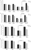
PKR kinase mediates the antiproliferative effects of TNFα and IFNγ on normal hematopoiesis. A and B, normal CD34+ bone marrow-derived cells were incubated in the presence or absence of TNFα (A) or IFNγ (B), in the presence of a PKR inhibitor or a corresponding control-inactive inhibitor, in clonogenic assays in methylcellulose, as indicated. CFU-GM and BFU-E progenitor colonies were scored after 14 days in culture. The data are expressed as percentages of control colony formation from untreated cells and represent the means ± S.E. of five independent experiments for A and four independent experiments for B. p values for the indicated paired t test analyses are shown. C and D, normal human bone marrow-derived CD34+ cells were transfected with control siRNA or PKR siRNA, as indicated and incubated in clonogenic assays in methylcellulose in the presence or absence of TNFα (C) or IFNγ (D), as indicated. CFU-GM and BFU-E progenitor colonies were scored after 14 days in culture. The data are expressed as percentages of colony formation from control siRNA transfected cells and represent the means ± S.E. of five experiments for C and four experiments for D. p values for the indicated paired t test analyses are shown. CTRL, control.
FIGURE 9.
FIGURE 9.
Effects of PKR knockdown on the generation of the antiproliferative effects of TNFα and IFNγ on normal hematopoiesis. A, U937 cells nucleofected with control siRNA or PKR siRNA2, as described under “Materials and Methods.” Expression of PKR mRNA was assessed by real time RT-PCR. The data are expressed as percentages of mRNA expression for control siRNA-treated cells and represent the means ± S.E. of two experiments. B, normal human bone marrow-derived CD34+ cells were transfected with control siRNA or PKR siRNA2, as indicated and incubated in clonogenic assays in methylcellulose in the presence or absence of TNFα or IFNγ, as indicated. CFU-GM and BFU-E progenitor colonies were scored after 14 days in culture. The data are expressed as percentages of colony formation from control siRNA transfected cells and represent the means ± S.E. of four experiments. CTRL, control.
FIGURE 10.
FIGURE 10.
Inhibition of PKR enhances colony formation from MDS bone marrow or peripheral blood. A, mononuclear cells derived from peripheral blood or bone marrow of patients with MDS were incubated in clonogenic assays in methylcellulose in the presence of PKR inhibitor or inactive inhibitor, as indicated. CFU-GM and BFU-E progenitor colonies were scored after 14 days in culture. p values for the indicated paired t test analyses are shown. B, mononuclear cells derived from peripheral blood or bone marrow of patients with MDS. The cells were nucleofected with control siRNA or PKR siRNA, as incubated, and CFU-GM and BFU-E progenitor colony formation was assessed in clonogenic assays in methylcellulose. The data are expressed as percentages of colony formation from control siRNA transfected cells and represent the means ± S.E. of four independent experiments. p values for the indicated paired t test analyses are shown. CTRL, control.

Similar articles

Cited by

References

    1. Platanias L. C. (2005) Nat. Rev. Immunol. 5, 375–386 - PubMed
    1. Borden E. C., Sen G. C., Uze G., Silverman R. H., Ransohoff R. M., Foster G. R., Stark G. R. (2007) Nat. Rev. Drug Discov. 6, 975–990 - PMC - PubMed
    1. Schoenborn J. R., Wilson C. B. (2007) Adv. Immunol. 96, 41–101 - PubMed
    1. Joshi S., Kaur S., Kroczynska B., Platanias L. C. (2010) Cytokine 52, 123–127 - PubMed
    1. Stark G. R. (2007) Cytokine Growth Factor Rev. 18, 419–423 - PMC - PubMed

Publication types