. 2011 Sep;90(3):599-611.
doi: 10.1189/jlb.0610376. Epub 2011 Jun 7.

Protein kinase Cδ is a critical component of Dectin-1 signaling in primary human monocytes

Affiliations

Protein kinase Cδ is a critical component of Dectin-1 signaling in primary human monocytes

Deena H Elsori et al. J Leukoc Biol. 2011 Sep.

Abstract

Zymosan, a mimic of fungal pathogens, and its opsonized form (ZOP) are potent stimulators of monocyte NADPH oxidase, resulting in the production of O(2)(.-), which is critical for host defense against fungal and bacterial pathogens and efficient immune responses; however, uncontrolled O(2)(.-) production may contribute to chronic inflammation and tissue injury. Our laboratory has focused on characterizing the signal transduction pathways that regulate NADPH oxidase activity in primary human monocytes. In this study, we examined the involvement of various pattern recognition receptors and found that Dectin-1 is the primary receptor for zymosan stimulation of O(2)(.-) via NADPH oxidase in human monocytes, whereas Dectin-1 and CR3 mediate the activation by ZOP. Further studies identified Syk and Src as important signaling components downstream of Dectin-1 and additionally identified PKCδ as a novel downstream signaling component for zymosan-induced O(2)(.-) as well as phagocytosis. Our results show that Syk and Src association with Dectin-1 is dependent on PKCδ activity and expression and demonstrate direct binding between Dectin-1 and PKCδ. Finally, our data show that PKCδ and Syk but not Src are required for Dectin-1-mediated phagocytosis. Taken together, our data identify Dectin-1 as the major PRR for zymosan in primary human monocytes and identify PKCδ as a novel downstream signaling kinase for Dectin-1-mediated regulation of monocyte NADPH oxidase and zymosan phagocytosis.


Figures

Figure 1.
Figure 1.. TLR4 and TLR2 are not involved in zymosan or ZOP-induced O2.– production by primary human monocytes.
(A) Zymosan and ZOP-induced O2.– production are not regulated by TLR4 in activated monocytes. Primary human monocytes were plated in 24-well plates (500 μl; 1×10⁶/ml). Monocytes were pretreated for 30 min in the presence or absence of the TLR4 antagonist and then activated with zymosan (100 μg/ml), ZOP (100 μg/ml), or LPS (1 μg/ml). O2.– production was measured during the first hour of activation. (B) TLR2 is also not involved in zymosan-induced O2.– production. Primary monocytes were plated as in A. Monocytes were left untreated or were treated with a TLR2-blocking antibody (10 μg/ml) for 1 h, followed by stimulation with zymosan (100 μg/ml) or E. coli 0111:B4 PGN (5 μg/ml). O2.– production was measured as described in Materials and Methods. (C) TLR2 is not required for ZOP-induced O2.– release. Monocytes (500 μl; 1×10⁶/ml) were treated or left untreated with a TLR2-blocking antibody (clone TL2.1) for 1 h and then activated with ZOP or PGN. Following activation for 1 h O2.–, production was measured. (A–C) Data represent the mean ± sd (n=3) from a representative experiment of four independent experiments that were performed. Results were similar in all experiments.
Figure 2.
Figure 2.. Dectin-1 and CR3 regulate O2.– production in activated human monocytes.
(A) Zymosan and ZOP signal through Dectin-1 and CR3 for O2.– production in human monocytes, which were plated as in Fig. 1A. Monocytes were left untreated or were treated with laminarin (500 μg/ml) or CR3-blocking antibody (10 μg/ml) for 1 h. Monocytes were then activated with ZOP or zymosan (100 μg/ml), and O2.– was measured during the first hour of activation. (B) In ZOP-induced cells, CR3-blocking antibody inhibited more than laminarin, and the two together caused additive inhibition. Cells were activated with ZOP in the presence or absence of laminarin, CR3-blocking antibody, or a combination of both. Superoxide was measured as described previously. (A and B) Data show the mean ± sd (n=3) from a representative experiment of three that were performed.
Figure 3.
Figure 3.. Zymosan triggers Dectin-1 phosphorylation.
Human monocytes were plated in six-well plates at a concentration of 5 × 10⁶ cells/2 ml/well. Monocytes were activated using 2 mg/ml zymosan for 15 min or left untreated. Postnuclear cell lysates were prepared, and Dectin-1 was immunoprecipitated using a mouse mAb against Dectin-1. Immune complexes were subjected to 10% SDS-PAGE and then transferred to PVDF membranes. Dectin-1 immune complexes were first immunoblotted with an antiphosphotyrosine antibody (PY99) and then stripped and reprobed with goat polyclonal antibody against human Dectin-1 to check IP and equal loading. Data were collected from three independent experiments and shown as the mean ± sd. WB, Western blot; p, phosphorylation.
Figure 4.
Figure 4.. Zymosan induces tyrosine phosphorylation of Src kinase through Dectin-1, and Src regulates NADPH oxidase activity.
(A) Zymosan induces Src tyrosine phosphorylation in activated monocytes, and this event is regulated by Dectin-1 and Syk. Human monocytes were plated as in Fig. 3. The cells were then left unactivated or were activated with zymosan (2 mg/ml) for 30 min. The phospho-Src protein was detected by Western blotting using a rabbit anti-human polyclonal phospho-Tyr416-Src antibody. In lane 3, the cells were pretreated for 30 min with laminarin (500 μg/ml), followed by zymosan activation. In lanes 4 and 5, cells were pretreated with two selective Syk inhibitors. Lane 6 represents cells pretreated with DMSO alone as vehicle control. The membrane was then stripped, and Src protein was detected using a rabbit anti-human polyclonal antibody (left, lower panel). Data represent results from two independent experiments and are shown as the mean ± data range. (B) PP2 and SU6656 block zymosan-induced O2.– production. Human monocytes plated as described in Fig. 1A were activated with zymosan. In some groups, cells were pretreated with Src inhibitors PP2, SU6656, or PP3, an inactive analog of PP2 at dose-dependent manner prior to zymosan activation. Pretreatment with the inhibitors or vehicle solution (2% DMSO final concentration) was done for 1 h before zymosan activation. O2.– production was measured in the first hour of activation. Data are from a representative experiment of four that were performed showing mean ± sd; n = 3.
Figure 5.
Figure 5.. Syk is tyrosine-phosphorylated in a Dectin-1-dependent manner following monocyte activation, and Syk activity is required for O2.– production.
(A) Laminarin blocks Syk tyrosine phosphorylation in zymosan-activated monocytes. Phospho-Syk protein was detected using a rabbit phospho-Syk Tyr525/526 mAb. In lane 3 (left, upper panel), monocytes were pretreated with laminarin (500 μg/ml) for 30 min and then activated with zymosan (2 mg/ml) for 30 min. After activation, cells were lysed, and lysates were resolved by SDS-PAGE, electrophoretically transferred to a PVDF membrane, and analyzed by Western blotting. The blot was stripped and reprobed with a Syk mAb to check for equal protein loading (left, lower panel). Data were collected from two independent experiments and shown as the mean ± data range. (B) Tyrosine phosphorylation of Syk kinase is regulated by Src activity. In experiments similar to those in A, phospho-Syk protein was detected using a rabbit polyclonal phospho-Syk Tyr525/526 antibody. In lanes 3 and 4 (left, upper panel), monocytes were pretreated with PP2 (25 μM) and PP3 (25 μM) for 30 min and then activated with zymosan (2 mg/ml) for 30 min. The blot was stripped and reprobed with a Syk mAb to check for equal protein loading (left, lower panel). Data were collected from two independent experiments and shown as the mean ± data range. (C) The oxindole Syk inhibitor blocks zymosan-induced O2.– production in a dose-dependent manner. Monocytes were plated as described in Fig. 1A. Monocytes were pretreated with the oxindole Syk inhibitor for 30 min prior to zymosan stimulation. Data are from a representative experiment of four that were performed showing similar results. Data are the mean ± sd (n=3).
Figure 6.
Figure 6.. PKCδ, Syk, and Src association with Dectin-1 is dependent on PKCδ activity.
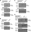
(A) Zymosan induces PKCδ/Dectin-1 complex formation, and Rottlerin inhibits this interaction. Monocytes (5×10⁶/2 ml) were left untreated or treated with Rottlerin (5 μM) for 30 min prior to zymosan (2 mg/ml) stimulation for an additional 15 min. Monocyte lysates were immunoprecipitated with a mouse mAb against human Dectin-1. Immune complexes were subjected to 10% SDS-PAGE and then transferred to PVDF membranes. Dectin-1 immune complexes were first immunoblotted with rabbit polyclonal antibody against phospho-Ser/Thr and then stripped and reprobed with anti-PKCδ antibody. Finally, the blot was reprobed with a goat polyclonal antibody against human Dectin-1. (B) Syk/Dectin-1 complex formation is regulated by PKCδ. Monocytes were treated with Rottlerin as in A. Lysates (100 μg protein) were immunoprecipated with anti-Dectin-1 antibody and blotted with Syk antibody. The blots were reprobed with anti-Dectin-1 polyclonal antibody to assess loading (lower panel). (C) Src association with Dectin-1 is reduced by pretreating with the PKCδ inhibitor, Rottlerin. Monocytes were plated as in A and were left untreated or treated with Rottlerin (5 μM) for 30 min prior to zymosan stimulation for an additional 10 min. Monocyte lysates were immunoprecipitated with anti-Dectin-1 antibody and blotted with Src antibody. The blots were reprobed with anti-Dectin-1 polyclonal antibody to assess loading (lower panel). (D) Src regulates phospho-Syk association with Dectin-1. Monocytes (5×10⁶/2 ml) were left untreated or treated with the oxindole Syk inhibitor, PP2, or PP3 (20 μM) for 30 min prior to zymosan (2 mg/ml) stimulation for an additional 15 min. Monocyte lysates were immunoprecipitated with a mouse mAb against human Dectin-1. Immune complexes were subjected to 10% SDS-PAGE and then transferred to PVDF membranes. Dectin-1 immune complexes were first immunoblotted with rabbit mAb against phospho-Syk and then stripped and reprobed with anti-Syk antibody. Finally, the blot was reprobed with a goat polyclonal antibody against human Dectin-1 to assess equal loading.
Figure 7.
Figure 7.. Down-regulation of PKCδ protein expression by PKCδ AS-ODN suppresses the association of Syk and Src with Dectin-1.
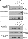
Monocytes (5×10⁶/group) were left untreated or treated with PKCδ AS- or S-ODN (10 μM) for 48 h, as described in Materials and Methods, prior to stimulation with zymosan (2 mg/ml) for 15 min. Cells were lysed and immunoprecipitated with a mouse mAb against human Dectin-1. Immune complexes were resolved by 10% SDS-PAGE and immunoblotted with Syk (A) and Src (B) antibodies (top panels). The blots were reprobed with anti-Dectin-1 goat polyclonal antibody to assess equal IP (A and B, middle panels). Finally, the blots were stripped and reprobed with anti-PKCδ antibody (A and B, bottom panels) to evaluate the effect of AS-ODN on the association of PKCδ with Dectin-1. Monocyte lysates (50 μg) and human rDectin-1 (rh-Dectin1; 1 μg) were used as positive controls.
Figure 8.
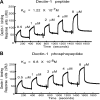
Figure 8.. Direct binding was observed between Dectin-1 and PKCδ by SPR using Biacore.
PKCδ was captured on a CM5 sensor chip, and serial concentrations of nonphospho- and phospho-Dectin-1 ITAM-motif peptides were passed through the flow cell over the immobilized PKCδ. The lines indicate the actual curves calculated after correction for the buffer effect. (A and B) Binding curves of nonphospho- and phospho-Dectin-1 peptides with immobilized PKCδ. Kd of 1.22 × 10⁻⁷ M and 6.8 × 10⁻⁸ M were observed, respectively.
Figure 9.
Figure 9.. PKCδ and Syk are required for Dectin-mediated phagocytosis.
(A) Anti-Dectin antibody inhibits the uptake of zymosan by primary human monocytes, which were pretreated with 10 μg/ml anti-Dectin antibody and fed pHrodo SE-labeled zymosan, and zymosan uptake was detected by flow cytometry. Cells treated with only zymosan, dark blue line; cells pretreated with anti-Dectin antibody, green line; or IgG isotype control, red line. Results are typical of three experiments. (B) Syk is required for zymosan phagocytosis by human monocytes, which were pretreated with oxindole Syk inhibitor (25 μM) and Src inhibitor PP2 (25 μM), fed pHrodo SE-labeled zymosan, and analyzed by flow cytometry. Cells treated with zymosan alone, dark blue line; cells pretreated with the oxindole Syk inhibitor, green line; cells pretreated with PP2, red line. Results are typical of five experiments. (C) PKCδ AS-ODN inhibit PKCδ protein expression. Monocytes were treated with PKCδ AS- or S-ODN, as described in Materials and Methods. The cells were then lysed and analyzed by Western blot analysis to detect PKCδ protein expression. The same blot was then stripped and reprobed with β-tubulin antibody to assess equal loading. (D) PKCδ AS-ODN inhibits Dectin-mediated phagocytosis. After AS-ODN treatment, monocytes were fed pHrodo SE-labeled zymosan and analyzed by flow cytometry. Nonlabeled control cells, dashed line; zymosan-treated cells, red line; cells pretreated with AS-ODN, dark blue line; or S-ODN, green line. Data are from a representative experiment of three with similar results. (E) Microscopic analysis of zymosan phagocytosis. Monocytes treated with zymosan alone (top panel), PKCδ S-ODN-treated cells (middle panel), and PKCδ AS-ODN-treated cells (bottom panel) were collected after pHrodo SE-labeled zymosan phagocytosis and plated on poly-L-lysine-coated microscope slides. To visualize the cell membrane and nucleus, monocytes were incubated with FITC-conjugated CD11b (CR3α) antibody and DAPI. The bright red particles indicate the intracellular ingestion of zymosan, whereas the dim red particles show the extracellular zymosan. Data are from a representative experiment of three with similar results.
Figure 10.
Figure 10.. Zymosan and ZOP activation of human monocyte NADPH oxidase and phagocytosis.
This model summarizes the receptors and related signaling pathways that regulate the assembly and the activation of NADPH oxidase activity and zymosan phagocytosis in primary human monocytes. Previously, we have shown that several pathways, including calcium influx, phosphorylation of cPLA2 by PKCα, arachidonic acid release, PKCδ-dependent phosphorylation of p47phox/p67phox, and Rac-1 translocation to the membrane, regulate NADPH oxidase activity in activated human monocytes [, , , , –13]. This model includes the results reported in this manuscript. The model shows that Dectin-1 is the predominant receptor for zymosan and that Dectin-1 and CR3 participate as receptors for ZOP for regulating NADPH oxidase activation in primary monocytes. Zymosan, acting primarily via Dectin-1, induces the phosphorylation/activation of Syk and Src kinases, two proteins that signal downstream of Dectin-1 in many cell systems. Src and Syk kinase regulate the activity of monocyte NADPH oxidase. Zymosan induces Dectin-1-dependent PKCδ phosphorylation, and PKCδ regulates its own association with Dectin-1, as well as regulating the recruitment of Syk and Src to this receptor complex. PKCδ is a newly identified participant in Dectin-1 signaling and was shown to regulate not only Dectin-1-dependent NADPH activity but also phagocytosis of zymosan particles. In contrast to NADPH oxidase activation, Src activity is not required for zymosan phagocytosis.

Similar articles

Cited by

References

    1. Cathcart M. K., McNally A. K., Morel D. W., Chisolm G. M., III (1989) Superoxide anion participation in human monocyte-mediated oxidation of low-density lipoprotein and conversion of low-density lipoprotein to a cytotoxin. J. Immunol. 142, 1963–1969 - PubMed
    1. Bey E. A., Cathcart M. K. (2000) In vitro knockout of human p47phox blocks superoxide anion production and LDL oxidation by activated human monocytes. J. Lipid Res. 41, 489–495 - PubMed
    1. Li Q., Subbulakshmi V., Fields A. P., Murray N. R., Cathcart M. K. (1999) Protein kinase Cα regulates human monocyte O-2 production and low density lipoprotein lipid oxidation. J. Biol. Chem. 274, 3764–3771 - PubMed
    1. Li Q., Cathcart M. K. (1997) Selective inhibition of cytosolic phospholipase A2 in activated human monocytes. Regulation of superoxide anion production and low density lipoprotein oxidation. J. Biol. Chem. 272, 2404–2411 - PubMed
    1. Cathcart M. K. (2004) Regulation of superoxide anion production by NADPH oxidase in monocytes/macrophages: contributions to atherosclerosis. Arterioscler. Thromb. Vasc. Biol. 24, 23–28 - PubMed

Publication types

MeSH terms