Review
. 2012 Jan;83(1):31-44.
doi: 10.1016/j.mvr.2011.05.004. Epub 2011 May 20.

Heterotrimeric G proteins, focal adhesion kinase, and endothelial barrier function

Affiliations
Review

Heterotrimeric G proteins, focal adhesion kinase, and endothelial barrier function

Tracy Thennes et al. Microvasc Res. 2012 Jan.

Abstract

Ligands by binding to G protein coupled receptors (GPCRs) stimulate dissociation of heterotrimeric G proteins into Gα and Gβγ subunits. Released Gα and Gβγ subunits induce discrete signaling cues that differentially regulate focal adhesion kinase (FAK) activity and endothelial barrier function. Activation of G proteins downstream of receptors such as protease activated receptor 1 (PAR1) and histamine receptors rapidly increases endothelial permeability which reverses naturally within the following 1-2 h. However, activation of G proteins coupled to the sphingosine-1-phosphate receptor 1 (S1P1) signal cues that enhance basal barrier endothelial function and restore endothelial barrier function following the increase in endothelial permeability by edemagenic agents. Intriguingly, both PAR1 and S1P1 activation stimulates FAK activity, which associates with alteration in endothelial barrier function by these agonists. In this review, we focus on the role of the G protein subunits downstream of PAR1 and S1P1 in regulating FAK activity and endothelial barrier function.


Figures

Figure 1
Figure 1. Role of G proteins in regulating endothelial barrier function
A. Endothelial specific depletion of Gαq/Gα11 in mice prevents PAR1-induced increase in endothelial permeability. Evans blue extravasation was determined in mice skin implants lacking indicated Gα subunits in endothelium. In control and endothelial depleted Gα12/13 mice, PAR1-activating peptide increased Evans blue accumulation. Deletion of endothelial Gαq/11 inhibited local Evans blue extravasation in response to PAR1 activation, as well as other GPCR ligands including histamine, LPA, and PAF (data not shown). (© Korhonen et al., 2009. Originally published in The Journal of Experimental Medicine. doi:10.1084/jem.20082150) B. Gβγ restores basal endothelial barrier function following thrombin-induced increase in endothelial permeability. Human pulmonary artery endothelial cells (HPAEC) transducing control siRNA (SiSc) or Gβ1 siRNA (SiGβ1) were stimulated with 50 nM thrombin and transendothelial electrical resistance (TER) across the endothelial monolayer was recorded. Thrombin induced a rapid decrease in HPAEC transducing control siRNA, indicating an increase in intercellular junction permeability. The permeability naturally reverses to the basal level within next 120 min. However, thrombin induced a persistent increase in endothelial monolayer permeability in Gβ1 depleted cells, indicating that Gβ1 is required for restoring basal endothelial permeability. (© Knezevic et al., 2009. Originally published in Journal of Experimental Medicine. doi: 10.1084/jem.20090652).
Figure 2
Figure 2. Model of G protein activation of FAK
Intramolecular interactions between the FAK FERM domain and its Kinase domain keep FAK in an inactive conformation by sterically blocking Y397 residues from potential substrates. Dissociation of FAK FERM domain from the kinase domain leads to rapid autophosphorylation of FAK at Y397 residue. Phospho-Y397 residue forms a high affinity binding site for Src family kinases including p60cSrc and Fyn, which by phosphorylating FAK at Y576/577 fully activate FAK. Src family kinases can also induce FAK phosphorylation at Y861 and Y925 at the C-terminus. FAK may also be phosphorylated at serine residues and their proximity to proline-rich regions may influence FAK-mediated protein-protein interactions and thereby endothelial barrier function.
Figure 3
Figure 3. FAK regulation of endothelial barrier function
A. Thrombin induces sustained activation of FAK. HPAE cells were stimulated with 50 nM thrombin for indicated times and lysates were immunoblotted with Y397 or Y576 residue specific anti-phospho-FAK antibodies or pan anti-FAK as control to determine the phosphorylation of FAK. FAK phosphorylation was increased within 10 min, and remained elevated at this level for up to 120 min (© Knezevic et al., 2009. Originally published in Journal of Experimental Medicine. doi: 10.1084/jem.20090652). B. Impairment of FAK function causes persistent increase in endothelial permeability. HPAEC infected with control virus (Adv-GFP) or dominant-negative FAK viruses (Ad-FRNK) were stimulated with 50 nM thrombin and TER across the endothelial monolayer was recorded. Thrombin also decreased TER to the same extent in dominant negative FAK (GFP-FRNK) expressing cells, but in contrast to control cells, the monolayer did not recover within 2 hr (This research was originally published in the Journal of Biological Chemistry. Holinstat et al. JBC. 2005; Vol: 281, NO.4, pp2296-2305. © The American Society for Biochemistry and Molecular Biology.) C. Gβγ depletion prevents FAK activation. HPAE cells were transfected with control (Sc) or Gβ1 siRNA and after 48 hr post transfection cells were stimulated with 50 nM thrombin for indicated times. Lysates were immunoblotted with Y397 or Y576 residue specific anti-phospho-FAK antibodies or pan anti-FAK as control to determine the phosphorylation of FAK. Thrombin failed to induce FAK activity in Gβ1 depleted transfected cells (© Knezevic et al., 2009. Originally published in Journal of Experimental Medicine. doi: 10.1084/jem.20090652).
Figure 4
Figure 4. RACK1 negatively regulates Gβ1 function
A. Depletion of RACK1 promotes Gβ1 interaction with FAK and Fyn. HPAEC were transfected with control siRNA (Sc) or RACK1 siRNA (SiRACK1) and 48 hr post transfection cells were stimulated with 50 nM thrombin for indicated times. Cell lysates were immunoprecipitated with anti-Fyn antibody and probed with anti-Gβ1 or anti-FAK antibodies to assess interaction. RACK1 knockdown basally potentiated the interaction between Gβ1, Fyn and FAK, which did not further increase following stimulation with thrombin. B. Knockdown of RACK1 increases FAK tyrosine phosphorylation. siSc and siRACK1 expressing cells were assessed for FAK tyrosine phosphorylation following thrombin challenge using site-specific FAK phosphor-antibodies. Results indicate that suppression of endogenous RACK1 basally activated FAK and the phosphorylation did not increase further after thrombin challenge. C. Knockdown of RACK1 accelerates adherens junction assembly. HPAE cells transfected with siSc or SiRACK1 were immuno-stained with anti-VE-cadherin antibody to quantify interendothelial gap formation following thrombin stimulation. Results showed that suppression of RACK1 potentiated adherens junction re-annealing after thrombin challenge indicating RACK1 prevents endothelial barrier recovery by blocking the interaction between Gβ1, Fyn and FAK. (© Knezevic et al., 2009. Originally published in Journal of Experimental Medicine. doi: 10.1084/jem.20090652).
Figure 5
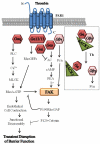
Figure 5. PAR1 signaling inducing FAK activity and reversible disruption of endothelial barrier function
Upon ligation of PAR1 by thrombin, the Gα subunit of the heterotrimeric G protein dissociates from Gβγ. Gαq increases intracellular Ca2+ concentration, which increases endothelial permeability by activating MLCK and RhoA. RhoA also induces FAK activation. FAK negatively regulates RhoA-GTP by activating p190RhoGAP, thus turning off endothelial cell contraction. Gβγ mediates activation of FAK through Fyn kinase leading to interaction of FAK with p120-catenin that facilitates re-annealing of adherens junction and thereby restores normal barrier function. Inset shows the release of Gβγ from RACK1/Gα complex upon thrombin stimulation.
Figure 6
Figure 6. S1P enhances endothelial barrier function
A. Gi increases transendothelial electrical resistance in response to S1P. HPAEC seeded on TER electrodes were stimulated with 1μM S1P in the absence or presence of pertussis toxin (Ptx), which inhibits Gi. S1P alone caused a rapid, sustained increase in TER values, which was prevented in cells pre-treated with Ptx, indicating S1P strengthens endothelial barrier function through at Gi dependent pathway for (This research was originally published in the Journal of Biological Chemistry. Mehta et al. Sphingosine 1-Phosphate-induced Mobilization of Intracellular Ca2+ Mediates Rac Activation and Adherens Junction Assembly in Endothelial Cells. JBC. 2002; Vol: 280, NO.17, pp17321-17328. © the American Society for Biochemistry and Molecular Biology.) B. S1P induced tyrosine phosphorylation of FAK is sensitive to inhibitors of Gi and PLC. HUVECs were preincubated with various inhibitors for 30 min after which these cells were stimulated with 5 μM S1P for 2 min. Cell lysates were immunoprecipitated after which they were immunoblotted with anti-phosphotyrosine antibody to assess FAK activation. Results showed that S1P-induced FAK phosphorylation via Gi-PLC pathway independent of PKC activity (Reprinted from Lee et al., Sphingosine 1-phosphate stimulates tyrosine phosphorylation of focal adhesion kinase and chemotactic motility of endothelial cells via the G(i) protein-linked phospholipase C pathway, 5;268, 47-53, 2000 with permission from Elsevier).
Figure 7
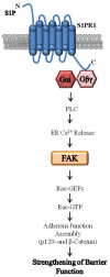
Figure 7. S1P1 signaling leading to FAK activation and enhancement of endothelial barrier function
Upon ligation of S1P1 by S1P, the Gαi (or) Gβγ subunit mediates an increase in intracellular Ca2+ leading to FAK activation. FAK regulates Rac1 to modulate adherens junction assembly which is required for strengthening of the endothelial barrier.
Figure 8
Figure 8. FAK serves as an essential node between PAR1 and S1P1 signaling that maintains normal endothelial permeability
Thrombin activation of PAR1 causes the dissociation of the dissociation of the Gα subunit of the heterotrimeric G protein dissociates from Gβγ. Gαq-mediated increase in intracellular Ca2+ concentration increases endothelial permeability by activating MLCK and RhoA. RhoA also induces FAK activation. FAK negatively regulates RhoA-GTP by activating p190RhoGAP to turn off endothelial cell contraction. P190RhoGAP also interacts with p120-catenin to facilitate re-annealing of adherens junctions thereby restoring normal barrier function downstream of PAR1. S1P activation of S1P1 stimulates Gαi (Gβγ) -mediated PLC activation and increase in intracellular Ca2+ that activates Rac1 and FAK to facilitate strengthening of adherens junctions and the endothelial barrier integrity. Importantly, PAR1 by inducing SPHK1 activity increases S1P levels that in a paracrine manner activate S1P1 signaling cascade to reverse the endothelial barrier permeability increase by thrombin.

Similar articles

Cited by

References

    1. Abedi H, Zachary I. Vascular endothelial growth factor stimulates tyrosine phosphorylation and recruitment to new focal adhesions of focal adhesion kinase and paxillin in endothelial cells. J Biol Chem. 1997;272:15442–51. - PubMed
    1. Adam AP, et al. Src-induced tyrosine phosphorylation of VE-cadherin is not sufficient to decrease barrier function of endothelial monolayers. J Biol Chem. 2010;285:7045–55. - PMC - PubMed
    1. Ahmmed GU, Malik AB. Functional role of TRPC channels in the regulation of endothelial permeability. Pflugers Arch. 2005;451:131–42. - PubMed
    1. Alessandro R, et al. Endothelial cell spreading on type IV collagen and spreading-induced FAK phosphorylation is regulated by Ca2+ influx. Biochem Biophys Res Commun. 1998;248:635–40. - PubMed
    1. Allende ML, et al. G-protein-coupled receptor S1P1 acts within endothelial cells to regulate vascular maturation. Blood. 2003;102:3665–7. - PubMed

Publication types

MeSH terms

Substances

LinkOut - more resources