. 2011 Jul;121(7):2898-910.
doi: 10.1172/JCI44925.

CCL17-expressing dendritic cells drive atherosclerosis by restraining regulatory T cell homeostasis in mice

Affiliations

CCL17-expressing dendritic cells drive atherosclerosis by restraining regulatory T cell homeostasis in mice

Christian Weber et al. J Clin Invest. 2011 Jul.

Abstract

Immune mechanisms are known to control the pathogenesis of atherosclerosis. However, the exact role of DCs, which are essential for priming of immune responses, remains elusive. We have shown here that the DC-derived chemokine CCL17 is present in advanced human and mouse atherosclerosis and that CCL17+ DCs accumulate in atherosclerotic lesions. In atherosclerosis-prone mice, Ccl17 deficiency entailed a reduction of atherosclerosis, which was dependent on Tregs. Expression of CCL17 by DCs limited the expansion of Tregs by restricting their maintenance and precipitated atherosclerosis in a mechanism conferred by T cells. Conversely, a blocking antibody specific for CCL17 expanded Tregs and reduced atheroprogression. Our data identify DC-derived CCL17 as a central regulator of Treg homeostasis, implicate DCs and their effector functions in atherogenesis, and suggest that CCL17 might be a target for vascular therapy.


Figures
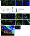
Figure 1
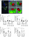
Figure 1. CCL17-expressing DCs accumulate within atherosclerotic lesions.
(A) A network of EYFP+CD11c+ DCs (green) can be detected by immunofluorescence in the aortic root (left 2 panels, scale bars: 50 μm) and by multiphoton microscopy in the en face prepared aorta (middle right panel, scale bar: 20 μm) of CD11c-EYFP reporter mice; representative maximum intensity projection of a 30-μm-thick volume is shown. Multiphoton microscopy of an en face prepared aorta of a wild-type mouse stained for MHC-II+ (red); collagen was visualized by SHG (blue, far right panel, scale bar: 20 μm). (B) Quantification of EGFP+CD11c+ DCs in aortas of healthy Ccl17E/+Apoe–/– and Ccl17E/EApoe–/– mice or after high-fat diet (HFD) feeding using enzymatic digestion and FACS analysis; representative dot blots and percentages within quadrants are shown (n = 6 each). *P < 0.05. ctrl, control. (C and D) EGFP+ DCs (green, indicated by arrows) in the aortic root of Ccl17E/+Apoe–/– and Ccl17E/EApoe–/– mice after 12 weeks of high-fat diet feeding (C) and after 6 months of normal chow (D); cell nuclei are counterstained by DAPI (blue); scale bars, 50 μm. (E) Maximum intensity projection of a z-stack (left) and high-magnification z-slice (right) with EGFP+ DCs (bright green cytoplasmic staining; arrows) in the atherosclerotic aortic root of a Ccl17E/+Apoe–/– mouse on normal chow. Nuclei are counterstained with PI (red); PI staining can also appear yellow due to background fluorescence. Collagen is visible due to SHG (blue). Scale bars: 40 μm.
Figure 2
Figure 2. Ccl17 deficiency reduces atherosclerosis.
(A) Atherosclerotic lesions were quantified in the aortic root and thoraco­abdominal aorta after staining with oil red O in Ccl17+/+Apoe–/– and Ccl17E/EApoe–/– mice on normal chow for 6 months; individual data points represent average plaque area per mouse; horizontal bars denote mean. Representative images of the aortic root (scale bars: 500 μm) and the thoracoabdominal aorta are shown. (B and C) The relative content of MOMA-2+ macrophages (scale bars: 200 μm) and CD3+ T cells (C) per plaque area was analyzed by quantitative immunofluorescence. Representative images of macrophage staining (green) are shown (B); cell nuclei are counterstained by DAPI (blue); dashed lines indicate the internal elastic lamina. *P < 0.05.
Figure 3
Figure 3. Ccl17 deficiency affects T cell distributions.
(A) Quantification of PE-CD4+ T cells and APC-Tregs accumulating in atherosclerotic aortas of high-fat diet–fed Ccl17+/+Apoe–/– and Ccl17E/EApoe–/– mice 3 days after adoptive transfer, using enzymatic digestion and FACS analysis. Representative dot plots depict frequencies of labeled cells among CD45+ aortic cells. Data points represent frequencies of transferred aortic cells in individual mice; horizontal bars denote mean of all mice. (B) Transwell migration of CD4+ T cells toward Ccl17E/+ or Ccl17E/E BMDCs was quantified by FACS analysis (n = 5). (C) Absolute numbers of CD4+ T cells recruited to air pouches injected with PBS (ctrl, n = 8) or recombinant mouse CCL17 (50 ng/ml, n = 9) were quantified in lavage fluid after 4 hours. (D and E) Flow cytometric analysis of CD3+CD4+ T cells (D) and CD4+Foxp3+CD25+ Tregs (E) in LNs of Ccl17+/+ and Ccl17E/E mice employing indicated surface markers. Representative dot plots as well as relative and absolute numbers of Tregs are shown; numbers in dot plots are percentage of CD4+ events. Data points represent frequencies of cells in individual mice; horizontal bars, mean of all mice. (F) Foxp3 mRNA expression in LNs of Ccl17+/+Apoe–/– and Ccl17E/EApoe–/– mice fed a high-fat diet (n = 3 each). (G) Detection of Foxp3+GFP+ cells (arrows, both panels) in atherosclerotic plaques of Ldlr–/– mice reconstituted with Foxp3gfp.KI BM after 9 weeks of high-fat diet; cell nuclei were counterstained by DAPI (blue, lower panel); dotted lines demarcate lesional area. Scale bars: 50 μm. (H) Foxp3 mRNA expression in atherosclerotic aortas of Ccl17+/+Apoe–/– and Ccl17E/EApoe–/– mice on high-fat diet (n = 3 each). (I) Quantification of CD4+CD25+Foxp3+ Tregs in atherosclerotic aortic segments of Ccl17+/+Apoe–/– and Ccl17E/EApoe–/– mice on high-fat diet using enzymatic digestion and FACS analysis. *P < 0.05.
Figure 4
Figure 4. CCL17 controls the maintenance of Tregs.
(A) CFSE-labeled CD4+CD25 T cells were transferred into Ccl17+/+ or Ccl17E/E mice, and frequencies of CFSE+Foxp3+ Tregs and CFSE+CD4+ cells among T cells in LNs were analyzed by flow cytometry after 10 days. Representative dot plots and percentages within gates are shown (n = 10 per group). (B) Sorted unpulsed or OVA-2–pulsed EGFP or EGFP+ Ccl17E/+ or Ccl17E/E BMDCs were incubated with OT-II T cells in vitro (n = 5 independent experiments). T cell proliferation was quantified by CSFE dilution and FACS analysis after 3 days. OT-II T cells were back-sorted, and Foxp3 mRNA expression was analyzed by real-time PCR. Frequencies of apoptotic annexin V+ cells were quantified by FACS analysis. *P < 0.05 versus unpulsed; #P < 0.05 versus OVA-pulsed EGFP+Ccl17E/+ BMDCs. Phosphorylation of Stat5 (pStat5) was assessed by flow cytometry; representative histograms are shown. Fluorescence-minus-one measurements served as control. (C) OVA-2–pulsed Ccl17+/+ or Ccl17E/E BMDCs were incubated with OT-II Tregs (n = 3 independent experiments). Frequencies of Foxp3+CD25+CD4+ and of annexin V+ Tregs among Foxp3+CD25+CD4+ Tregs were quantified by FACS analysis after 3 days; representative dot plots and percentage of Foxp3+CD25+ Tregs among CD4+ T cells within gates are shown. *P < 0.05 versus Ccl17+/+ BMDCs. pStat5 was assessed by flow cytometry in Tregs; representative histograms are shown. (D) FACS analysis of annexin V+CD4+Foxp3+ Tregs in LNs of Ccl17+/+ and Ccl17E/E mice. Data points represent frequencies of cells in individual mice; horizontal bars denote mean of all mice. *P < 0.05.
Figure 5
Figure 5. CCL17+ DCs mediate atherogenesis by priming T cells.
(A) Multiphoton microscopy was used to obtain 2D optical slices (left images) and 3D projections of matching zoomed areas (middle and right images) revealing EGFP+ DCs (green cytoplasmic staining) in close contact with DiI-labeled CD4+ T cells (red) in atherosclerotic vessel walls at the carotid artery bifurcation (upper panels, ×10 μm) and the aortic root (lower panels, ×25 μm) of Ccl17E/+Apoe–/– mice fed a high-fat diet. Dashed lines demarcate lesional area; L, lumen. (B) CD4+ T cells sorted from 6-month-old Ccl17+/+Apoe–/– and Ccl17E/EApoe–/– mice on normal chow were adoptively transferred into 8-week-old Ccl17+/+Apoe–/– and Ccl17E/EApoe–/– mice depleted of CD4+ cells. Undepleted Ccl17+/+Apoe–/– and Ccl17E/EApoe–/– mice served as controls. After 6 weeks of high-fat diet, atherosclerotic lesions were quantified in the aortic root and aorta after staining with oil red O. Individual data points represent average plaque area per mouse; horizontal bars denote mean. *P < 0.05. (C) Atherosclerotic lesions were quantified in aortas stained with oil red O in Ccl17+/+Apoe–/– and Ccl17E/EApoe–/– mice treated with depleting anti-CD25 antibody or isotype control and fed a high-fat diet for 4 weeks. Individual data points represent average plaque area per mouse; horizontal bars denote mean. *P < 0.05.
Figure 6
Figure 6. CCL17 as a potential therapeutic target.
(A) Apoe–/– mice fed a high-fat diet for 4 weeks were injected with blocking antibody to CCL17 or isotype control for an additional 4 weeks. Atherosclerotic lesions were quantified in the aortic root (scale bars: 500 μm) and aorta after oil red O staining. Individual data points represent average plaque area per mouse; horizontal bars denote mean; representative images are shown. (B) Frequencies of CD4+Foxp3+ Tregs among CD3+ cells in LNs were determined by flow cytometry; Foxp3 mRNA levels in LNs by real-time PCR analysis. (C) Real-time PCR analysis of Foxp3 mRNA expression in atherosclerotic aortas. *P < 0.05.

Similar articles

Cited by

References

    1. Hansson GK, Libby P. The immune response in atherosclerosis: a double-edged sword. Nat Rev Immunol. 2006;6(7):508–519. doi: 10.1038/nri1882. - DOI - PubMed
    1. Weber C, Zernecke A, Libby P. The multifaceted contributions of leukocyte subsets to atherosclerosis: lessons from mouse models. Nat Rev Immunol. 2008;8(10):802–815. - PubMed
    1. Shortman K, Naik SH. Steady-state and inflammatory dendritic-cell development. Nat Rev Immunol. 2007;7(1):19–30. doi: 10.1038/nri1996. - DOI - PubMed
    1. Zenke M, Hieronymus T. Towards an understanding of the transcription factor network of dendritic cell development. Trends Immunol. 2006;27(3):140–145. doi: 10.1016/j.it.2005.12.007. - DOI - PubMed
    1. Millonig G, et al. Network of vascular-associated dendritic cells in intima of healthy young individuals. Arterioscler Thromb Vasc Biol. 2001;21(4):503–508. - PubMed

Publication types

Substances