. 2011;6(5):e20420.
doi: 10.1371/journal.pone.0020420. Epub 2011 May 24.

VPS29 is not an active metallo-phosphatase but is a rigid scaffold required for retromer interaction with accessory proteins

Affiliations

VPS29 is not an active metallo-phosphatase but is a rigid scaffold required for retromer interaction with accessory proteins

James D Swarbrick et al. PLoS One. 2011.

Abstract

VPS29 is a key component of the cargo-binding core complex of retromer, a protein assembly with diverse roles in transport of receptors within the endosomal system. VPS29 has a fold related to metal-binding phosphatases and mediates interactions between retromer and other regulatory proteins. In this study we examine the functional interactions of mammalian VPS29, using X-ray crystallography and NMR spectroscopy. We find that although VPS29 can coordinate metal ions Mn(2+) and Zn(2+) in both the putative active site and at other locations, the affinity for metals is low, and lack of activity in phosphatase assays using a putative peptide substrate support the conclusion that VPS29 is not a functional metalloenzyme. There is evidence that structural elements of VPS29 critical for binding the retromer subunit VPS35 may undergo both metal-dependent and independent conformational changes regulating complex formation, however studies using ITC and NMR residual dipolar coupling (RDC) measurements show that this is not the case. Finally, NMR chemical shift mapping indicates that VPS29 is able to associate with SNX1 via a conserved hydrophobic surface, but with a low affinity that suggests additional interactions will be required to stabilise the complex in vivo. Our conclusion is that VPS29 is a metal ion-independent, rigid scaffolding domain, which is essential but not sufficient for incorporation of retromer into functional endosomal transport assemblies.


Conflict of interest statement

Competing Interests: The authors have declared that no competing interests exist.

Figures

Figure 1
Figure 1. Interactions of VPS29 examined in this study.
VPS29 has structural similarity to phosphatase enzymes and has the potential to bind two divalent cations . VPS29 binds VPS35 with an interface that incorporates the metal-binding pocket, the α3 helix and the Phe63 side-chain . The α3 helix is known to adopt different conformations in previous crystal structures , , , and Phe63 is known to adopt different conformations upon metal binding to VPS29 . A conserved hydrophobic surface lies opposite to the VPS35 interface, and is known to be required for binding to TBC1D5 in mammalian cells , and to the heterodimeric SNX complex in yeast . Critically mutation of Leu152 to Glu is known to abolish these interactions. Note, the SNX proteins are also thought to form contacts directly with VPS35 , .
Figure 2
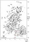
Figure 2. NMR assignement of VPS29.
VPS29 [1H,15N]-HSQC spectrum. Assigned peaks are labelled by residue number.
Figure 3
Figure 3. VPS29 binds Zn2+ in solution as determined by NMR spectroscopy.
(A) A number of residues in the [1H,15N]-HSQC spectra of VPS29 show specific perturbations on addition of Zn2+. Spectra are shown of VPS29 in the apo state (black) or in the presence of 200 µM ZnCl2 (red). Resonances showing significant chemical shift changes are indicated. (B) Zn2+ binds to VPS29 in solution within the same major pocket identified by X-ray crystallography. Residues showing significant chemical shift upon Zn titration are highlighted on the structure in red (Δδ greater than 2 SD). Each monomer from the asymmetric unit of the Zn2+-bound mouse VPS29 crystal structure has been mapped, with crystallographically identified Zn2+ ions indicated in blue (see Fig. 3 for details). Note however, that VPS29 is monomeric in solution, not dimeric. No significant evidence is seen for binding to the low occupancy Zn2+ site coordinated by Asp55, His57 and His33 (dashed circle). (C) Titration of VPS29 with Zn2+ can be followed by observing the change in intensity for the Gly133 NH resonance in bound and unbound states. (D) Plot of the bound and unbound intensity ratio for the Gly133 NH resonance as a function of Zn2+ concentration. The blue line shows the fit to the Hill equation. The estimated affinity is low with an overall K d>250 µM. (E) Binding of VPS29 to either Mn2+ or Zn2+ cannot be measured by ITC, confirming the low affinity of interaction. A weak endothermic signal is observed upon metal titration at 25°C but the binding affinity cannot be estimated. Top panels show raw data and bottom panels show integrated normalised data. No significant binding signals were observed under these conditions (0.04 mM protein, 2.5 mM metals, 25°C). Other metals including Mg2+, Ca2+ and Ni2+, and temperature regimes from 10–37°C produced similar negative results. (F) Mn2+ and Zn2+ bind to EDTA exothermically and with high affinity under identical conditions by ITC (0.2 mM EDTA, 2.5 mM metals, 25°C). The binding affinity is too high to be determined at the concentrations used.
Figure 4
Figure 4. Crystal structures of VPS29 bound to Mn2+ and Zn2+.
Crystal structure of VPS29 determined by X-ray crystallography bound to either Mn2+ (A) or Zn2+ (B). Top panels show overall protein structures as ribbon diagrams with anomalous difference maps contoured at 3σ shown in red. Each monomer from the asymmetric unit is indicated in green and blue. Mn2+ ions are shown as magenta spheres, and Zn2+ ions are shown as salmon spheres. Middle panels (i) show zoomed in regions of the putative active site residues and bound metal ions. Bottom panels (ii) show enlarged regions for minor low occupancy sites distal to the major binding pocket.
Figure 5
Figure 5. Metals do not affect VPS29 phosphatase activity or interaction with VPS35.
(A) SDS-PAGE gel showing purified VPS29 and trimeric retromer proteins used for phosphatase assays stained with Coomassie Blue. (B) No detectable phosphatase activity was measured for VPS29 alone or in complex with VPS35 and VPS26. Phosphatase assays used the CI-MPR peptide CSSTKLVSFHDD(pS)DEDLLHI. The release of phosphate was measured using Biomol Green reagent and colorimetric assay at 620 nm. Calf intestinal alkaline phosphatase (CIAP) is shown for comparison. (C) When VPS29 has bound metal, the conformation of Phe63 is altered such that it may clash with VPS35 and inhibit binding. The diagram shows a close up of the interaction between VPS29 and VPS35 . The Mn2+-bound VPS29 structure (green ribbon, and yellow side-chains) is overlayed with VPS35-bound VPS29 (blue ribbon and cyan side-chain). VPS35 is shown in surface representation. (D) No significant difference is observed in binding to VPS35 in the presence of EDTA or MnCl2 indicating that metals do not influence complex formation. VPS29 interaction with VPS35 was analysed by ITC; top panels show raw data and bottom panels show integrated normalised data.
Figure 6
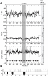
Figure 6. 15N NMR relaxation data for VPS29 indicates a generally rigid structure with no large-scale mobility with well defined secondary structural elements.
(A) Longitudinal T1 and transverse T2 relaxation times as well as the {1H}15N heteronuclear NOEs are shown as a function of protein sequence. Residues within helix α3 are highlighted in grey. Six N-terminal (non-native) resides are shown and are labelled as residues 201–206. The protein secondary structure is indicated at the bottom of the figure. Data was recorded using a 600 MHz spectrometer. (B) The TALOS+ artificial neural network (ANN)-predicted secondary structural elements of VPS29. Length of bars corresponds to probability of a residue to be helix (black) or β-strand (grey).
Figure 7
Figure 7. The α3 helix of VPS29 adopts a compact conformation in solution.
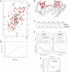
(A) Comparison of previous VPS29 crystal structures reveals differences in the orientations of the α3 helix. VPS29 adopts an extended α3 orientation in the mouse apo VPS29 crystal structure (PDB 1Z2X; green), and a compact orientation in the VPS35-bound human VPS29 structure (PDB 2R17; blue). Ala100, Gln103 and Tyr129 are shown for each structure, to show residues for which long-range NOEs are observed. In the apo mouse VPS29 structure these residues would be too far apart to observe these NOE contacts. (B) RDC correlation plots indicate the apo mouse VPS29 protein adopts a compact structure in solution, where the α3 helix is similar to the VPS35-bound conformation, but not the previous mouse VPS29 crystal structure. Shown are 133 1DHN RDCs fitted to the apo human VPS29 structure (PDB 1W24), human VPS29 in complex with VPS35 (PDB 2R17) and the mouse VPS29 structure (PDB 1Z2X). The residues of helix α3 are highlighted in red (95–107). Residue 99 was not included due to severe overlap in the 2D 15N IPAP spectra. Single value decomposition analysis of the HN-N RDCs to the apo human, VPS35-bound human or apo mouse X-ray structures yielded the following: The largest component of the alignment tensor (Szz) were 1.31e−3, 1.35e−3, 1.09e−3 and Rhombicities (Syy−Sxy/Szz) were 0.53, 0.53 and 0.48. (C) 2D 1HN-1H strips from the 3D 15N NOESY-HSQC show the NOE connectivities along helix α3 (residues 97–107). Diagonal peaks for each strip are labelled with an asterisk and proximal protons that give rise to observable NOEs are annotated. Medium range i, i+2 1HN-1HN NOES are underlined while medium range i, i+3 and i, i+4 1Hα-1HN NOES are identified with arrows. Both are diagnostic of a regular alpha helix. Two long range NOEs to the Hε protons of Tyr129 from the 1HN of Ala100 and Gln103 are labelled as well as the long-range (cross sheet) 1HN-1HN NOE between Y129 and F122. (D) 1DHN RDCs observed (black) compared to those calculated for each residue for mouse VPS29 structure (PDB 1Z2X, green), apo human VPS29 structure (PDB 1W24, red) and human VPS29 in complex with VPS35 (PDB 2R17, blue). (E) Detail comparing the close fit of the 1DHN RDCs to the α3 orientation in the apo human VPS29 compared to the poor fit to the mouse α3 orientation.
Figure 8
Figure 8. VPS29 binds specifically to SNX1 but with low affinity in vitro.
(A) Immunoprecipitations from HeLa cells do not detect association of retromer with SNX1 even in the presence of increased levels of VPS29 expression. Cells expressing GFP-VPS29 or GFP-VPS29(L152E) mutant were subjected to immunoprecipitation with either VPS26 or SNX1 antibodies. VPS26 (and thus VPS29-containing retromer) associates readily with the effector complex containing strumpellin , confirming that known binding partners can be detected in the immuno-isolates. However, no SNX1 is detected, and furthermore, in reverse experiments SNX1 does not precipitate retromer indicating that their association in vivo is relatively weak or transient. (B) Titration of VPS29 with SNX1 in NMR experiments reveals specific but weak association in vitro. A selected region is shown for the 15N-HSQC spectra of VPS29 in the presence of increasing concentrations of SNX1. (C) Chemical shift perturbations are shown for VPS29 in the presence of SNX1. Inset shows a plot of the chemical shift perturbation for Leu26 NH as a function of SNX1 concentration. (D) SNX1 binds to VPS29 via the conserved hydrophobic surface on the opposite face to the metal-binding pocket and VPS35 binding interface. Residues that show the largest perturbations on SNX1 binding (>2 standard deviations) are mapped on the VPS29 structure in blue. The structure of VPS29 (surface, and green ribbons) is shown in complex with VPS35(476–780) (red ribbons) . The side-chains of the VPS29 hydrophobic surface are indicated. (E) Mutation of the hydrophobic surface of VPS29 (L152E) prevents VPS29-SNX1 association. The [1H,15N]-HSQC spectra for VPS29(L152E) in the absence (black) and presence (red) of SNX1 indicates no significant association is occurring.

Similar articles

Cited by

References

    1. Bonifacino JS, Hurley JH. Retromer. Curr Opin Cell Biol. 2008;20:427–436. - PMC - PubMed
    1. Collins BM. The structure and function of the retromer protein complex. Traffic. 2008;9:1811–1822. - PubMed
    1. Seaman MN. Recycle your receptors with retromer. Trends Cell Biol. 2005;15:68–75. - PubMed
    1. Seaman MN. Endosome protein sorting: motifs and machinery. Cell Mol Life Sci. 2008;65:2842–2858. - PMC - PubMed
    1. Arighi CN, Hartnell LM, Aguilar RC, Haft CR, Bonifacino JS. Role of the mammalian retromer in sorting of the cation-independent mannose 6-phosphate receptor. J Cell Biol. 2004;165:123–133. - PMC - PubMed

Publication types

MeSH terms