. 2011 Jul;32(7):1392-9.
doi: 10.1016/j.peptides.2011.05.011. Epub 2011 May 17.

Effects of cell-type specific leptin receptor mutation on leptin transport across the BBB

Affiliations

Effects of cell-type specific leptin receptor mutation on leptin transport across the BBB

Hung Hsuchou et al. Peptides. 2011 Jul.

Abstract

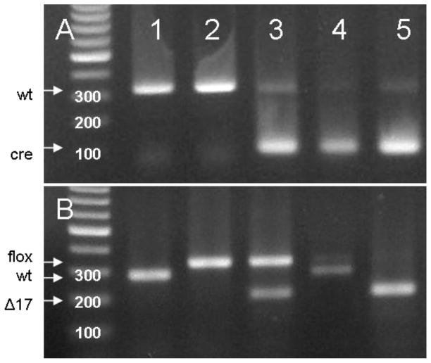
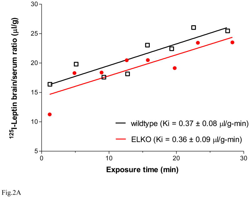
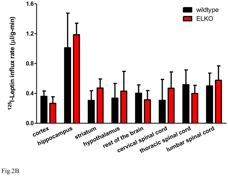
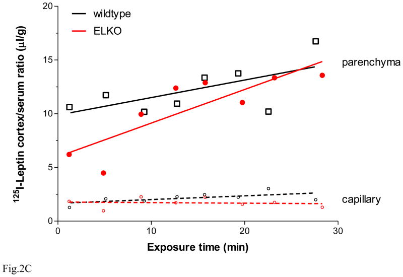
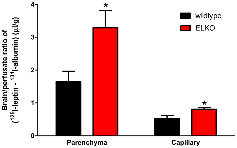
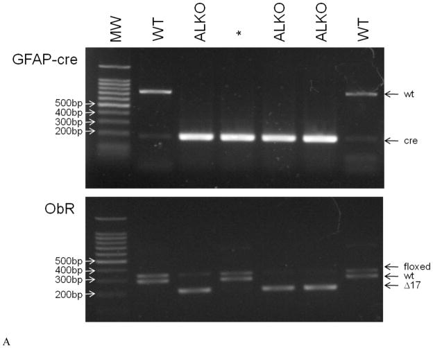
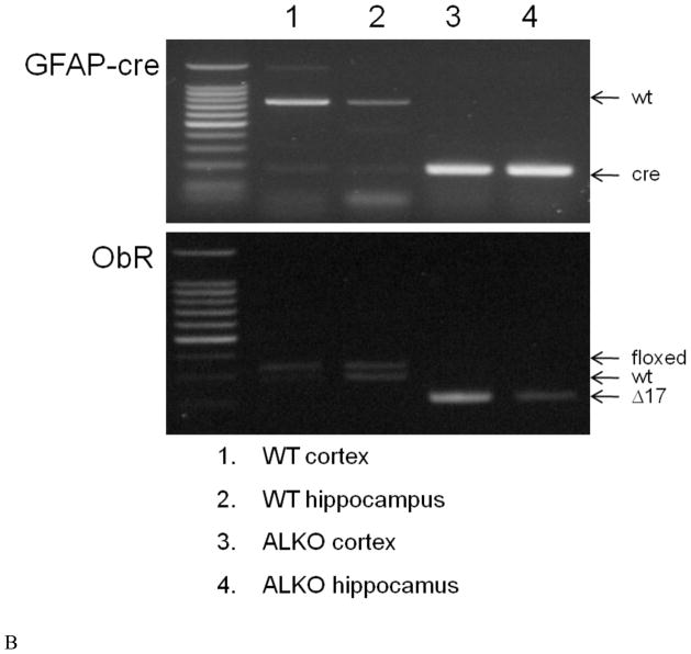
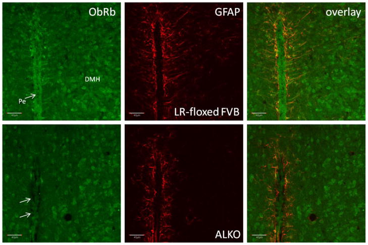
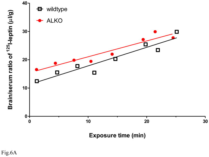
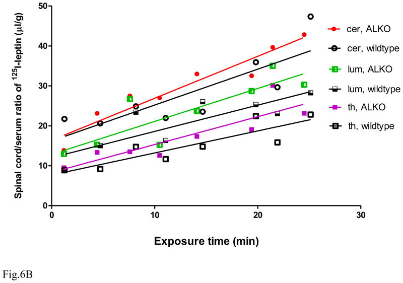
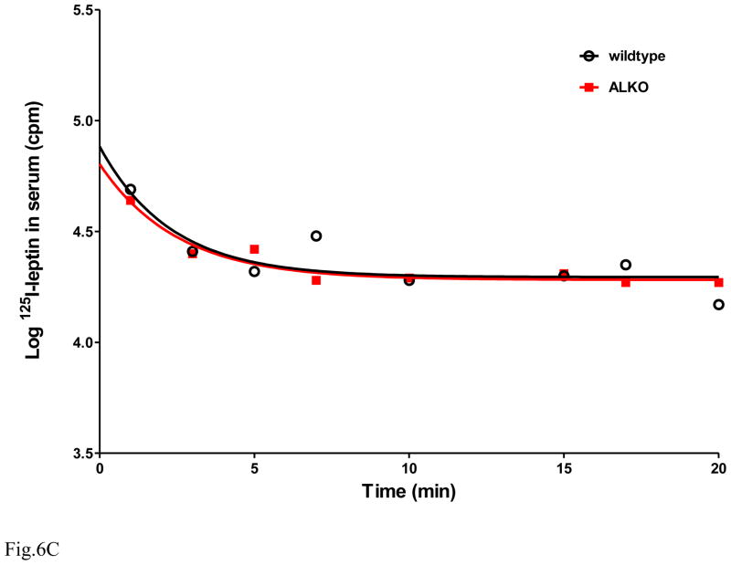
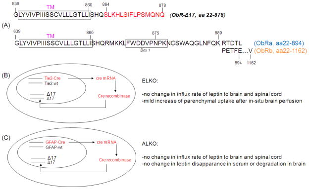
The functions of leptin receptors (LRs) are cell-type specific. At the blood-brain barrier, LRs mediate leptin transport that is essential for its CNS actions, and both endothelial and astrocytic LRs may be involved. To test this, we generated endothelia specific LR knockout (ELKO) and astrocyte specific LR knockout (ALKO) mice. ELKO mice were derived from a cross of Tie2-cre recombinase mice with LR-floxed mice, whereas ALKO mice were generated by a cross of GFAP-cre with LR-floxed mice, yielding mutant transmembrane LRs without signaling functions in endothelial cells and astrocytes, respectively. The ELKO mutation did not affect leptin half-life in blood or apparent influx rate to the brain and spinal cord, though there was an increase of brain parenchymal uptake of leptin after in situ brain perfusion. Similarly, the ALKO mutation did not affect blood-brain barrier permeation of leptin or its degradation in blood and brain. The results support our observation from cellular studies that membrane-bound truncated LRs are fully efficient in transporting leptin, and that basal levels of astrocytic LRs do not affect leptin transport across the endothelial monolayer. Nonetheless, the absence of leptin signaling at the BBB appears to enhance the availability of leptin to CNS parenchyma. The ELKO and ALKO mice provide new models to determine the dynamic regulation of leptin transport in metabolic and inflammatory disorders where cellular distribution of LRs is shifted.


Figures

Fig. 1
Fig. 1
DNA genotyping of the offspring of LR loxP/+/Tie2cre/wt and LRloxP/loxP mice was performed for Tie2-cre (A) and LR-flox (B). The left lane shows molecular weight (bp). Lane 1: wildtype (+/+, +/+); lane 2: LR-flox (+/+, f/f); lane 3: LR-flox, Tie2(tek)-cre (cre/+, f/f), the true ELKO mouse; lane 4: LR-flox heterozygote, Tie2-cre (cre/+, f/+); lane 5: LR-flox with general Exon 17 deletion, Tie2-cre (cre/+, Δ17/Δ17).
Fig. 2
Fig. 2
The effect of the ELKO mutation on permeability of 125I-leptin across the BBB was tested in 3 m old mice by intravenous delivery of the tracer and multiple time-regression analysis (n = 8/group). (A) The influx rate and volume of distribution of 125I-leptin in the brain did not differ significantly between the ELKO and wildtype mice 1 – 20 min after intravenous delivery. (B) The influx rate of 125I-leptin in different brain and spinal cord regions did not differ between the ELKO and wildtype mice. (C) Cortical samples were subjected to the capillary depletion procedure, which showed that parenchymal uptake was significantly (p < 0.005) higher than that of the enriched microvessels in both ELKO and wildtype mice; however, there was no significant increase of influx rate of leptin to brain parenchyma in the ELKO mice.
Fig. 2
Fig. 2
The effect of the ELKO mutation on permeability of 125I-leptin across the BBB was tested in 3 m old mice by intravenous delivery of the tracer and multiple time-regression analysis (n = 8/group). (A) The influx rate and volume of distribution of 125I-leptin in the brain did not differ significantly between the ELKO and wildtype mice 1 – 20 min after intravenous delivery. (B) The influx rate of 125I-leptin in different brain and spinal cord regions did not differ between the ELKO and wildtype mice. (C) Cortical samples were subjected to the capillary depletion procedure, which showed that parenchymal uptake was significantly (p < 0.005) higher than that of the enriched microvessels in both ELKO and wildtype mice; however, there was no significant increase of influx rate of leptin to brain parenchyma in the ELKO mice.
Fig. 3
Fig. 3
The effect of the ELKO mutation on brain uptake of 125I-leptin was determined by 5 min of in-situ brain perfusion, followed by capillary depletion. The ELKO mice (n = 5) showed a higher uptake in both parenchymal and capillary fractions than the wildtype controls (n = 6). *: p < 0.05.
Fig. 4
Fig. 4
Genotyping of ALKO mice. (A) DNA genotyping from tail samples of a representative litter of mice that contains wildtype (floxed LR, wildtype LR, wildtype GFAP-cre) and ALKO (LR-Δ17, GFAP-cre) PCR products. The asterisk (*) represents the mouse with GFAP-cre and LR-flox heterozygote, which was not used in the experiments. (B) PCR from cultured primary astrocytes from different brain regions in the wildtype and ALKO mice. In the wildtype cortex (lane 1) and hippocampus (lane 2), wildtype GFAP-cre, floxed LR, and wildtype LR products were present. In the ALKO cortex (lane 3) and hippocampus (lane 4), GFAP-cre and LR-Δ17 were present.
Fig. 4
Fig. 4
Genotyping of ALKO mice. (A) DNA genotyping from tail samples of a representative litter of mice that contains wildtype (floxed LR, wildtype LR, wildtype GFAP-cre) and ALKO (LR-Δ17, GFAP-cre) PCR products. The asterisk (*) represents the mouse with GFAP-cre and LR-flox heterozygote, which was not used in the experiments. (B) PCR from cultured primary astrocytes from different brain regions in the wildtype and ALKO mice. In the wildtype cortex (lane 1) and hippocampus (lane 2), wildtype GFAP-cre, floxed LR, and wildtype LR products were present. In the ALKO cortex (lane 3) and hippocampus (lane 4), GFAP-cre and LR-Δ17 were present.
Fig. 5
Fig. 5
Immunohistochemistry verifying the successful and specific deletion of ObRb in the ALKO mice. Absence of ObRb immunofluorescence was seen in α1 tanycytes lining the third ventricle adjacent to the dorsal medial hypothalamus (arrows). In the wildtype mouse, confocal microscopy showed co-localization of ObRb and GFAP in this region. Neuronal ObRb was unchanged by ALKO mutation. The GFAP(+) tanycyte processes showed a tortuous and abbreviated morphology in the ALKO mice, indicative of mild astrogliosis. Scale bar: 40 μm.
Fig. 6
Fig. 6
BBB transport of leptin in the ALKO mice (n = 8/group). (A) In whole brain, the ALKO and wildtype groups did not differ in the rate of influx or volume of distribution of 125I-leptin. (B) In different regions of the spinal cord (cervical, thoracic, and lumber segments), the ALKO and wildtype controls did not differ in the blood-to-spinal cord transport of 125I-leptin. (C) The disappearance of 125I-leptin in serum followed a 2-phase exponential decay. There was no difference between the wildtype and ALKO groups.
Fig. 6
Fig. 6
BBB transport of leptin in the ALKO mice (n = 8/group). (A) In whole brain, the ALKO and wildtype groups did not differ in the rate of influx or volume of distribution of 125I-leptin. (B) In different regions of the spinal cord (cervical, thoracic, and lumber segments), the ALKO and wildtype controls did not differ in the blood-to-spinal cord transport of 125I-leptin. (C) The disappearance of 125I-leptin in serum followed a 2-phase exponential decay. There was no difference between the wildtype and ALKO groups.
Fig. 7
Fig. 7
Lack of effect of ALKO mutation on degradation patterns of leptin in serum and brain of mice at different times after intravenous delivery of 125I-leptin. Acid precipitation showed that 125I-leptin remained mostly intact within the first 7 min, but had a linear degradation over time. There was no difference in the % intact leptin in the brain homogenates of the wildtype and ALKO mice (n = 8/group).
Fig. 8
Fig. 8
Diagram of the structure of mutant ObR in the ALKO mice. (A) The LR-flox mice contain two loxP sites flanking exon 17, and recombination with human GFAP-driven Cre-recombinase gene leads to excision of exon 17 and a premature stop codon at aa878. The resultant mutant LR-Δ17 does not contain box 1 shared by the short-forms of membrane-bound LR (a, c, d) or box 2 and 3 unique to ObRb. (B) Features of ELKO mice. (C) Features of ALKO mice. DNA genotyping for ELKO mice. Lane 1: wildtype (+/+, +/+); lane 2: LR-flox (+/+, f/f); lane 3: LR-flox, Tie2(tek)-cre (cre/+, f/f), the true ELKO mouse; lane 4: LR-flox heterozygote, Tie2-cre (cre/+, f/+); lane 5: LR-flox with general Exon 17 deletion, Tie2-cre (cre/+, Δ17/Δ17).

Similar articles

Cited by

References

    1. Abbott NJ. Astrocyte-endothelial interaction and blood-brain barrier permeability. J Anat. 2002;200:629–38. - PMC - PubMed
    1. Banks WA, Coon AB, Robinson SM, Moinuddin A, Shultz JM, Nakaoke R, et al. Triglycerides induce leptin resistance at the blood-brain barrier. Diabetes. 2004;53:1253–60. - PubMed
    1. Banks WA, Farrell CL. Impaired transport of leptin across the blood-brain barrier in obesity is acquired and reversible. Am J Physiol. 2003;285:E10–E15. - PubMed
    1. Banks WA, Kastin AJ. Physiological consequences of the passage of peptides across the blood-brain barrier. Rev Neurosci. 1993;4:365–72. - PubMed
    1. Banks WA, Kastin AJ, Huang W, Jaspan JB, Maness LM. Leptin enters the brain by a saturable system independent of insulin. Peptides. 1996;17:305–11. - PubMed

Publication types

MeSH terms