. 2011 Jun 21;108(25):E222-9.
doi: 10.1073/pnas.1102231108. Epub 2011 May 23.

The DISABLED protein functions in CLATHRIN-mediated synaptic vesicle endocytosis and exoendocytic coupling at the active zone

Affiliations

The DISABLED protein functions in CLATHRIN-mediated synaptic vesicle endocytosis and exoendocytic coupling at the active zone

Fumiko Kawasaki et al. Proc Natl Acad Sci U S A. .

Abstract

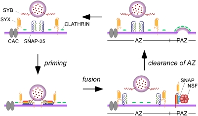
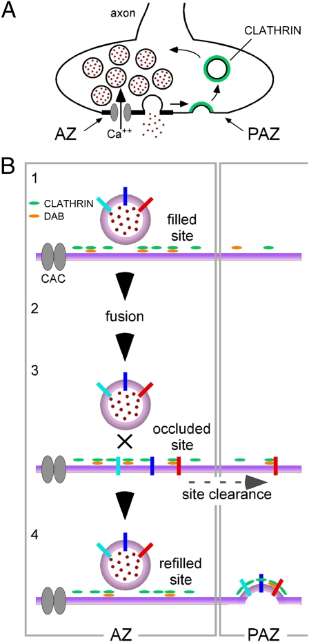
Members of the DISABLED (DAB) family of proteins are known to play a conserved role in endocytic trafficking of cell surface receptors by functioning as monomeric CLATHRIN-associated sorting proteins that recruit cargo proteins into endocytic vesicles. Here, we report a Drosophila disabled mutant revealing a novel role for DAB proteins in chemical synaptic transmission. This mutant exhibits impaired synaptic function, including a rapid activity-dependent reduction in neurotransmitter release and disruption of synaptic vesicle endocytosis. In presynaptic boutons, Drosophila DAB and CLATHRIN were highly colocalized within two distinct classes of puncta, including relatively dim puncta that were located at active zones and may reflect endocytic mechanisms operating at neurotransmitter release sites. Finally, broader analysis of endocytic proteins, including DYNAMIN, supported a general role for CLATHRIN-mediated endocytic mechanisms in rapid clearance of neurotransmitter release sites for subsequent vesicle priming and refilling of the release-ready vesicle pool.


Conflict of interest statement

The authors declare no conflict of interest.

Figures

Fig. 1.
Fig. 1.
dab mutant. (A) Schematic of the dDAB protein and interaction domains in comparison with mDAB-2 [adapted from Maurer and Cooper (54)]. Binding partners are indicated in parentheses. (B) dabEC1 mutation. Alignment of PTB/DH domain sequences from mDAB-2, dDAB, and ceDAB-1. Amino acid identities (black) and similarities (gray) are shaded. A nonsense mutation in dabEC1 (indicated in red) truncates the protein within the conserved N-terminal PTB domain. Protein sequence accession numbers: mDAB-2 (NP_075607), dDAB (AAF49424), and ceDAB-1 (NP_495731). (C) Western blot analysis indicates that full-length dDAB protein is absent in dabEC1 homozygotes and hemizygotes. Flies heterozygous for either dabEC1 or Df(3L)Exel6130, which removes the dab locus, exhibit a reduced level of dDAB relative to WT flies.
Fig. 2.
Fig. 2.
dDAB functions in synaptic transmission and exhibits a close spatial and functional relationship with CLATHRIN. (A) dabEC1 exhibits an activity-dependent reduction in neurotransmitter release. EPSCs were recorded from adult DLM neuromuscular synapses at 33 °C. Recordings were obtained from WT and dabEC1/Df(3L)Exel6130 (dabEC1/Df) synapses during 1-Hz train stimulation (100 s). All 100 EPSCs are superimposed. Initial EPSC amplitudes in WT and dabEC1/Df were 2.05 ± 0.08 μA (n = 19) and 2.15 ± 0.14 μA (n = 12), respectively, and not significantly different. (B) Peak EPSC amplitudes normalized to the initial amplitude are plotted as a function of stimulus number (n = 5). Here and in subsequent figures, data points represent the mean ± SEM. At 33 °C, marked enhancement of steady-state depression was observed in dabEC1/Df relative to WT. This phenotype was rescued (dab rescue) by neural (presynaptic) expression of a UAS transgene encoding WT dDAB fused with EGFP at its C terminus (UAS-dab-EGFP). (CV) dDAB and CLATHRIN are colocalized at AZs. Confocal immunofluorescence and native EGFP fluorescence images of DLM neuromuscular synapses expressing UAS-dab (CG), UAS-dab-EGFP (HL), UAS-EGFP-clc (MQ), or both the UAS-dab and UAS-EGFP-clc transgenes (RV). Anti-HRP and anti-BRUCHPILOT label the neuronal plasma membrane and presynaptic AZs, respectively, and native EGFP fluorescence is denoted as GFP. dDAB and CLATHRIN show strong colocalization. Both proteins are distributed in relatively bright puncta adjacent to AZs (non-AZ; arrows in D, I, N, and SV) as well as dim puncta located at AZs (co-AZ; arrowheads in D, I, N, and SV). A similar distribution of co-AZ and non-AZ puncta was observed for endogenous dDAB (Fig. S4A). BRP, BRUCHPILOT. Comparisons of pixel intensity profiles for dDAB (W) or CLC (X) with those for the AZ marker BRP. (Insets) Images correspond to F and P, respectively. A white line shown in each image designates a line of pixels whose intensities were normalized to the maximum pixel intensity and plotted as a function of distance. Similar activity-dependent synaptic phenotypes were observed at dab mutant and CLATHRIN CHC-RNAi synapses in response to 1-Hz (Y) or 5-Hz (Z) stimulation (n = 4–5). The initial EPSC amplitude at CHC-RNAi synapses was 2.12 ± 0.13 μA (n = 11) and not significantly different from WT.
Fig. 3.
Fig. 3.
Ultrastructural analysis reveals disruption of synaptic vesicle endocytosis and persistence of AZ-associated and docked vesicles at dab mutant and CLATHRIN RNAi synapses. (A) TEM images of DLM neuromuscular synapses from WT, dabEC1/Df(3L)Exel6130 (dabEC1/Df), and CHC-RNAi preparations. Synapses were fixed after 20-Hz stimulation (1,200 pulses) at 33 °C. With respect to WT, dab mutant and CHC-RNAi exhibited an increase in synaptic vesicle size (B) and an increase in the prevalence of large cisternae-like membrane structures (C). SV, synaptic vesicle. (D) Both dab mutant and CHC-RNAi synapses exhibited persistence or possibly accumulation of the docked synaptic vesicle pool with respect to WT. The distribution of synaptic vesicles in relation to distance from the AZ is shown. Synaptic vesicle densities within areas 1–3 (E) are plotted in F. In both mutants, with respect to WT, AZ-associated synaptic vesicles (area 1) persisted or possibly increased, whereas vesicle density further from the AZ (area 3) was reduced. Asterisks denote statistically significant differences with respect to WT.
Fig. 4.
Fig. 4.
Rapid functional requirement for CLATHRIN-mediated endocytic mechanisms may reflect impaired refilling of the release-ready vesicle pool. (A) Rapid onset of activity-dependent synaptic phenotypes elicited by 5-Hz train stimulation in WT, shiTS1, dabEC1, and CHC-RNAi preparations (n = 4–8). The initial EPSC amplitude at shiTS1 synapses was 2.07 ± 0.16 μA (n = 8) and not significantly different from WT (Fig. 2). (B) TEM images of shiTS1 DLM neuromuscular synapses exposed to 33 °C and stimulated at 20 Hz for 1 min. CLATHRIN-coated intermediates (arrows) were observed forming from internal cisternae (i and ii) and from the plasma membrane (iii). (B and C) shiTS1 exhibited WT synaptic vesicle size. However, both the prevalence of membrane cisternae (B and D) and the number of docked vesicles (E) were markedly increased with respect to WT. (F) Distribution of synaptic vesicles in relation to distance from the AZ at WT and shiTS1 synapses. In shiTS1, AZ-associated synaptic vesicles (area 1) were markedly increased with respect to WT, whereas vesicle density further from the AZ (area 3) was greatly reduced. (G and H) PPD and analysis of recovery in PPD at WT and mutant DLM neuromuscular synapses. (H) Time course of recovery in PPD was fit with two exponential components. The amplitude of each component is shown in brackets. Slowing of recovery in PPD was observed in three endocytic mutants and was comparable to that reported previously in SNAP-25TS. Corresponding plots of recovery in PPD are provided in Fig. S7. WT and SNAP-25TS data are those published in a previous study (13). (I) Relationship of SNAP-25TS and shiTS1 synaptic phenotypes. Activity-dependent synaptic phenotypes elicited by 5-Hz train stimulation at 33 °C in shiTS1, SNAP-25TS, and double mutants (n = 4–8). The double-mutant phenotype was similar to that of either single mutant alone. Asterisks denote statistically significant differences with respect to WT.
Fig. 5.
Fig. 5.
Working model for CLATHRIN-mediated endocytic mechanisms in rapid clearance of neurotransmitter release sites. In this model, synaptic vesicle priming and refilling of the release-ready vesicle pool depend on rapid endocytic mechanisms operating at the AZ. For simplicity, synaptic vesicle proteins to be cleared from the release site are not shown and the endocytic apparatus is represented by CLATHRIN alone. The cis-SNARE complex at the PAZ reflects previous studies demonstrating redistribution of t-SNAREs from AZ to PAZ regions, where they likely reside in cis-SNARE complexes that are disassembled by NSF and SNAP (13). Because it appears that endocytic mechanisms (but not NSF) are required for fast recovery in PPD (as is SNAP-25–dependent vesicle priming) (13), release site clearance rather than availability of t-SNARES may limit synaptic vesicle priming after a single stimulus. SYB; v-SNARE protein SYNAPTOBREVIN; SYX; t-SNARE protein SYNTAXIN; CAC, presynaptic voltage-gated calcium channel. Synaptic protein representations were modeled after those reported previously (55).
Fig. P1.
Fig. P1.
There is a rapid role for CLATHRIN-mediated endocytic mechanisms in neurotransmitter release. (A) Spatial relationship of exocytic synaptic vesicle fusion at AZ regions of the presynaptic plasma membrane and CLATHRIN-mediated endocytic vesicle formation in surrounding periactive zone regions. Endocytic proteins [including DISABLED (DAB) and CLATHRIN] located at neurotransmitter release sites (B, 1) may mediate release site clearance after synaptic vesicle fusion and permit rapid refilling with another vesicle (B, 2–4). CAC, presynaptic calcium channel; PAZ, periactive zone.

Similar articles

Cited by

References

    1. Jung N, Haucke V. Clathrin-mediated endocytosis at synapses. Traffic. 2007;8:1129–1136. - PubMed
    1. Smith SM, Renden R, von Gersdorff H. Synaptic vesicle endocytosis: Fast and slow modes of membrane retrieval. Trends Neurosci. 2008;31:559–568. - PMC - PubMed
    1. Shupliakov O, Brodin L. Recent insights into the building and cycling of synaptic vesicles. Exp Cell Res. 2010;316:1344–1350. - PubMed
    1. Dittman J, Ryan TA. Molecular circuitry of endocytosis at nerve terminals. Annu Rev Cell Dev Biol. 2009;25:133–160. - PubMed
    1. Gertler FB, Bennett RL, Clark MJ, Hoffmann FM. Drosophila abl tyrosine kinase in embryonic CNS axons: A role in axonogenesis is revealed through dosage-sensitive interactions with disabled. Cell. 1989;58:103–113. - PubMed

Publication types

MeSH terms

LinkOut - more resources