. 2011 May 27;34(5):755-68.
doi: 10.1016/j.immuni.2011.02.020. Epub 2011 May 19.

Gain-of-function Pyrin mutations induce NLRP3 protein-independent interleukin-1β activation and severe autoinflammation in mice

Affiliations

Gain-of-function Pyrin mutations induce NLRP3 protein-independent interleukin-1β activation and severe autoinflammation in mice

Jae Jin Chae et al. Immunity. .

Abstract

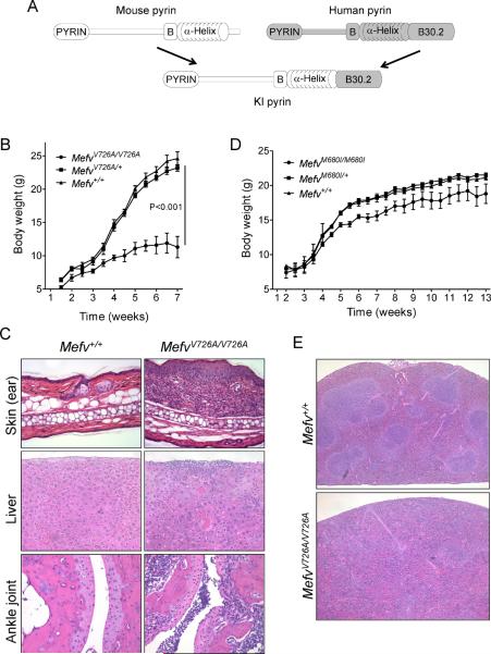
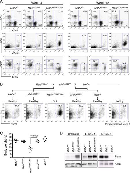
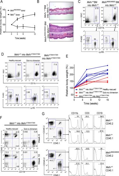
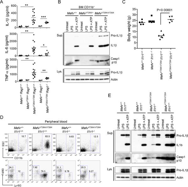
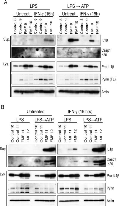
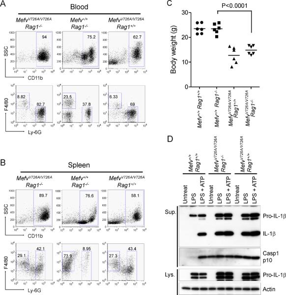
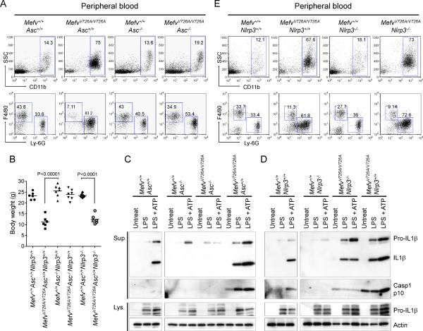
Missense mutations in the C-terminal B30.2 domain of pyrin cause familial Mediterranean fever (FMF), the most common Mendelian autoinflammatory disease. However, it remains controversial as to whether FMF is due to the loss of an inhibitor of inflammation or to the activity of a proinflammatory molecule. We generated both pyrin-deficient mice and "knockin" mice harboring mutant human B30.2 domains. Homozygous knockin, but not pyrin-deficient, mice exhibited spontaneous bone marrow-dependent inflammation similar to but more severe than human FMF. Caspase-1 was constitutively activated in knockin macrophages and active IL-1β was secreted when stimulated with lipopolysaccharide alone, which is also observed in FMF patients. The inflammatory phenotype of knockin mice was completely ablated by crossing with IL-1 receptor-deficient or adaptor molecule ASC-deficient mice, but not NLRP3-deficient mice. Thus, our data provide evidence for an ASC-dependent NLRP3-independent inflammasome in which gain-of-function pyrin mutations cause autoinflammatory disease.


Figures

Figure 1
Figure 1. Pyrin with an FMF-associated mutant B30.2 domain induces inflammation in mice
(A) Comparison of the schematic structure of pyrin proteins of human, mouse, and KI mice in which mouse pyrin is fused with the human B30.2 domains. Growth curves for (B) V726A mutant KI males (WT, n=5; heterozygotes, n=9; and homozygotes, n=4) and (D) M680I mutant KI females (WT, n=9; heterozygotes, n=10; and homozygotes, n=6). Data are as means ± s.d. (C) Hematoxylin and eosin (H&E) stained ear sections (200×) and liver sections (200×) from 8-week-old WT and MefvV726A/V726A mice, and H&E stained section (100×) (bottom) of ankle joints of 4-week-old MefvV726A/V726A mice. (E) H&E stained spleen sections (40×) from 8-week-old WT and MefvV726A/V726A mice. Additional information is provided in Figure S1 and Table S1.
Figure 2
Figure 2. Expansion of CD11b+ cells and gain of function B30.2 mutations with a gene dosage effect
(A) Flow cytometry analyses of spleens from 4- and 12-week-old MefvV726A/V726A mice. Total splenocytes were analyzed for the relative frequencies of lymphoid (CD3 and CD19 for T cells and B cells, respectively, first row) and myeloid cells (CD11b, second row). Within CD11b+-gated myeloid cells, Ly-6G+ neutrophils and F4/80+ monocytes or macrophages were analyzed (third row). Numbers indicate percentage of total cells in respective quadrants or gates. Data are representative of three independent experiments. (B) Generation of hemizygous (MefvV726A/−) KI mice for the mutant B30.2 domain. Peripheral blood cells were analyzed for CD11b+ cells from their 8-week-old offspring. Data are representative of three independent experiments. (C) Body weights of six 8-week-old males of Mefv+/+, MefvV726A/+, MefvV726A/V726A, MefvV726A/−, and Mefv−/−. (D) Expression of pyrin in peritoneal macrophages from M680I-KI and pyrin-deficient mice following 24 h of culture. Additional information is provided in Figure S2.
Figure 3
Figure 3. The inflammatory phenotypes of KI mice are induced from cells of the hematopoietic lineage
(A–C) BM cells from KI mice induce inflammatory phenotypes in WT mice. WT mice were lethally irradiated and given BM cells from either MefvM680I/M680I or WT mice intravenously. (A) Body weight after bone marrow transplantation (BMT). Data are shown in percent relative body weight changes as means ± s.d. of duplicate measurements from four of each recipient. (B) H&E stained ear sections (100×) from both recipients (WT BM cells into WT and MefvM680I/M680I BM cells into WT) after six weeks of BMT. (C) Flow cytometry analyses of peripheral blood cells from both recipients after 18 weeks of BMT. Data are representative of three independent experiments. (D–F) BM cells from congenic CD45.1 WT mice cure the inflammatory phenotypes of CD45.2 KI mice. MefvV726A/V726A mice were irradiated and given BM cells from either WT or MefvV726A/V726A mice intraperitoneally. (D) Flow cytometry analyses of peripheral blood cells after 10 weeks of BMT. (E) Body weight after BMT. Data are shown in percent relative body weight changes of individual recipients as indicated. (F) The degree of chimerism measured concomitantly by staining the congenic marker of donor cells (CD45.1). (G) The expansion of CD11b+ cell numbers is mediated by soluble factors. Congenic CD45.1+ WT BM cells were pre-mixed with either CD45.2+ WT BM cells (upper panels) or CD45.2+ MefvM680I/M680I BM cells (lower panels) and injected into lethally irradiated CD45.1+/CD45.2+ WT mice. Flow cytometry analyses of peripheral blood after 6 weeks of BMT for the chimerism (dot plot) and relative CD11b+, CD3+, and CD19+ cells according to the origin of each cell population (histogram). Data are representative of three independent experiments.
Figure 4
Figure 4. Inflammation in KI mice is induced by IL-1β
(A) Luminex analysis of serum cytokines of 8-week-old WT, MefvV726A/+, MefvV726A/V726A Rag1−/−, and MefvV726A/V726A Rag1−/− mice. *, P < 0.05; **, P < 0.0001; ***, P < 0.000001. (B) CD11b+ cells from BM of WT, MefvV726A/+, and MefvV726A/V726A mice were stimulated with LPS or no stimulus for 3 hr followed by treatment with or without ATP. Cell culture supernatants (Sup.) and cell lysates (Lys.) were analyzed by immunoblotting. (C–E) Analyses of MefvV726A/V726A mice on the Il1r1−/− background. (C) Body weights of 8-week-old males. (D) Flow cytometry analyses of peripheral blood cells from 8-week-old mice. Data are representative of three independent experiments. (E) Inflammasome activation in MefvV726A/V726A mice withIl1r1−/− background. Culture supernatants and lysates from BM CD11b+ cells were collected and subjected to immunoblot as described in (B). Additional information is provided in Figure S3.
Figure 5
Figure 5. Inflammasome activation in FMF patients
Macrophages differentiated from PBMCs of (A) two healthy donors (control 8 and 9), two FMF patients (FMF 9, homozygote of M694V; FMF 10, compound heterozygote of V726A/E148Q), (B) two healthy donors (control 10 and 11), and two FMF patients (FMF 11, heterozygote of V726A; FMF 12, compound heterozygote of V726A/P369S) were treated with IFN-γ or no stimulus for 16 hr. Macrophages were treated as described in Figure 4B, and cell culture supernatants (Sup.) and cell lysates (Lys.) were analyzed by immunoblotting. Additional information is provided in Figure S4.
Figure 6
Figure 6. Analyses ofMefvV726A/V726A mice on aRag1−/− background
(A and B) Flow cytometry analyses of peripheral blood cells (A) and splenocytes (B) from 8-week-old mice analyzed as described in Figure 3C. Data are representative of three independent experiments. (C) Body weights of 8-week-old males. (D) Inflammasome activation in MefvV726A/V726A mice with Rag1−/− background. Culture supernatants and lysates from BM CD11b+ cells were analyzed by immunoblotting. Additional information is provided in Figure S5.
Figure 7
Figure 7. FMF-associated B30.2 mutations activate an ASC-dependent but NALP3 independent pyrin inflammasome
(A–C) Analyses of the FMF KI (MefvV726A/V726A) mice with Asc−/− background. (A) Flow cytometry analyses of peripheral blood cells from 8-week-old mice analyzed as in Figure 3C. Data are representative of three independent experiments. (B) Body weights of 8-week-old males. (C) Inflammasome activation in MefvV726A/V726A mice with Asc−/− background. Culture supernatants and lysates from BM CD11b+ cells were analyzed by immunoblotting. (D–E) Analyses of the FMF KI (MefvV726A/V726A) mice with Nlrp3−/− background. . (D) Inflammasome activation in MefvV726A/V726A mice with Nlrp3−/− background. Culture supernatants and lysates from BM CD11b+ cells were analyzed by immunoblotting. (E) Flow cytometry analyses of peripheral blood cells from 8-week-old mice analyzed as in Figure 3C. Data are representative of three independent experiments Additional information is provided in Figure S6.

Comment in

Similar articles

Cited by

References

    1. Belkhir R, Moulonguet-Doleris L, Hachulla E, Prinseau J, Baglin A, Hanslik T. Treatment of familial Mediterranean fever with anakinra. Ann. Intern. Med. 2007;146:825–826. - PubMed
    1. Bilginer Y, Ayaz NA, Ozen S. Anti-IL-1 treatment for secondary amyloidosis in an adolescent with FMF and Behcet's disease. Clin. Rheumatol. 2009 - PubMed
    1. Booty MG, Chae JJ, Masters SL, Remmers EF, Barham B, Le JM, Barron KS, Holland SM, Kastner DL, Aksentijevich I. Familial Mediterranean fever with a single MEFV mutation: where is the second hit? Arthritis Rheum. 2009;60:1851–1861. - PMC - PubMed
    1. Brydges SD, Mueller JL, McGeough MD, Pena CA, Misaghi A, Gandhi C, Putnam CD, Boyle DL, Firestein GS, Horner AA, et al. Inflammasome-mediated disease animal models reveal roles for innate but not adaptive immunity. Immunity. 2009;30:875–887. - PMC - PubMed
    1. Calligaris L, Marchetti F, Tommasini A, Ventura A. The efficacy of anakinra in an adolescent with colchicine-resistant familial Mediterranean fever. Eur. J. Pediatr. 2008;167:695–696. - PMC - PubMed

Publication types