. 2011 Jul;25(7):1231-43.
doi: 10.1210/me.2011-0056. Epub 2011 May 12.

Adapter protein SH2B1beta binds filamin A to regulate prolactin-dependent cytoskeletal reorganization and cell motility

Affiliations

Adapter protein SH2B1beta binds filamin A to regulate prolactin-dependent cytoskeletal reorganization and cell motility

Leah Rider et al. Mol Endocrinol. 2011 Jul.

Abstract

Prolactin (PRL) regulates cytoskeletal rearrangement and cell motility. PRL-activated Janus tyrosine kinase 2 (JAK2) phosphorylates the p21-activated serine-threonine kinase (PAK)1 and the Src homology 2 (SH2) domain-containing adapter protein SH2B1β. SH2B1β is an actin-binding protein that cross-links actin filaments, whereas PAK1 regulates the actin cytoskeleton by different mechanisms, including direct phosphorylation of the actin-binding protein filamin A (FLNa). Here, we have used a FLNa-deficient human melanoma cell line (M2) and its derivative line (A7) that stably expresses FLNa to demonstrate that SH2B1β and FLNa are required for maximal PRL-dependent cell ruffling. We have found that in addition to two actin-binding domains, SH2B1β has a FLNa-binding domain (amino acids 200-260) that binds directly to repeats 17-23 of FLNa. The SH2B1β-FLNa interaction participates in PRL-dependent actin rearrangement. We also show that phosphorylation of the three tyrosines of PAK1 by JAK2, as well as the presence of FLNa, play a role in PRL-dependent cell ruffling. Finally, we show that the actin- and FLNa-binding-deficient mutant of SH2B1β (SH2B1β 3Δ) abolished PRL-dependent ruffling and PRL-dependent cell migration when expressed along with PAK1 Y3F (JAK2 tyrosyl-phosphorylation-deficient mutant). Together, these data provide insight into a novel mechanism of PRL-stimulated regulation of the actin cytoskeleton and cell motility via JAK2 signaling through FLNa, PAK1, and SH2B1β. We propose a model for PRL-dependent regulation of the actin cytoskeleton that integrates our findings with previous studies.


Figures

Fig. 1.
Fig. 1.
Schematic representation of WT and mutant forms of rat SH2B1β used in the study. Actin-binding domains (amino acids 150-200 and 615-670) are shown in gray. Filamin-binding domain (amino acids 200-260) is shown in black. PH is the plekstrin homology domain (amino acids 274-376), and SH2 is the SH2 domain (amino acids 527-620). Proline-rich regions (amino acids 13–24, 89-103, and 469-496) and dimerization domain (amino acids 24-85) are not shown.
Fig. 2.
Fig. 2.
JAK2 is tyrosyl phosphorylated in M2 and A7 cells overexpressing PRLR in response to PRL. Human PRLR was overexpressed in M2 and A7 cells. The cells were deprived of serum and treated with or without 400 ng/ml PRL for 15 min. JAK2 was immunoprecipitated with αJAK2. The immunoprecipitates and whole-cell lysates were immunoblotted with indicated antibodies. Each experiment was performed at least three times with similar results. IP, Immunoprecipitation.
Fig. 3.
Fig. 3.
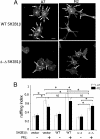
Actin-binding domains of SH2B1β and FLNa are required for maximal PRL-induced membrane ruffling. A, The FLNa-deficient human melanoma M2 cell line and its derivative cell line (A7), which stably expresses FLNa, were cotransfected with GFP-PRLR and myc-tagged WT SH2B1β or SH2B1β Δ-Δ mutant. Cells were serum deprived and treated with 400 ng/ml PRL for 15 min. Filamentous actin was visualized by staining with Alexa Fluor 350 phalloidin, and the transfected cells were visualized by both GFP fluorescence for PRLR and staining with αmyc for SH2B1β. Arrows indicate ruffles, and asterisks denote transfected cells. Scale bar, 20 μm. B, Ruffling index as a number of ruffles per cell was counted. White bars represent A7 cells, and black bars represent M2 cells. Bars represent mean ± se. *, P < 0.05. Each experiment was repeated three times. n = 300 for each experimental condition. NS, Not significant.
Fig. 4.
Fig. 4.
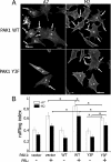
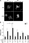
Tyrosyl-phosphorylated PAK1 and FLNa are required for maximal PRL-induced membrane ruffling. A, M2 and A7 cell lines were cotransfected with GFP-PRLR and myc-tagged WT PAK1 or Y3F PAK1 mutant. Cells were treated as in Fig. 3. Filamentous actin was visualized by staining with Alexa Fluor 350 phalloidin, and the transfected cells were visualized by both GFP fluorescence for PRLR and staining with αmyc for PAK1. Arrows indicate ruffles, and asterisks denote transfected cells. Scale bar, 20 μm. B, Ruffling index as a number of ruffles per cell was counted. White bars represent A7 cells, and black bars represent M2 cells. Bars represent mean ± se. *, P < 0.05. Each experiment was repeated three times. n = 300 for each experimental condition.
Fig. 5.
Fig. 5.
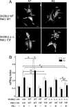
The actin-binding domains of SH2B1β, pTyr-PAK1, and FLNa are required for maximal PRL-induced membrane ruffling. A, A7 and M2 cell lines were cotransfected with GFP-PRLR, mRFP-tagged WT SH2B1β or SH2B1β Δ-Δ mutant, and myc-tagged WT PAK1 or PAK1 Y3F mutant. Cells were treated as in Fig. 3. Myc-PAK1 was visualized by αmyc, whereas PRLR was visualized by GFP fluorescence, and mRFP fluorescence for SH2B1β. PAK1 WT and/or SH2B1β WT or Δ-Δ localization in ruffles was used to count total number of ruffles per transfected cell. Arrows indicate ruffles, and asterisks denote transfected cells. Scale bar, 20 μm. B, Ruffling index as a number of ruffles per cells was counted. White bars represent A7 cells, and black bars represent M2 cells. Bars represent mean ± se. *, P < 0.05. Each experiment was repeated three times. n = 300 for each experimental condition. NS, Not significant; vctr, vector.
Fig. 6.
Fig. 6.
Endogenous SH2B1β associates with endogenous FLNa. Whole-cell lysates of 293T cells (lane 3) were immunoprecipitated with αSH2B1β antibody (lane 1) or with control IgG (lane 2) and immunoblotted with αFLNa antibody (upper). Endogenous FLNa was detected in the whole-cell lysate (lane 3) and in the αSH2B1β immunoprecipitate (lane 1) but not the control lane (lane 2). The same blot was reprobed with αSH2B1β (bottom). Several bands representing different levels of SH2B1β phosphorylation (indicated on the right) were detected in the cell lysate (lane 3) and αSH2B1β immunoprecipitate (lane 1) but not in the control lane (lane 2). Each experiment was performed at least three times with similar results.
Fig. 7.
Fig. 7.
FLNa repeats 17–23 is the site for SH2B1β interaction. A, Schematic depiction of FLNa truncations [modified from Cukier et al. (49)]. B, Different GST-tagged truncated FLNa mutants were purified from bacterial lysates and immobilized on glutathione-agarose beads. Myc-tagged SH2B1β was translated in vitro using TNT Coupled Reticulocyte Lysate System. The mixture of GST-tagged FLNa mutants and in vitro translated SH2B1β was rotated for 1 h. The glutathione-agarose beads were washed, GST-FLNa constructs-bound myc-SH2B1β was detected by IB with αmyc (upper panel). SH2B1β was detected only in lane 5, indicating that only FLNa-4 construct (repeats 17–23 and dimerization domain) directly binds to SH2B1β. Amount of GST or GST-FLNa mutants input was detected by IB with αGST (left bottom panel). Migration of myc- SH2B1β alone is shown on the bottom right panel. Each experiment was performed at least three times with similar results. Lane 1, GST; lane 2, FLNa-1 (actin-binding domain of FLNa); lane 3, FLNa-2 (repeats 1–10); lane 4, FLNa-3 (repeats 11–16); lane 5, FLNa-4 (repeats 17–23 and dimerization domain); and lane 6, FLNa-5 (dimerization domain).
Fig. 8.
Fig. 8.
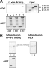
Amino acids 200–260 of SH2B1β is the site for FLNa interaction. A, Different in vitro translated myc-tagged truncated SH2B1β mutants were incubated with either GST (lanes 1, 3, and 5) or GST-tagged FLNa-4 (lanes 2, 4, and 6). Bound myc-SH2Bβ mutant was detected by IB with αmyc. Lanes 1 and 2, Amino acids 1-105 of SH2B1β; lanes 3 and 4, amino acids 1-200 of SH2B1β; and lanes 5 and 6, amino acids 1-260 of SH2B1β. B, mRFP-tagged (200–260) amino acids mutant of SH2B1β was translated in vitro in the presence of 35S methionine and incubated with either GST (lane 1) or GST-tagged FLNa-4 (lane 2). Bound mRFP-SH2Bβ mutant was detected by autoradiogram (48 h of film exposure). Equal amounts of 35S methionine-labeled SH2B1β input is shown on the right (24 h of film exposure). Amount of GST or GST-FLNa-4 was detected by IB with αGST. Each experiment was performed at least three times with similar results.
Fig. 9.
Fig. 9.
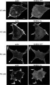
SH2B1β WT and SH2B1β 3Δ colocalize with actin in the ruffles of A7 and M2 cells. M2 and A7 cells overexpressing mRFP-tagged SH2B1β WT or 3Δ mutant were stained with Alexa Fluor 647 phalloidin for actin visualization. Arrows indicate ruffles, and asterisks denote blebs. Each experiment was performed at least three times with similar results. Scale bar, 20 μm.
Fig. 10.
Fig. 10.
The FLNa-binding domain of SH2B1β, pTyr-PAK1 and FLNa are required for PRL-induced membrane ruffling. A, A7 and M2 cell lines were cotransfected with GFP-PRLR, mRFP-tagged WT SH2B1β (data not shown) or SH2B1β 3Δ mutant (shown), and myc-tagged WT PAK1 (data not shown) or PAK1 Y3F mutant (shown). Cells were treated as in Fig. 3. Myc-PAK1 was visualized by αmyc, whereas PRLR was visualized by GFP fluorescence and mRFP fluorescence for SH2B1β. PAK1 and SH2B1β localization in ruffles was used to count total number of ruffles per transfected cell. Arrows indicate ruffles, and asterisks denote transfected cells. Scale bar, 2 0 μm. B, Ruffling index as a number of ruffles per cell was counted. White bars represent A7 cells, and black bars represent M2 cells. Bars represent mean ± se. *, P < 0.05. Each experiment was repeated three times. n = 300 for each experimental condition. vctr, Vector.
Fig. 11.
Fig. 11.
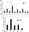
The FLNa-binding domains of SH2B1β, pTyr-PAK1, and FLNa are required for PRL-induced cell migration. A, A7 and M2 cell lines were cotransfected with GFP-PRLR, mRFP-tagged WT SH2B1β or SH2B1β 3Δ mutant, and myc-tagged WT PAK1 or PAK1 Y3F mutant. After deprivation, monolayers of cells were scarified and incubated in deprivation media with or without 400 ng/ml PRL. The percentage wound closure in 8 h was calculated for each condition. Bars represent mean ± se. *, P < 0.05. B, Clones of T47D cells stably expressing GFP, GFP-containing PAK1 WT, or PAK1 Y3F were transiently transfected with vector, WT SH2B1β, or SH2B1β 3Δ mutant. Monolayers of cells were wounded in the presence or absence of 200 ng/ml PRL and assessed after 18 h. Bars represent mean ± se. *, P < 0.05 compared with cells expressing vectors and treated with PRL. Each experiment was repeated three times. n = 30 for each experimental condition. vctr, Vector.
Fig. 12.
Fig. 12.
PRL-activated JAK2 promotes formation of multiprotein complex. Schematic representation of the proposed working model. PRL-activation of JAK2 leads to tyrosyl phosphorylation of PAK1 on tyrosines 153, 201, and 285, thereby increasing PAK1 activities (both the serine/threonine kinase activity and ability to create potential protein-protein interactions) and stimulating phosphorylation of FLNa. Phosphorylated FLNa stimulates the kinase activity of PAK1 and has increased actin-regulating activity. FLNa, which directly binds to SH2B1β, relocates SH2B1β to the JAK2-PAK1-FLNa complex. Because SH2B1β is the enhancer of the kinase activity of JAK2, the formation of the complex results in enhancement of JAK2 activation and further activation of the JAK2-PAK1-FLNa complex that leads to actin cytoskeleton reorganization via actin-regulating proteins PAK1, FLNa, and SH2B1β. Binding of FLNa to the plasma membrane is not shown.

Similar articles

Cited by

References

    1. Bole-Feysot C, Goffin V, Edery M, Binart N, Kelly PA. 1998. Prolactin (PRL) and its receptor: actions, signal transduction pathways and phenotypes observed in PRL receptor knockout mice. Endocr Rev 19:225–268 - PubMed
    1. Horseman ND. 1999. Prolactin and mammary gland development. J Mammary Gland Biol Neoplasia 4:79–88 - PubMed
    1. Goffin V, Bernichtein S, Touraine P, Kelly PA. 2005. Development and potential clinical uses of human prolactin receptor antagonists. Endocr Rev 26:400–422 - PubMed
    1. Bernichtein S, Touraine P, Goffin V. 2010. New concepts in prolactin biology. J Endocrinol 206:1–11 - PubMed
    1. Clevenger CV, Chang WP, Ngo W, Pasha TL, Montone KT, Tomaszewski JE. 1995. Expression of prolactin and prolactin receptor in human breast carcinoma. Evidence for an autocrine/paracrine loop. Am J Pathol 146:695–705 - PMC - PubMed

Publication types

MeSH terms