. 2011 Jul;7(7):2261-77.
doi: 10.1039/c1mb05061c. Epub 2011 May 6.

Functional diversification of the RING finger and other binuclear treble clef domains in prokaryotes and the early evolution of the ubiquitin system

Affiliations

Functional diversification of the RING finger and other binuclear treble clef domains in prokaryotes and the early evolution of the ubiquitin system

A Maxwell Burroughs et al. Mol Biosyst. 2011 Jul.

Abstract

Recent studies point to a diverse assemblage of prokaryotic cognates of the eukaryotic ubiquitin (Ub) system. These systems span an entire spectrum, ranging from those catalyzing cofactor and amino acid biosynthesis, with only adenylating E1-like enzymes and ubiquitin-like proteins (Ubls), to those that are closer to eukaryotic systems by virtue of possessing E2 enzymes. Until recently E3 enzymes were unknown in such prokaryotic systems. Using contextual information from comparative genomics, we uncover a diverse group of RING finger E3s in prokaryotes that are likely to function with E1s, E2s, JAB domain peptidases and Ubls. These E1s, E2s and RING fingers suggest that features hitherto believed to be unique to eukaryotic versions of these proteins emerged progressively in such prokaryotic systems. These include the specific configuration of residues associated with oxyanion-hole formation in E2s and the C-terminal UFD in the E1 enzyme, which presents the E2 to its active site. Our study suggests for the first time that YukD-like Ubls might be conjugated by some of these systems in a manner similar to eukaryotic Ubls. We also show that prokaryotic RING fingers possess considerable functional diversity and that not all of them are involved in Ub-related functions. In eukaryotes, other than RING fingers, a number of distinct binuclear (chelating two Zn atoms) and mononuclear (chelating one zinc atom) treble clef domains are involved in Ub-related functions. Through detailed structural analysis we delineated the higher order relationships and interaction modes of binuclear treble clef domains. This indicated that the FYVE domain acquired the binuclear state independently of the other binuclear forms and that different treble clef domains have convergently acquired Ub-related functions independently of the RING finger. Among these, we uncover evidence for notable prokaryotic radiations of the ZF-UBP, B-box, AN1 and LIM clades of treble clef domains and present contextual evidence to support their role in functions unrelated to the Ub-system in prokaryotes. In particular, we show that bacterial ZF-UBP domains are part of a novel cyclic nucleotide-dependent redox signaling system, whereas prokaryotic B-box, AN1 and LIM domains have related functions as partners of diverse membrane-associated peptidases in processing proteins. This information, in conjunction with structural analysis, suggests that these treble clef domains might have been independently recruited to the eukaryotic Ub-system due to an ancient conserved mode of interaction with peptides.


Figures
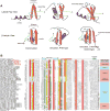
Fig. 1
Fig. 1
(A) Cartoon representations of mononuclear and PHD-type and C-terminal strand-containing binuclear treble clef domains. Two views are provided: one with the line of sight perpendicular to the lateral flap (top) and one with the line of sight perpendicular to the β-hairpin. β-Strands are depicted as arrows colored in orange with the arrowheads at the C-terminal end, while α-helices are depicted as coils, colored in purple. The lateral flap structure is colored in green. Zinc ion coordinating residues are depicted as short lines, colored in blue. The relative spatial location of zinc ions are marked with a black circle shaded in yellow. (B) Multiple sequence alignment of RING finger with a special emphasis on prokaryotic versions. Proteins are annotated by their gene names, species abbreviations and Genbank index (gi) numbers and are further grouped by their familial associations, shown to the right of the alignment. Secondary structure assignments are shown above the alignment, where the green arrow represents the β-strand and the orange cylinder the α-helix. Secondary structure was derived from a combination of crystal structures and alignment based predictions. Poorly conserved inserts are replaced by the corresponding number of residues. The alignment was colored based on 75% consensus and the coloring scheme and consensus abbreviations are as in Fig. 4. Species abbreviations are as follows: Aboo: Aciduliprofundum boonei; Acol: Anaerotruncus colihominis; Asp.: Acidobacterium sp.; Bmar: Blastopirellula marina; Bsp.: Bacteroides sp.; CCal: Candidatus Caldiarchaeum; Cmet: Clostridium methylpentosum; Cspu: Capnocytophaga sputigena; EHV1: Equid herpesvirus 1; Esir: Eubacterium siraeum; Fjoh: Flavobacterium johnsoniae; Fpra: Faecalibacterium prausnitzii; HHV8: Human herpesvirus 8; Hoch: Haliangium ochraceum; Hsap: Homo sapiens; Ipal: Isosphaera pallida; Kfla: Kribbella flavida; Mboo: Methanoregula boonei; Mhun: Methanospirillum hungatei; Mmar: Microscilla marina; Mmet: marine metagenome; Mmus: Mus musculus; Mpal: Methanocella paludicola; Mxan: Myxococcus xanthus; Ppac: Plesiocystis pacifica; Psp.: Prevotella sp.; Psta: Pirellula staleyi; Ralb: Ruminococcus albus; Rbro: Ruminococcus bromii; Rfla: Ruminococcus flavefaciens; Rsp.: Ruminococcus sp.; Scer: Saccharomyces cerevisiae; Sgri: Streptomyces griseus;Tbis: Thermobispora bispora; Umet: uncultured methanogenic archaeon RC-I.
Fig. 2
Fig. 2
Architectures and operons are grouped according to the treble-clef domain or ubiquitin domain that is contained in them. Operons that contain other proteins or domains involved in the ubiquitin system pathway are marked with a red asterisk. Genes that are not translated in the database are marked with an “untrans” prefix. Note that the Isosphaera RING domain does not co-occur with the ubiquitin pathway genes, but is in a distinct genome location with respect to the latter. Architectures and operons are labeled by the gene names, gis and species name (in brackets). Genes in conserved gene neighborhoods are shown as boxed arrows with the arrow head pointing in the 3′ direction. Standard abbreviations are used for most domains. Non-standard abbreviations include: Y: uncharacterized conserved domain, β-P: WD40-like betapropeller repeats, HISKIN: histidine kinase, REC: receiver, TM: transmembrane helix, and TGase: transglutaminase, ZnR: zinc ribbon.
Fig. 3
Fig. 3
(A) A scheme for the possible origin of the FYVE domain. (1) The precursor mononuclear domain. (2) Duplication of the precursor gives rise to a LIM-like intermediate. (3) Partial duplication of the LIM-like intermediate. (4) Circular permutation event gives rise to the FYVE domain. Metal-ion coordinating residue pairs are denoted by “C” and pairs which coordinate the same zinc ion are joined by black lines. In step 3 the linkages which were retained in the FYVE domain are colored in red. β-Strands are colored in blue and numbered in each internal duplicated treble clef domain (for ease of display, the lateral flap is here represented by two smaller β-strands). The conserved core α-helix is colored in brown and denoted with the letter “H”. In step 4, a differential coloring scheme is employed to emphasize the respective origins of the observed structural components. (B) Interaction modes of treble clef domains with their partner proteins. Cartoon representation of the core treble clef scaffold, with coloring scheme similar to (A) but with the lateral flap colored in green and the variable C-terminal region colored in grey. Identified interaction partners are labeled by arrows with the name of the treble clef domain and its cognate binding partner separated by “↔”. General regions corresponding roughly to the binding pockets mentioned in the text are denoted by dashed circles shaded in grey. The surface which stacks binding partners against the exposed β-strand is depicted by a dashed line, colored in grey. Zinc ion residues are labeled and shown as black circles, shaded in yellow. Approximate locations of binuclear coordinating residues are shown as red lines.
Fig. 4
Fig. 4
(A) Multiple sequence alignment of prokaryotic UBP and (B) B-box domains. Protein nomenclature and secondary structure assignments are as in Fig. 1. The highly conserved CD motif seen in prokaryotic UBP domains are marked with asterisks. Residue coloring is based on 90% consensus for the UBP domain and 80% consensus for the B-box domain. The coloring scheme and consensus abbreviations are as follows: h, hydrophobic (ACFILMVWY); l, aliphatic (LIV) and a, aromatic (FWY) residues shaded yellow; b, big residues (LIYERFQKMW), shaded gray; s, small residues (AGSVCDN) and u, tiny residues (GAS), shaded green; p, polar residues (STEDKRNQHC) shaded blue; –, acidic residues (DE), shaded magenta, o, alcohol (ST) group containing residues shaded orange, zinc coordinating residues and absolutely conserved residues are shaded red. Species abbreviations are as follows: Aaur: Arthrobacter aurescens; Acap: Acidobacterium capsulatum; Amar: Aeromicrobium marinum; Amed: Amycolatopsis mediterranei; Amir Actinosynnema mirum; Asp.: Acidobacterium sp.; Asp.: Arthrobacter sp.; BEll: bacterium Ellin514; Bcen: Burkholderia cenocepacia; Bfra: Bacteroides fragilis; Bphy: Burkholderia phytofirmans; CKor: Candidatus Koribacter; CSol: Candidatus Solibacter; Cagg: Chloroflexus aggregans; Caka: Coraliomargarita akajimensis; Caur: Chloroflexus aurantiacus; Cfla: Chthoniobacter flavus; Cmic: Clavibacter michiganensis; Cseg: Caulobacter segnis; Dalk: Desulfatibacillum alkenivorans; Dfer: Dyadobacter fermentans; Faln: Frankia alni; Fsp.: Frankia sp.; Fsp.: Fusobacterium sp.; Glov: Geobacter lovleyi; Gvio: Gloeobacter violaceus; Hche: Hahella chejuensis; Hsap: Homo sapiens; Ical: Intrasporangium calvum; Krac: Ktedonobacter racemifer; Krad: Kineococcus radiotolerans; Ksed: Kytococcus sedentarius; Kset: Kitasatospora setae; Lhof: Leptotrichia hofstadii; Mace: Methanosarcina acetivorans; Mbar: Methanosarcina barkeri; Mlot: Mesorhizobium loti; Mlut: Micrococcus luteus; Mmar: Methanothermobacter marburgensis; Mmar: Mycobacterium marinum; Mpal: Methanocella paludicola; Mrum: Methanobrevibacter ruminantium; Msme: Mycobacterium smegmatis; Msmi: Methanobrevibacter smithii; Msp.: Marinobacter sp.; Msp.: Mycobacterium sp.; Mthe: Methanothermobacter thermautotrophicus; Mvan: Mycobacterium vanbaalenii; Mxan: Myxococcus xanthus; Ndas: Nocardiopsis dassonvillei; Nfar: Nocardia farcinica; Nmar: Nitrosopumilus maritimus; Nsp.: Nocardioides sp.; Nthe: Natranaerobius thermophilus; Obac: Opitutaceae bacterium; Rmuc: Rothia mucilaginosa; Rpal: Rhodopseudomonas palustris; Rsal: Renibacterium salmoninarum; Saur: Stigmatella aurantiaca; Sbin: Streptomyces bingchenggensis; Sery: Saccharopolyspora erythraea; Shyg: Streptomyces hygroscopicus; Ssp.: Streptomyces sp.; Sthe: Symbiobacterium thermophilum; Svio: Streptomyces violaceusniger; Syn: Synechococcus sp.; Tcur: Thermomonospora curvata; Umar: uncultured marine; Umet: uncultured methanogenic archaeon; Vpar: Variovorax paradoxus; Xcam: Xanthomonas campestris; Xcel: Xylanimonas cellulosilytica; Xlae: Xenopus laevis.

Similar articles

Cited by

References

    1. Hochstrasser M. Nature. 2009;458:422–429. - PMC - PubMed
    1. Hershko A, Ciechanover A. Annu Rev Biochem. 1998;67:425–479. - PubMed
    1. Glickman MH, Ciechanover A. Physiol Rev. 2002;82:373–428. - PubMed
    1. Ardley HC, Robinson PA. Essays Biochem. 2005;41:15–30. - PubMed
    1. Pickart CM. Annu Rev Biochem. 2001;70:503–533. - PubMed

Publication types