. 2011 May 3:6:20.
doi: 10.1186/1749-8104-6-20.

Prospero and Pax2 combinatorially control neural cell fate decisions by modulating Ras- and Notch-dependent signaling

Affiliations

Prospero and Pax2 combinatorially control neural cell fate decisions by modulating Ras- and Notch-dependent signaling

Mark Charlton-Perkins et al. Neural Dev. .

Abstract

Background: The concept of an equivalence group, a cluster of cells with equal potential to adopt the same specific fate, has served as a useful paradigm to understand neural cell type specification. In the Drosophila eye, a set of five cells, called the 'R7 equivalence group', generates a single photoreceptor neuron and four lens-secreting epithelial cells. This choice between neuronal versus non-neuronal cell fates rests on differential requirements for, and cross-talk between, Notch/Delta- and Ras/mitogen-activated protein kinase (MAPK)-dependent signaling pathways. However, many questions remain unanswered related to how downstream events of these two signaling pathways mediate distinct cell fate decisions.

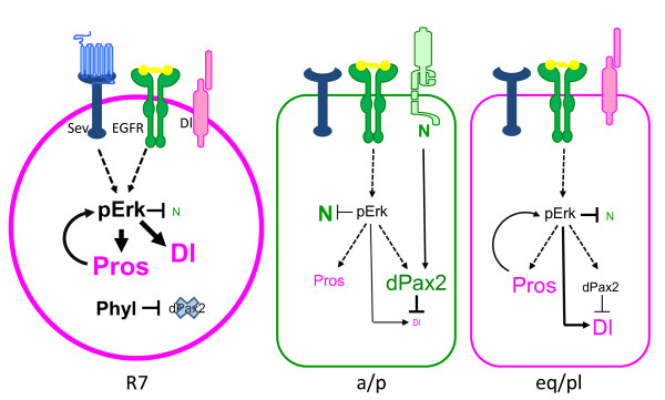
Results: Here, we demonstrate that two direct downstream targets of Ras and Notch signaling, the transcription factors Prospero and dPax2, are essential regulators of neuronal versus non-neuronal cell fate decisions in the R7 equivalence group. Prospero controls high activated MAPK levels required for neuronal fate, whereas dPax2 represses Delta expression to prevent neuronal fate. Importantly, activity from both factors is required for proper cell fate decisions to occur.

Conclusions: These data demonstrate that Ras and Notch signaling are integrated during cell fate decisions within the R7 equivalence group through the combinatorial and opposing activities of Pros and dPax2. Our study provides one of the first examples of how the differential expression and synergistic roles of two independent transcription factors determine cell fate within an equivalence group. Since the integration of Ras and Notch signaling is associated with many developmental and cancer models, these findings should provide new insights into how cell specificity is achieved by ubiquitously used signaling pathways in diverse biological contexts.


Figures

Figure 1
Figure 1
Pros and dPax2 together control cone cell development and lens formation. (A,B,D-K) Scanning electron micrographs (SEMs) of adult eyes (A,D,F,H,J) or immunostaining of 45% pupal retinas for the CC-specific marker Cut (green) and the membrane marker E-cadherin (blue) (B,E,G,I,K) were analyzed from the following genotypes: yw67; Sp/CyO; TM2/TM6B (A,B), eyflp3.5; Sp/CyO; FRT82ubi-GFPnls, RpS3/FRT82B-pros17 (D,E), yw67; Sp/CyO; TM2/TM6B; spapol/spapol(F,G), yw67; Sp/CyO; FRT82B-pros17/TM6B; spapol/spapol(H,I), and UAS-dPax2RNAi; sev-GAL4/CyO; UAS-Pros RNAi/TM6B (J,K). A slightly more severe phenotype from that shown in (J,K) is apparent in pros17/spapoldouble mutants, but these eyes collapsed during SEM and were particularly difficult to dissect. Panels (A',D',F',H',J') represent the boxed areas in the corresponding larger SEM. Circles represent CC clusters within individual ommatidia, and the dotted circle in (E) represents a rare yet significant loss of one CC observed in pros mutants. Control eyes have regular lens facets by SEM (A) and four CCs per ommatida by Cut and E-cadherin double-staining (B). (C) Quantification of CC numbers shows a strong genetic interaction and dual requirement for Pros and dPax2 in CC specification. *P < 0.05, **P < 0.001, n.d. = none detected. Error bars represent standard deviation. (D,E) pros17Minute LOF eyes (see Materials and methods) show mild roughening by SEM (D), and a rare yet significant loss of one CC (E, dotted circle). (F,G) spapoleyes show some roughening (F), consistent with a regular loss in PPCs [28] and at least one CC (G,G', circles). Removing one copy of pros from spapolmutants causes further perturbation of lens morphogenesis, with holes in the center of the lenses frequently observed (H), and CC number is reduced to approximately two per ommatidia (I). sev>dPax2RNAi + ProsRRNAi lack lens facets by SEM (J) and Cut-positive cells are not detected (K).
Figure 2
Figure 2
Distinct subsets of cone cells are present in the eye imaginal disc. (A,C,E-G) In wild-type late third instar eye imaginal discs, Pros (A,E,F, magenta), dPax2 (C,F, blue), and Cut (A,C,E, green; G, white) were analyzed by immunostaining. Pros is higher in the equatorial (Eq) and polar (Pl) CCs, while the expression of Cut (E,G, green) and dPax2 (G, blue) are elevated in the anterior (A) and posterior (P) CCs. (B,D) These expression patterns represented in diagrams. Individual ommatidia are circled; anterior is left. (E) In spapoleye imaginal discs, Cut is mostly absent until the last few most posterior rows, while Pros expression (E, magenta; E', white) initiates in all CC precursors (circles). (F) In pros17mitotic clones, dPax2 expression (F, blue; F', white) is unchanged between pros mutant (Pros-negative, -/-) and wild-type (Pros-positive, magenta, +/+) tissue. (G) Cut expression is specifically delayed in Eq/Pl CCs (dotted circles) in pros17mutant tissue (-/-) (individual ommatidial CC clusters are highlighted with dashed circles), whereas in control tissue (+/+), a normal complement of four CCs is observed in comparably positioned ommatidia (solid circles).
Figure 3
Figure 3
Pros and dPax2 are sufficient to recruit cone cells, but from different cell populations. (A,B,E,F,I,J,M) Retinas from 45% pupa (A,B,E,F,I,J,M) were immunostained for CC nuclei (Cut, green in (A,A',E,E',I,I',M,M')), PR nuclei (Otd, magenta in (A,A'E,E'I,I',M,M')), and PPC nuclei (BarH1, green in (B,F,J) or white in (B',F',J')), and cell outlines were revealed by Dlg staining (black in (B,F,J)). (C,D,G,H,K,L) Adult eyes were stained with toluidine blue (C,G,K) or immunostained (D,H,L) with the R7 opsins Rh3 (magenta) and Rh4 (blue) and fluorescently labeled phalloidin was used to mark the actin-rich apical surfaces of photoreceptors (green). (N) Quantification of PRs (magenta), CCs (green), and PPCs (blue). (O) Summary of phenotypes observed with the gain-of-function (GOF) experiments (top) and wild-type Pros and dPax2 expression patterns (bottom). MC, mystery cells [35], a potential source of ectopic CCs in the double GOF experiments. (A-D) Control pupal eyes form eight PRs (A,A'), four CCs (A,A'), and two PPCs (B,B'), and adult eyes form a trapezoid of six large outer PR rhabdomeres surrounding a smaller, central R7 PR (C) expressing Rh3 and Rh4 (D). (E,F) In sev>Pros pupal retinas, ectopic Cut-positive CCs are observed in ommatidia with a full complement of PRs (E, circle), and only one PPC is commonly observed (F,F', circle). Ommatidia with a complement of four CCs occasionally form an extra R7 (E, dotted circle; G,H). Ectopic CCs are also observed in sev>dPax2 pupal retinas (I,I'). PPCs are disorganized in sev>dPax2 retinas, but do not change significantly in number (J,J',N). PR number is reduced by one in sev>dPax2 pupal retinas (I,I') and R7s are frequently absent from adult eyes (K,L). (M) In sev>pros+dPax2 pupal eyes, one ectopic CC per ommatidia is frequently observed (circle), but PR and PPC numbers remain unchanged (N) (also see Additional file 1C,D). *P < 0.05, **P < 0.001. Error bars represent standard deviation.
Figure 4
Figure 4
Pros is necessary and sufficient for high pERK levels. (A,B,D-G) Eye imaginal discs from control (A,E), pros17mitotic clones (B,F) and sev>Pros eyes (D,G) were immunostained for Pros (A,B, green; E,G, magenta), pERK (A,B,D, magenta), and Yan (E,G, green; F, magenta). GFP was used to mark wild-type (versus pros17mutant) tissue (B,F, green), and nuclei were visualized with DAPI (E,F,G, blue). (C) A diagram representing Pros regulation by Ras/MAPK signaling previously reported [26,27,29], and the positive feedback onto pERK described here. In optical sections at the level of the R7 and CCs, high Pros expression correlates with high pERK expression (A,C, arrows), whereas a cell autonomous reduction in pERK is observed in pros mutant tissue (B, non-GFP positive cells). No change in pERK is observed in more basal optical sections where Pros is not expressed (data not shown). (E) In wild-type imaginal discs, Yan expression (green) becomes reduced as Pros expression (magenta) increases in specified CCs. (F) In pros mutants, Yan (magenta) is only present in nuclei in GFP-negative (that is, pros-negative) tissue, whereas it is both nuclear and cytoplasmic in surrounding GFP-positive wild-type tissue (green). (G) In sev>Pros imaginal discs, Yan (green) is reduced throughout the disc, being almost undetectable in cells expressing particularly high levels of Pros (magenta).
Figure 5
Figure 5
dPax2 effects Delta/Notch signaling. (A-F) Eye imaginal discs from control (A,C), spapolmutants (B,C), mirr>GFP (E), and mirr>GFP,dPax2RNAi were immunostained for Delta (A,B, green), E-cadherin (A,B, blue), LacZ (C,D, green), Pros (C,D, magenta), GFP (E,F, green), or E(spl) (E,F, magenta). All discs are oriented with anterior left. In wild-type discs, Delta expression is high in the first four rows of ommatidia after the morphogenetic furrow (MF) and then decreases thereafter (A). In spapolmutants, however, Delta is up-regulated again by row 7. Delta-LacZ expression in wild-type discs is down-regulated at the more posterior rows of the disc and do not co-localize with Pros in CC precursors (C), whereas in spapol mutants, Delta-LacZ expression is maintained at high levels throughout the disc and co-localizes with Pros in the most posterior rows (D), indicating that dPax2 transcriptionally represses Delta expression in CC precursors. E(spl) (E,F) is equally expressed when a UAS-GFP transgene is misexpressed in the dorsal half of the eye imaginal disc with mirr-GAL4 (E), but is significantly reduced where UAS-dPax2RNAi/UAS-GFP are co-expressed (F), revealing that dPax2 is important for maintaining high Notch activity.
Figure 6
Figure 6
dPax and Pros control the neuronal to non-neuronal switch in the R7 equivalence group. (A-D) In eyes overexpressing Pros in the absence of dPax2 (sev>Pros; spapoleyes), lenses are almost absent by SEM (A,B), no Cut-positive CCs are observed (C, green), and R7 number is significantly increased, with three to four often present in individual ommatidia (D, circles). (E-H) In eyes overexpressing dPax2 in the absence of Pros (sev>dPax2+prosRNAieyes), lens formation is mildly disrupted (E,F), an average of five CCs/ommatidia are formed (G, circles), and R7 PRs are rarely observed (H). (I) Quantification of PR (magenta) and CC (green) numbers in pros and dPax2 LOF and GOF experiments (see Table 1 for values, and Materials and methods for specific genotypes). Error bars represent standard deviation
Figure 7
Figure 7
Proposed model for the roles of Pros and dPax2 during cell fate decisions in the R7 equivalence group. Model depicting the outcome of Ras/MAPK and Notch (N/Delta (Dl signaling in the R7, anterior (a)/posterior (p) CCs, and equatorial (eq)/polar (pl) CCs based on the differences in Pros and dPax2 observed in these different cell populations. Phyllopod (Phyl) has been previously suggested to prevent dPax2 expression in the presumptive R7 [34]. In this model, the a/p CCs would be dominated by dPax2/Cut/Notch activity, whereas the eq/pl CCs would be dominated by Pros/pERK/Delta. Based on our LOF analysis, we propose that a/p and eq/pl CCs represent two distinct CCs that are inter-convertible. Moreover, in pros mutants, we postulate that four 'a/p' CCs form, whereas in dPax2 mutants, 'eq/pl' CCs form.

Similar articles

Cited by

References

    1. Kumar JP. Signalling pathways in Drosophila and vertebrate retinal development. Nat Rev Genet. 2001;2:846–857. doi: 10.1038/35098564. - DOI - PubMed
    1. Simon MA, Bowtell DD, Dodson GS, Laverty TR, Rubin GM. Ras1 and a putative guanine nucleotide exchange factor perform crucial steps in signaling by the sevenless protein tyrosine kinase. Cell. 1991;67:701–716. doi: 10.1016/0092-8674(91)90065-7. - DOI - PubMed
    1. Frankfort BJ, Mardon G. R8 development in the Drosophila eye: a paradigm for neural selection and differentiation. Development. 2002;129:1295–1306. - PubMed
    1. Voas MG, Rebay I. Signal integration during development: insights from the Drosophila eye. Dev Dyn. 2004;229:162–175. doi: 10.1002/dvdy.10449. - DOI - PubMed
    1. Wolff T, Ready DF. Pattern Formation in the Drosophila Retina. Cold Spring Harbor, NY: Cold Spring Harbor Laboratory Press; 1993.

Publication types

MeSH terms