. 2011 Apr 21;6(4):e19183.
doi: 10.1371/journal.pone.0019183.

Mutations in protocadherin 15 and cadherin 23 affect tip links and mechanotransduction in mammalian sensory hair cells

Affiliations

Mutations in protocadherin 15 and cadherin 23 affect tip links and mechanotransduction in mammalian sensory hair cells

Kumar N Alagramam et al. PLoS One. .

Abstract

Immunocytochemical studies have shown that protocadherin-15 (PCDH15) and cadherin-23 (CDH23) are associated with tip links, structures thought to gate the mechanotransducer channels of hair cells in the sensory epithelia of the inner ear. The present report describes functional and structural analyses of hair cells from Pcdh15(av3J) (av3J), Pcdh15(av6J) (av6J) and Cdh23(v2J) (v2J) mice. The av3J and v2J mice carry point mutations that are predicted to introduce premature stop codons in the transcripts for Pcdh15 and Cdh23, respectively, and av6J mice have an in-frame deletion predicted to remove most of the 9th cadherin ectodomain from PCDH15. Severe disruption of hair-bundle morphology is observed throughout the early-postnatal cochlea in av3J/av3J and v2J/v2J mice. In contrast, only mild-to-moderate bundle disruption is evident in the av6J/av6J mice. Hair cells from av3J/av3J mice are unaffected by aminoglycosides and fail to load with [(3)H]-gentamicin or FM1-43, compounds that permeate the hair cell's mechanotransducer channels. In contrast, hair cells from av6J/av6J mice load with both FM1-43 and [(3)H]-gentamicin, and are aminoglycoside sensitive. Transducer currents can be recorded from hair cells of all three mutants but are reduced in amplitude in all mutants and have abnormal directional sensitivity in the av3J/av3J and v2J/v2J mutants. Scanning electron microscopy of early postnatal cochlear hair cells reveals tip-link like links in av6J/av6J mice, substantially reduced numbers of links in the av3J/av3J mice and virtually none in the v2J/v2J mice. Analysis of mature vestibular hair bundles reveals an absence of tip links in the av3J/av3J and v2J/v2J mice and a reduction in av6J/av6J mice. These results therefore provide genetic evidence consistent with PCDH15 and CDH23 being part of the tip-link complex and necessary for normal mechanotransduction.


Conflict of interest statement

Competing Interests: The authors have declared that no competing interests exist.

Figures

Figure 1
Figure 1. Mutations in av6J, av3J and v2J mice.
(A) Schematic representation of the mouse PCDH15 and CDH23 proteins. The location of the av6J and av3J mutations in Pcdh15, and the v2J mutation in Cdh23 are indicated. (B) Changes in the amino acid sequences of PCDH15 and CDH23 resulting from the mutations.
Figure 2
Figure 2. Bundle morphology in phalloidin stained av6J, av3J and v2J cochlear hair cells at P3.
Images are from homozygous av6J/av6J (A–C), av3J/av3J (D–F), v2J/v2J (G–I) mice and a heterozygous +/av3J control (J–L) mouse. Mild to moderate disruption of the hair bundles was observed in the cochlea of av6J/av6J mice (A–C). Severe damage to all regions was observed in both the av3J/av3J (D–F) and the v2J/v2J (G–I) mutants. Scale bar = 10 µm.
Figure 3
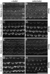
Figure 3. Distributions of PCDH15-CD1, -CD2, -CD3 and -ectodomain (ECD) antigen HL5614 in the organ of Corti of av6J mice at P3.
Hair bundles from +/av6J (A, C, E, G) and av6J/av6J (B, D, F, H) mice were stained with antibody PB303 to PCDH15-CD1 (A, B), antibody PB464-2B to PCDH15-CD2 (C, D), antibody PB375 to PCDH15-CD3 (E, F) and antibody HL5614 to PCDH15-ectodmain (G, H). Images are from the basal (A, B, E, F) and apical (C, D, G, H) coils. Staining seen with antibodies to the CD1 (A, B), CD2 (C, D) and CD3 (E, F) isoforms of PCDH15 is similar in +/av6J (A, C, E) and av6J/av6J (B, D, F) mice. Staining seen with HL5614 to the PCDH15 ectodomain in av6J/av6J (H) hair bundles is weak in comparison to that seen in +/av6J hair bundles (G) and is most prominent in the region of the kinocilium (arrows). I, Inner hair cell; O1, O2, O3, outer hair cells in rows 1, 2, and 3 respectively. Scale bar = 20 µm.
Figure 4
Figure 4. FM1-43 uptake in av6J, av3J and v2J cochleae.
Images are from +/av6J (A, C), av6J/av6J (B, D), +/av3J (E, G), av3J/av3J (F, H), +/v2J (I, K) and v2J/v2J (J, L) cochlear hair cells at the equivalent of P3. In the apical turn and basal coils of the av6J/av6J mouse (B, D) uptake is diminished compared to the control (A, C). In contrast to hair cells from av6J/av6J mice, hair cells from av3J/av3J (F, H) and v2J/v2J (J, L) mice completely fail to load with FM1-43 in both apical (F, J) and basal (H, L) coils. Uptake in heterozygous +/av6J (A, C), +/av3J (E, G) and +/v2J mice (I, K) is comparable. Scale bar = 20 µm.
Figure 5
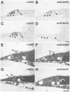
Figure 5. [3H]-gentamicin labelling and gentamicin toxicity in av6J and av3J cochlear cultures.
(A–D) Autoradiographs of [3H]-gentamicin uptake in +/av6J (A), av6J/av6J (B), +/av3J (C) and av3J/av3J (D) apical-coil hair cells. Arrowheads indicate outer hair cells, arrows indicate inner hair cells. [3H]-gentamicin uptake observed in the av6J/av6J mouse (B) is reduced relative to that in the +/av6J control (A). In contrast, av3J/av3J hair cells (D) completely fail to load with [3H]-gentamicin whilst +/av3J control loading (C) is similar to that observed for +/av6J hair cells (A). (E–H) Toluidine blue stained light micrographs of gentamicin treated cochlear cultures from +/av6J (E), av6J/av6J (F), +/av3J (G) and av3J/av3J (H) mice. Arrows indicate extruded hair cells. Whilst +/av6J (E), av6J/av6J (F) and +/av3J hair cells (G) are all sensitive to gentamicin, av3J/av3J hair cells (H) are resistant to this antibiotic. Scale bars = 20 µm.
Figure 6
Figure 6. Mechano-electrical transduction in av6J, av3J and v2J outer hair cells.
(A, B) Transducer currents from heterozygous +/av6J and homozygous av6J/av6J P4 OHCs in response to 45 Hz sinusoidal force stimuli. Holding potential was −84 mV and the membrane potential was stepped, in 20 mV increments, between −124 mV and +136 mV. For clarity only responses to every third voltage step are shown. Driver voltage (DV, amplitude 40 V) to the fluid jet is shown above the traces. Positive DVs move the hair bundle towards the kinocilium. Membrane potentials are shown next to some of the traces. Recordings shown are single traces and are offset so that the zero-transducer current levels are equally spaced. (A): Cm 2.9 pF; Rs 1.9 MΩ. (B): Cm 3.4 pF; Rs 1.4 MΩ. (C) Averaged current-voltage curves, measured peak-to-peak from two +/av6J OHCs (one P4 and one P5) and three av6J/av6J OHCs (one P4 and two P5). (D, E) Transducer currents from heterozygous +/av3J and homozygous av3J/av3J P4 OHCs. Cells were held at −84 mV and the membrane potential was stepped, in 20 mV increments, between −104 mV and +96 mV. Only responses to every other voltage step are shown. Driver voltage was 35 V. Recordings in (D) and (E) are averages of 4 repetitions each, (D): Cm 6.2 pF; Rs 1.9 MΩ. (E): Cm 5.8 pF; Rs 1.5 MΩ. (F) Average current-voltage curves, measured peak-to-peak, for five +/av3J (P2+2) and five av3J/av3J (P2+2) OHCs. (G, H) Transducer currents from heterozygous +/v2J (P0+3) and homozygous v2J/v2J (P1+2) OHCs, stimulus protocol and data presentation as in (D, E). Recordings averaged from 3 (G) and 2 (H) repetitions. (G): Cm 5.9 pF; Rs 4.3 MΩ. (H): Cm 4.9 pF; Rs 4.0 MΩ. (I) Averaged current-voltage curves, measured peak-to-peak from two +/v2J OHCs (P0+3) and three v2J/v2J (P0+3 and P1+2) OHCs.
Figure 7
Figure 7. Mechanoelectrical transduction of v2J outer hair cells in response to force steps.
(A, B) Transducer currents recorded at membrane potentials of −84 mV and +86 mV from a +/v2J (A) and a v2J/v2J (B) OHC. The +/v2J OHC (same cell as Fig. 6G) responds with transducer currents (inward at −84 mV, outward at +84 mV) to force steps in the positive direction (elicited by positive driver voltages to the fluid jet as shown above the current traces). Force steps in the negative direction (elicited by negative driver voltages) shut off the small fraction of transducer channels open at rest. Transducer currents of the v2J/v2J OHC are elicited by force steps in the negative direction and adapt strongly at −84 mV but not at +86 mV. There is no transducer current activated at rest. (C, D) Peak transducer current as a function of driver voltage to the fluid jet. Smooth curves are 2nd order Boltzmann functions: I = Imax/(1+exp(a2(DV2-DV))*(1+exp(a1(DV1-DV)))). Fits for +/v2J are at −84 mV, a1 = 0.617 V−1, a2 = 0.127 V−1, DV1 = 3.42 V, DV2 = 11.5 V, and at +86 mV, a1 = 0.230 V−1, a2 = 0.095 V−1, DV1 = 2.15 V, DV2 = 5.67 V. Fits for v2J/v2J are at −84 mV, a1 = −0.663 V−1, a2 = −0.318 V−1, DV1 = −16.7 V, DV2 = −22.6 V, and at +86 mV, a1 = −0.972 V−1, a2 = −0.196 V−1, DV1 = −7.00 V, DV2 = −7.69 V.
Figure 8
Figure 8. Tip link morphology in av6J cochlear hair cells.
Tip links from +/av6J (A, B) and av6J/av6J (C–G) hair cells at P9. (A, B) Heterozygous +/av6J control specimens show some single (white arrow in A), forked (white arrowhead and inset in A) and multiple tip links (double white arrows in A and B). Lateral links are also present (black arrowheads in A). (C, D) av6J/av6J stereocilia showing a single tip link (white arrow in C) and a forked tip link (white arrowhead in D). (E–G) av6J/av6J profiles showing various forms of link occur near the tips. These include single tip-link filaments on adjacent stereocilia (white arrows in E and G), several lateral links (black arrowheads in E and G), and two separate ‘tip links’ from one stereocilium tip (white arrows in F). Scale bars = 100 nm.
Figure 9
Figure 9. Tip link morphology in av3J cochlear hair cells.
Links from +/av3J (A–C) and av3J/av3J (D–F) mice at P3. (A) In the heterozygote, bundles have a regular appearance, similar to those of wild type mice, with stereocilia in precisely aligned rows. Additional short stereocilia, typical of this stage of development, can be seen, all of approximately the same height. Note multiple links can be seen on all the stereocilia. Scale bar = 1 µm. (B) Detail of a bundle from the heterozygote showing links emanating from the tips of the shorter stereocilia and connecting to the sides of the taller stereocilia behind. As is also typical of immature animals, there are sometimes two such links diverging from the same tip onto adjacent taller stereocilia (double white arrows). Scale bar = 500 nm. (C) Detail of the bundle shown in B at higher magnification. Some forked tip links (white arrowhead) are evident and some single filament ones (arrow). Scale bar = 300 nm. (D) Example of a homozygote bundle showing the disrupted bundle structure, with misalignments and gaps in the rows, but also many short stereocilia as in the heterozygote. Scale bar = 1 µm. (E) Detail of a homozygote hair bundle. Although numerous potential tip link sites are evident, and some tip links may be present (arrows), the majority lack tip links or tip-link like filaments. There is also in general fewer links of any kind on these bundles. Scale bar = 500 nm. (F) Detail of an area from E showing several fine tip-link like structures (arrows). Scale bar = 300 nm.
Figure 10
Figure 10. Tip link morphology in v2J cochlear hair cells.
Links from +/v2J (A–C) and v2J/v2J (D–F) mice at P4. (A) Heterozygous +/v2J OHC row 1 hair bundle. The normal row structure is evident at this stage and the stereocilia are well organised. (B) Detail from the bundle shown in A. Normal tip links (white arrows) and lateral links (black arrowheads) are evident near the tips of the stereocilia. (C) Heterozygous +/v2J IHC bundle showing well defined tip links (white arrows), including some that are forked (white arrowhead). (D) Hair bundle from a homozygous v2J/v2J apical row 1 OHC. The bundle is distorted by a central split and there is unevenness in the height of the stereocilia within a row, especially the tallest row. (E) Detail of the bundle shown in D. There is little evidence of robust, full-length tip links (compare B and E). (F) IHC from a v2J/v2J mouse. Occasional stumpy links are evident near the tips of the stereocilia (white arrows). Scale bars = 1 µm in A and D; 200 nm in B, C, E and F.
Figure 11
Figure 11. Transmission electron microscopy of vestibular hair bundles in av6J, av3J and v2J mice.
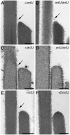
Representative TEM images from utricular hair bundles in +/av6J (A), av6J/av6J (B), +/av3J (C), av3J/av3J (D), +/v2J (E) and v2J/v2J (F) mice at P26 (A–B), P15 (C–D) and P11 (E–F). Arrows point to tip links. Scale bars = 200 nm.

Similar articles

Cited by

References

    1. Leibovici M, Safieddine S, Petit C. Mouse models for human hereditary deafness. Curr Top Dev Biol. 2008;84:385–429. - PubMed
    1. Dror AA, Avraham KB. Hearing loss: mechanisms revealed by genetics and cell biology. Annu Rev Genet. 2009;43:411–437. - PubMed
    1. Petit C, Richardson GP. Linking genes underlying deafness to hair-bundle development and function. Nat Neurosci. 2009;12:703–710. - PMC - PubMed
    1. Friedman LM, Dror AA, Avraham KB. Mouse models to study inner ear development and hereditary hearing loss. Int J Dev Biol. 2007;51:609–631. - PubMed
    1. Pickles JO, Comis SD, Osborne MP. Cross-links between stereocilia in the guinea pig organ of Corti, and their possible relation to sensory transduction. Hear Res. 1984;15:103–112. - PubMed

Publication types