Review
. 2011 Jun;9(6):427-39.
doi: 10.1038/nrmicro2574. Epub 2011 Apr 27.

Subversion of the actin cytoskeleton during viral infection

Affiliations
Review

Subversion of the actin cytoskeleton during viral infection

Matthew P Taylor et al. Nat Rev Microbiol. 2011 Jun.

Abstract

Viral infection converts the normal functions of a cell to optimize viral replication and virion production. One striking observation of this conversion is the reconfiguration and reorganization of cellular actin, affecting every stage of the viral life cycle, from entry through assembly to egress. The extent and degree of cytoskeletal reorganization varies among different viral infections, suggesting the evolution of myriad viral strategies. In this Review, we describe how the interaction of viral proteins with the cell modulates the structure and function of the actin cytoskeleton to initiate, sustain and spread infections. The molecular biology of such interactions continues to engage virologists in their quest to understand viral replication and informs cell biologists about the role of the cytoskeleton in the uninfected cell.


Figures

Figure 1
Figure 1. Actin filament dynamics
a | Actin filaments (known as filamentous actin (F-actin)) are formed by two parallel strands of head–tail polymers of actin monomers (globular actin (G-actin)). Actin polymerization is initiated by the ARP2/3 complex and stimulated by cofactors such as profilin. The direction of actin filaments is determined by the orientation of the monomers, with the positive end being defined as opposite the ATP-binding pocket. Actin depolymerization can occur at either end of the filament. Cofilin interacts with actin dimers to promote disassembly, which can be initiated by the activity of gelsolin. b | Actin filaments that were polymerized in vitro and visualized under an electron microscope. Image is reproduced, with permission, from REF. © (2009) American Society for Biochemistry and Molecular Biology.
Figure 2
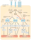
Figure 2. RHO-family GTPase-mediated modelling of the actin cytoskeleton
E-cadherins, integrins or guanylyl-nucleotide-binding protein (G protein)-coupled receptors (GPCRs) activate SRC, which phosphorylates focal adhesion kinase 1 (FAK1). FAK1 promotes the formation of protrusive actin structures by activating RHO-family GTPases such as RAC1 and cell division cycle 42 (CDC42). The downstream effectors of RHO-family GTPases are: Wiscott–Aldrich syndrome protein (WASP)–WASP-family verprolin-homologous protein (WAVE) proteins, Diaphanous-related formins (mDIA proteins; also known as DRF or DIAPH proteins) and kinases such as PAKs and RHO-associated protein kinases (ROCKs). WASP–WAVE proteins stimulate the activation of the ARP2/3 complex. PAKs and ROCKs contribute to the formation of actin filaments by inactivating cofilin via phosphorylation of LIM domain kinases (LIMKs). mDIA stimulates the nucleation and extension of parallel actin filaments. RHOA is initially inactivated by integrin signalling via paxillin. Phosphoinositide 3-kinase (PI3K) is phosphorylated by SRC and then activates RHOA, leading to the formation of stress fibres. ABL tyrosine kinases negatively regulate the RHO–ROCK signalling pathway while activating RAC1 and WASP–WAVE (for reviews, see REFS 10,11). ROCK proteins also stimulate the phosphorylation of myosin regulatory light chain (MLC), thus contributing to the contractility of actin–myosin. Several viral proteins interact with this signalling machinery at multiple levels (see main text for details). Black arrows indicate direct phosphorylation or stimulation of the downstream molecule. Inhibitory or indirect interactions are shown as red or dotted lines, respectively. Stimulatory or modulatory interactions of viral proteins are indicated by black lines. Ad, adenoviruses; E4ORF4, early region ORF4 protein; EBNA2, EBV nuclear antigen 2; EBV, Epstein–Barr virus; F, fusion protein; HBV, hepatitis B virus; HPV, human papillomavirus; HSV, herpes simplex virus; HPIV-3, human parainfluenza virus 3; PRV, pseudorabies virus; RSV, Rous sarcoma virus; SV40, simian virus 40; VV, vaccinia virus;
Figure 3
Figure 3. Manifestations of actin rearrangement
Within the cell, actin filaments can be arranged to form multiple structures. Stress fibres are large assemblies of actin filaments that can span the length of the cell. The presence of myosin in stress fibres enables contractility. Underneath the plasma membrane is the loosely organized network of actin filaments that is termed cortical actin. Actin filaments also can be organized to produce a range of cellular extensions, including podosomes, lamellipodia, filopodia, microvilli and large membrane ruffles. Podosomes contain several actin-binding proteins, signalling molecules and metalloproteinases (black dots). ER, endoplasmic reticulum.
Figure 4
Figure 4. Models of entry
Virions enter cells through a range of pathways, some of which are depicted here. a | Viral surfing. Myosin II at the base of the filopodium pulls the actin filaments, while the constant actin turnover at the tip pushes them. This movement makes virions ‘surf’ down the filopodium to the sites of entry. In some cases, virions bind glycophosphatidylinositol (GPI)-anchored complement decay-accelerating factor (DAF), resulting in cytoskeletal reorganization that enables the virions to rapidly reach their specific receptors. b | Actin-enhanced clathrin-mediated endocytosis. Clathrin initiates endocytosis, and actin filaments (possibly cortical actin) are recruited to increase the size of the endocytic structure. c | The suggested involvement of cortical actin in regulating formation of the fusion pore; this pore formation is mediated by viral fusion proteins such as the human parainfluenza virus 3 (HPIV-3) fusion protein (F). CAR, coxsackie virus and adenovirus receptor.
Figure 5
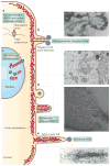
Figure 5. Actin involvement in viral replication and egress
a | The cytoplasmic replication complexes of some negative-strand RNA viruses associate with actin. A putative replication complex of a paramyxovirus is shown, with globular actin stimulating RNA polymerase activity. b | Measles virions bud off the plasma membrane at the ends of microvillus-like structures, often associating with the ends of negative-oriented actin filaments. As seen in the myosin-labelled electron micrograph, budding virions are indicated by arrowheads, extracellular virions are dense structures at the membrane, and the directionality of the microfilament (arrows) is determined by the orientation of myosin barbs. c | Actin fibres interact with assemblies of DNA virus capsids within the nucleus. An electron micrograph of a pseudorabies virus (PRV)-infected nucleus is shown. The high-contrast spots are capsids, and the long, fibrous projections are actin fibres. d | Retroviruses also bud off the plasma membrane, presumably by stimulating the cortical actin network. As virion budding progresses, small actin-filled microvilli form underneath the virions, as observed under cryoelectron microscopy. e | Vaccinia virus (VV)-infected cells produce dense actin comet tails underneath virions. These comet tails push the virus long distances away from the cell to enhance viral dissemination and spread. HRSV, human respiratory syncytial virus; HSV, herpes simplex virus; MMTV, mouse mammary tumour virus. Part b electron micrograph is reproduced, with permission, from REF. © (1986) Elsevier. Part c electron micrograph is reproduced from REF. . Part d electron micrograph is reproduced from REF. . Part e electron micrograph is reproduced, with permission, from REF. © (1996) The Company of Biologists.

Similar articles

Cited by

References

    1. Pollard TD, Cooper JA. Actin, a central player in cell shape and movement. Science. 2009;326:1208–1212. - PMC - PubMed
    1. Weaver AM, Young ME, Lee WL, Cooper JA. Integration of signals to the Arp2/3 complex. Curr Opin Cell Biol. 2003;15:23–30. - PubMed
    1. Moreau V, et al. A complex of N-WASP and WIP integrates signalling cascades that lead to actin polymerization. Nature Cell Biol. 2000;2:441–448. - PubMed
    1. Weisswange I, Newsome TP, Schleich S, Way M. The rate of N-WASP exchange limits the extent of ARP2/3-complex-dependent actin-based motility. Nature. 2009;458:87–91. - PubMed
    1. Carlier MF, et al. Actin depolymerizing factor (ADF/cofilin) enhances the rate of filament turnover: implication in actin-based motility. J Cell Biol. 1997;136:1307–1322. - PMC - PubMed

Publication types