Review
. 2011 May;11(5):318-29.
doi: 10.1038/nri2971.

Illuminating viral infections in the nervous system

Affiliations
Review

Illuminating viral infections in the nervous system

Dorian B McGavern et al. Nat Rev Immunol. 2011 May.

Abstract

Viral infections are a major cause of human disease. Although most viruses replicate in peripheral tissues, some have developed unique strategies to move into the nervous system, where they establish acute or persistent infections. Viral infections in the central nervous system (CNS) can alter homeostasis, induce neurological dysfunction and result in serious, potentially life-threatening inflammatory diseases. This Review focuses on the strategies used by neurotropic viruses to cross the barrier systems of the CNS and on how the immune system detects and responds to viral infections in the CNS. A special emphasis is placed on immune surveillance of persistent and latent viral infections and on recent insights gained from imaging both protective and pathogenic antiviral immune responses.


Conflict of interest statement

statement The authors declare no competing financial interests.

Figures
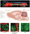
Figure 1
Figure 1. Anatomy of the brain
The outer lining of the brain sits beneath the skull bone and is collectively referred to as the meninges. The meninges are composed of the dura mater, arachnoid mater and pia mater. Cerebral spinal fluid (CSF) flows between the arachnoid and the pia mater through the subarachnoid space. This space also contains meningeal macrophages, stromal cells, trabeculae that physically connect the arachnoid to the pia mater and blood vessels that traverse the meninges and penetrate down into the brain parenchyma (not shown). The meninges are considered part of the blood–CSF barrier. The glial limitans lies beneath the pia mater, and is comprised of basal lamina and astrocytic endfeet. This layer keeps the meninges separate from the underlying brain parenchyma, which contains neurons, astrocytes, oligodendrocytes and microglial cells (see Supplementary information S2 (movie)). Although not depicted, the glial limitans can also be found around post-capillary venules within the brain parenchyma. Microglial cells continually scan the brain parenchyma for damage and foreign materials such as infectious agents (see Supplementary information S1 (movie)). The anatomical features presented in this figure are illustrated with three-dimensional projections captured in a living mouse brain by intravital two-photon laser scanning microscopy (TPLSM).
Figure 2
Figure 2. CNS viral entry and spread
a | The blood–brain barrier is a multi-layered barrier to free diffusion of vascular components into the brain parenchyma. Specialized endothelial cells (red) have junctional proteins that restrict movement through inter-endothelial gaps and secrete a laminin basement membrane (light pink). Both perivascular macrophages and pericytes lie in close apposition to the vessel wall. Astrocytes form the glial limitans that is comprised of a laminin barrier and astrocytic endfeet. In addition, parenchymal juxtavascular microglial cells have processes that extend along blood vessels and even down into the perivascular space towards the basal lamina, allowing for sampling of this space (not shown). Although the blood–brain barrier normally protects the central nervous system (CNS) from pathogens, viruses have adapted strategies to enter through both haematological and axonal routes. Some viruses, such as HIV, use a ‘Trojan horse’ method of entry by travelling in monocytes. Infected monocytes pass through the blood–brain barrier during normal turnover of perivascular macrophages or as a result of the production of pro-inflammatory mediators, such as CC-chemokine ligand 2 (CCL2), which compromise the barrier. Other viruses, including human T-lymphotropic virus type 1 (HTLV1), bind to endothelial receptors such as glucose transporter type 1 (GLUT1), allowing for infection of endothelial cells and release of pro-inflammatory mediators (for example, CCL2). b | Viral CNS entry also occurs through peripheral neurons. Herpes simplex virus 1 (HSV 1) entry into sensory neurons is facilitated by nectin 1 that is expressed on axons. Viral spread to the neuronal cell body is then expedited by hijacking of the fast axonal retrograde transport system (solid black arrow). c | Cell-to-cell transport of viruses can be conducted using various strategies. Rabies virus, pseudorabies virus (PRV) and HSV-1 are released at a synapse and use a retrograde trans-synaptic pathway to infect neighbouring neurons. Measles virus dissemination between neurons is thought to occur through microfusions between neighbouring cells. d | In the case of HSV-1, anterograde transport (from cell body to axon) can lead to infection of neighbouring cells when the virus exits via axonal varicosities before reaching the axon termini. During HSV-1 reactivation, the virus uses the anterograde system (dashed black arrow) to reach axon termini and reinfect epithelial cells by binding to nectin 1 or herpesvirus entry mediator (HVEM) receptors .
Figure 3
Figure 3. Imaging antiviral immune responses in the brain
Three approaches are commonly used to image immune cells and resident cells of the central nervous system (CNS) in living brain tissue. a | To perform intravital imaging, mice are anaesthetized and immobilized with a metal brace to prevent movement artefacts. A section of the skull bone is then surgically thinned to a thickness of 20–30 μm or removed entirely. Skull thinning is the most physiological preparation method because the underlying meninges and brain parenchyma are not disturbed. Following a craniotomy (skull removal procedure), a glass viewing window is usually inserted. This surgical procedure disrupts the meninges and causes a severe brain injury response. After skull thinning or removal, four-dimensional time-lapse imaging can be performed using an upright two-photon microscope. The microscope objective is dipped into artificial cerebral spinal fluid (CSF), which is added to ensure that the normal CSF composition is maintained. Depending on the exact preparation and microscope used, imaging depths of up to 200–400 μm beneath the skull can be achieved. b | Another approach used to image deeper structures relies on acute brain slices. For this preparation, the brain is removed and sliced in ice-cold artificial CSF using a vibratome. The slice is then equilibrated in artificial CSF and later placed in an imaging chamber through which warm, oxygenated artificial CSF flows.
Figure 4
Figure 4. Viral meningitis in real time
Immune responses to central nervous system (CNS) viral infections can be visualized in real time using intravital two-photon laser scanning microscopy (TPLSM). A representative maximal projection of a three-dimensional (3D) z stack is shown for a symptomatic mouse 6 days following infection with lymphocytic choriomeningitis virus (LCMV; upper panels) (see Supplementary information S4 (movie)). The skull (blue) was surgically thinned to maintain a physiological setting for the intravital imaging experiment. Quantum dots (red) were injected intravenously just before imaging to visualize blood vessels, and naive LCMV-specific CD8+ T cells tagged with green fluorescent protein (GFP; green) were injected 1 day before infection to provide a traceable representative of the pathogenic cytotoxic T lymphocyte (CTL) response. GFP+ LCMV-specific CTLs invade and begin patrolling the meningeal space 5–6 days post infection. This is associated with reduced vascular flow and integrity. The lower panels show representative 3D projections from a time-lapse experiment in which the contents of a blood vessel (red) first slow and then begin leaking into the subarachnoid space (white arrowhead). This leakage represents a breach in the blood–cerebral spinal fluid barrier (see Supplementary information S5 (movie)).

Similar articles

Cited by

References

    1. Ginhoux F, et al. Fate mapping analysis reveals that adult microglia derive from primitive macrophages. Science. 2010;330:841–845. This study demonstrated that microglial cells are derived from primitive myeloid precursors rather than haematopoietic cells. - PMC - PubMed
    1. Nimmerjahn A, Kirchhoff F, Helmchen F. Resting microglial cells are highly dynamic surveillants of brain parenchyma in vivo. Science. 2005;308:1314–1318. This study used TPLSM to show that microglial processes are highly dynamic and continually scan the naive brain parenchyma. - PubMed
    1. Bulloch K, et al. CD11c/EYFP transgene illuminates a discrete network of dendritic cells within the embryonic, neonatal, adult, and injured mouse brain. J Compar Neurol. 2008;508:687–710. - PubMed
    1. Chinnery HR, Ruitenberg MJ, McMenamin PG. Novel characterization of monocyte-derived cell populations in the meninges and choroid plexus and their rates of replenishment in bone marrow chimeric mice. J Neuropathol Exp Neurol. 2010;69:896–909. - PubMed
    1. Tyler KL. Emerging viral infections of the central nervous system: part 2. Arch Neurol. 2009;66:1065–1074. - PMC - PubMed

Publication types

MeSH terms