. 2011 Apr 19:8:26.
doi: 10.1186/1742-4690-8-26.

Tetherin does not significantly restrict dendritic cell-mediated HIV-1 transmission and its expression is upregulated by newly synthesized HIV-1 Nef

Affiliations

Tetherin does not significantly restrict dendritic cell-mediated HIV-1 transmission and its expression is upregulated by newly synthesized HIV-1 Nef

Christopher M Coleman et al. Retrovirology. .

Abstract

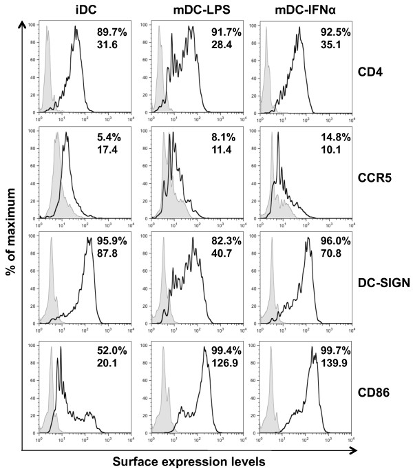
Background: Dendritic cells (DCs) are among the first cells to encounter HIV-1 and play important roles in viral transmission and pathogenesis. Immature DCs allow productive HIV-1 replication and long-term viral dissemination. The pro-inflammatory factor lipopolysaccharide (LPS) induces DC maturation and enhances the efficiency of DC-mediated HIV-1 transmission. Type I interferon (IFN) partially inhibits HIV-1 replication and cell-cell transmission in CD4+ T cells and macrophages. Tetherin is a type I IFN-inducible restriction factor that blocks HIV-1 release and modulates CD4+ T cell-mediated cell-to-cell transmission of HIV-1. However, the role of type I IFN and tetherin in HIV-1 infection of DCs and DC-mediated viral transmission remains unknown.

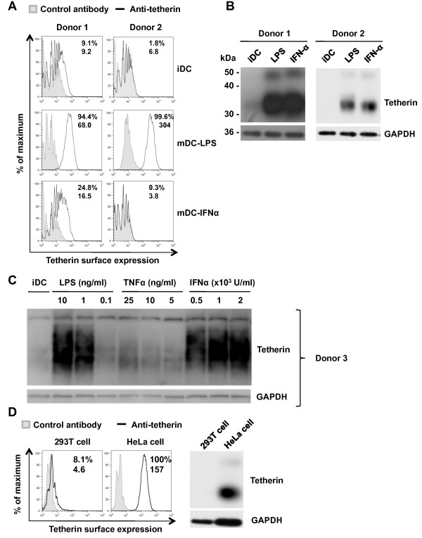
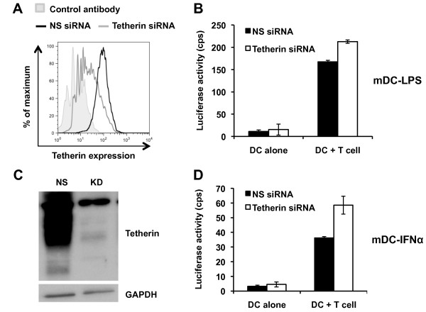
Results: We demonstrated that IFN-alpha (IFNα)-induced mature DCs restricted HIV-1 replication and trans-infection of CD4+ T cells. Tetherin expression in monocyte-derived immature DCs was undetectable or very low. High levels of tetherin were transiently expressed in LPS- and IFNα-induced mature DCs, while HIV-1 localized into distinct patches in these DCs. Knockdown of induced tetherin in LPS- or IFNα-matured DCs modestly enhanced HIV-1 transmission to CD4+ T cells, but had no significant effect on wild-type HIV-1 replication in mature DCs. Intriguingly, we found that HIV-1 replication in immature DCs induced significant tetherin expression in a Nef-dependent manner.

Conclusions: The restriction of HIV-1 replication and transmission in IFNα-induced mature DCs indicates a potent anti-HIV-1 response; however, high levels of tetherin induced in mature DCs cannot significantly restrict wild-type HIV-1 release and DC-mediated HIV-1 transmission. Nef-dependent tetherin induction in HIV-1-infected immature DCs suggests an innate immune response of DCs to HIV-1 infection.


Figures

Figure 1
Figure 1
IFNα induces DC maturation but does not alter the expression of HIV-1 receptors. iDC, mDC-LPS and mDC-IFNα were stained for cell surface expression of CD4, CCR5, DC-SIGN and CD86. On each histogram, the filled peaks are the controls of isotype or secondary antibody alone and the black peaks represent the staining of specific markers. Top and bottom numbers shown in plots are % positive and the geometric mean values of fluorescence intensity, respectively. Results shown are from DCs from a single donor representative of two independent experiments on DCs from different donors.
Figure 2
Figure 2
Transmission and replication of HIV-1 is restricted in mDC-IFNα. Transmission of HIV-1 by DCs was assessed by incubating DCs with either the single-cycle luciferase reporter HIV-1 or replication-competent HIV-1 NL(AD8) for 2 h, then co-cultured with Hut/CCR5 target cells for 3 or 2 days, respectively; transmission was assessed by whole-cell luciferase assay or release of p24 in supernatants. (A) mDC-IFNα do not enhance transmission of the single-cycle luciferase reporter virus to CD4+ T cells over iDC transmission levels. cps, counts per second. Mock, mock infected iDCs. Data represent mean ± SEM of three independent experiments performed on DCs from three different donors. U.D., undetectable (lower than detection limit). (B) mDC-IFNα do not enhance transmission of HIV-1 NL(AD8) to CD4+ T cells at 2 dpi (days post-infection) relative to iDC transmission levels. Graph represents mean data ± SEM from three independent experiments performed with DCs from three different donors. DCs were infected with WT NL(AD8) and p24 production in the cell lysates (C) or supernatants (D) was monitored after 2 h or 3-7 dpi using a p24 ELISA. AZT was used to assess productive HIV-1 infection. Data are from one experiment and representative of at least two independent experiments.
Figure 3
Figure 3
Pro-inflammatory stimuli upregulate tetherin expression in DCs. Tetherin expression on iDCs, mDC-LPS and mDC-IFNα from two different donors was assessed by (A) flow cytometry and (B) immunoblotting. (C) TNF-α treatment of DCs modestly upregulates tetherin expression compared with mDC-LPS and mDC-IFNα. Tetherin expression was detected by immunoblotting. (D) HEK293T and HeLa cells were used as negative and positive controls, respectively. Numbers shown in flow cytometry plots are % positive (top) and the geometric mean values of fluorescence intensity (bottom) for each histogram.
Figure 4
Figure 4
HIV-1 localizes with CD81 and tetherin in mature DCs. Localization of HIV-1 with cellular markers within mature DCs was assessed by confocal microscopy. (A) Representative confocal images of localization characteristics of HIV-GFP-Vpr in mDC-LPS; HIV-GFP-Vpr co-localizes with CD81 and tetherin, but not LAMP-1 in mDC-LPS. (B) Pearson's correlation coefficient analysis of mDC-LPS images. (C) Representative confocal images of localization characteristics of HIV-GFP-Vpr in mDC-IFNα; HIV-GFP-Vpr co-localizes with CD81 and tetherin, but not LAMP-1 in mDC- IFNα. (D) Pearson's correlation coefficient analysis of mDC-IFNα images. Numbers on graphic bars indicate the number of cells analyzed. Data presented are the mean ± SEM. Scale bars are 10 μm.
Figure 5
Figure 5
Tetherin knockdown in mature DCs modestly enhances HIV-1 transmission to CD4+ T cells. Due to the differential localization of tetherin in matured DCs (Figure 3), tetherin knockdown was verified in (A) mDC-LPS by flow cytometry and in (C) mDC-IFNα by immunoblotting. Each plot is representative of three independent experiments performed. NS, non-silencing scramble siRNA control; KD, knockdown using tetherin siRNA. Tetherin knockdown in (B) mDC-LPS and (D) mDC-IFNα significantly enhanced transmission of single-cycle luciferase HIV-1 to Hut/CCR5 cells. Each graph represents mean results ± SEM of two independent experiments performed on DCs from different donors.
Figure 6
Figure 6
Induced tetherin in mature DCs has different effects on WT and Vpu-deleted HIV-1 replication and DC-mediated HIV-1 transmission to CD4+ T cells. (A and B) The effect of tetherin on HIV-1 replication in mature DCs was assessed by tetherin knockdown and infection with WT NL(AD8) or NL(AD8)ΔVpu. Supernatant p24 in mDC-LPS (A) or mDC-IFNα (B) nucleofected with tetherin-specific siRNA or a non-silencing (NS) scramble siRNA control were assessed by p24 ELISA at 5 days post-infection. (C and D) The effect of tetherin on cell-to-cell transmission of WT NL(AD8) or NL(AD8)ΔVpu from tetherin-specific or NS siRNA nucleofected mDC-LPS (C) or mDC-IFNα (D) to Hut/CCR5 cells. Supernatants were collected after 2 days of co-culture and p24 concentration was assessed by ELISA. Graphs represent data from one donor representative of at least two experiments performed on DCs from different donors. Data are presented as mean ± SEM of duplicate samples.
Figure 7
Figure 7
HIV-1 replication in iDCs upregulates tetherin independently of Vpu. (A) Supernatants from DCs infected with WT HIV-1 NL(AD8) or NL(AD8)ΔVpu were assessed for p24 concentration to quantify HIV-1 release. Cell lysates from iDC (B), mDC-LPS (C), and mDC-IFNα (D) infected with HIV-1 or mock infected were detected by immunoblotting for the expression of HIV-1 Gag (p55 and p24), tetherin, and GAPDH at 2 h, 3, 5 and 7 days post-infection (dpi). M, mock infection; W, WT NL(AD8); Δ, NL(AD8)ΔVpu. (E) Flow cytometry analyses of cell surface tetherin expression in iDCs infected with WT NL(AD8), NL(AD8)ΔVpu or mock infected at 3 and 5 dpi. Similar results have been observed in at least three independent experiments using DCs from different donors.
Figure 8
Figure 8
HIV-1 replication in iDCs upregulates tetherin in a Nef-dependent manner. (A) Cell lysates from iDCs infected with WT NL(AD8), NL(AD8)ΔNef with (+) or without (-) AZT or mock infected were evaluated at 5 dpi for the expression of HIV-1 Gag (p55 and p24), tetherin and GAPDH by immunoblotting. (B) Flow cytometry analysis of surface tetherin expression in infected DCs at 5 dpi. M, Mock infection; WT, wild-type NL(AD8); ΔNef, NL(AD8)ΔNef. One representative experiment out of three is shown.
Figure 9
Figure 9
Nef enhances the expression levels of IFIT-1 mRNA in HIV-1-infected iDCs. The levels of IFIT-1 mRNA in iDCs mock infected or infected with WT NL(AD8) or NL(AD8)ΔNef were quantified using real-time RT-PCR. The IFIT-1 expression levels were normalized by the relative GAPDH expression levels. The results of mock-infected controls were assigned as 1 and the fold enhancement of IFIT-1 expression in HIV-1 infected iDCs is shown. WT, wild-type NL(AD8); ΔNef, NL(AD8)ΔNef. One representative experiment out of three is presented.

Similar articles

Cited by

References

    1. Mogensen TH, Melchjorsen J, Larsen CS, Paludan SR. Innate immune recognition and activation during HIV infection. Retrovirology. 2010;7:54. doi: 10.1186/1742-4690-7-54. - DOI - PMC - PubMed
    1. Wu L, KewalRamani VN. Dendritic-cell interactions with HIV: infection and viral dissemination. Nat Rev Immunol. 2006;6:859–868. doi: 10.1038/nri1960. - DOI - PMC - PubMed
    1. Coleman CM, Wu L. HIV interactions with monocytes and dendritic cells: viral latency and reservoirs. Retrovirology. 2009;6:51. doi: 10.1186/1742-4690-6-51. - DOI - PMC - PubMed
    1. Hladik F, Sakchalathorn P, Ballweber L, Lentz G, Fialkow M, Eschenbach D, McElrath MJ. Initial events in establishing vaginal entry and infection by human immunodeficiency virus type-1. Immunity. 2007;26:257–270. doi: 10.1016/j.immuni.2007.01.007. - DOI - PMC - PubMed
    1. Hladik F, McElrath MJ. Setting the stage: host invasion by HIV. Nat Rev Immunol. 2008;8:447–457. doi: 10.1038/nri2302. - DOI - PMC - PubMed

Publication types

MeSH terms

Substances