. 2011 Apr 19;20(4):455-68.
doi: 10.1016/j.devcel.2011.03.017.

Establishment of medial fates along the proximodistal axis of the Drosophila leg through direct activation of dachshund by Distalless

Affiliations

Establishment of medial fates along the proximodistal axis of the Drosophila leg through direct activation of dachshund by Distalless

Matt W Giorgianni et al. Dev Cell. .

Abstract

The proximodistal (PD) axis of the Drosophila leg is thought to be established by the combined gradients of two secreted morphogens, Wingless (Wg) and Decapentaplegic (Dpp). According to this model, high [Wg+Dpp] activates Distalless (Dll) and represses dachshund (dac) in the distal cells of the leg disc, while intermediate [Wg+Dpp] activates dac in medial tissue. To test this model we identified and characterized a dac cis-regulatory element (dac RE) that recapitulates dac's medial expression domain during leg development. Counter to the gradient model, we find that Wg and Dpp do not act in a graded manner to activate RE. Instead, dac RE is activated directly by Dll and repressed distally by a combination of factors, including the homeodomain protein Bar. Thus, medial leg fates are established via a regulatory cascade in which Wg+Dpp activate Dll and then Dll directly activates dac, with Wg+Dpp as less critical, permissive inputs.


Figures

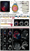
Figure 1
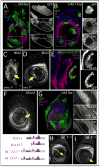
Figure 1. Identification of the Dac Ring Enhancer (RE)
(A) Left: Wg and Dpp gradients in the leg disc shown by staining for Wg (red) and the activated form of the downstream effector of Dpp signaling, phospho-Mad (blue). The middle and right panels show a schematic of a 3rd instar disc and the corresponding proximodistal fates in the adult leg. (B) Vista plot alignment of D. melanogaster dac locus compared to D. pseudobscura (dac coding region shown in blue). Yellow and orange boxes represent cloned regions tested for the ability to drive reporter gene expression. Pink boxes represent enhancers active in the eye identified previously (Pappu et al., 2005). dac7 is a deletion allele that begins in dac’s last exon, and extends 3′ to the gene, but its 3′ endpoint has not been mapped (Pappu et al., 2005). HI was further subdivided based upon sequence homology (Vista alignments show D. melanogaster HI compared to D. pseudobscura (top) and D. virilis (bottom)). (C) Leg discs stained for dac RE-lacZ (green), Dac (red), and Dll (blue). lacZ expression was first apparent in the 2nd instar, slightly before Dac protein was detectable. dac RE maintains a ringed pattern throughout development. RE expression is weaker in the Dac-only domain (bracket), compared to the Dac+Dll domain. dac RE is active at high levels at its distal edge where Dac protein is only weakly detected (asterisk).
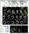
Figure 2
Figure 2. dac RE is regulated in a manner similar to dac
(A–H) Leg discs stained for RE-lacZ (red), Dac (blue), and Gal4 expressing clones (A, D, G, marked by GFP) or mutant clones (B, C, E, F, H; marked by the absence of GFP). For this and subsequent panels, smaller images show the individual staining patterns of the boxed regions. All clones were examined after growth for 48 hrs at 25°C unless otherwise indicated. arr and mad clones were generated in the early 2nd instar. (A) Clones expressing activated Arm, ArmΔN, create new domains of dac and dac RE activation in lateral tissue (asterisks) and repress both dac and dac RE in medial tissue where high levels of Dpp signaling are present (arrow). (B) Clones expressing an activated form of the Dpp receptor, TkvQD, repress both dac and dac RE medially (arrow) while activating both dac and dac RE in proximal clones (asterisk). (C) Distal clones mutant for arrow derepress both dac and dac RE. (D) Medial clones mutant for arrow have no effect on either dac or dac RE (arrow). (E) Distal clones mutant for mad derepress both dac and dac RE. (F) Medial clones mutant for mad have no effect on Dac protein but dac RE activity is absent. (G) UAS-Dac expression clones. The extent of dac RE repression is variable (compare arrow and asterisks). Discs stained 24 hrs post heatshock. (H) dac null clones. (I) dac7/7 mutant disc stained for dac RE-lacZ (green), Dac (red), and Dll (blue). In dac7 mutant discs dac RE activity is still present in a ring-like pattern, while Dac protein is virtually undetectable. See also Figure S1.
Figure 3
Figure 3. Normal dac RE activation when direct inputs by Mad or Pan are compromised
(A) Schematic of dac RE showing binding sites for Mad (green), Pan (blue), and TAAT sequences (pink). Binding sites that show specificity in EMSAs have darker shading. Brk binding is shown in boxes dropped down from the Mad boxes. (B–G) Leg discs at 2nd (B–G), early 3rd (B′–G′), and late 3rd (B″–G″) instar stages stained for the activities of dac RE and mutant versions of dac RE. Mutant elements are schematized above with the mutant sites represented by open bars. (B) dac RE. The inset shows RE (green) and Dac (red). (C) dac RE Mall. RE with seven Mad sites mutated. (D) dac RE P2,3. Mutating two Pan binding sites gives initial expression in some cells distal to the normal Dac domain (arrow). (E) dac RE P4,5,6. Mutation of the three weak Pan binding sites results in little effect on the RE ring pattern. (F) dac RE P1,2,3,7. Mutating these four binding sites results in distal expression in the 2nd instar (arrow), in addition to RE’s normal expression domain. A ring-like pattern is eventually formed, but distal expression remains into the 3rd instar. The inset shows RE (green) and Dac (red). (G) dac RE Pall. Mutation of seven candidate Pan sites results in distal expression in early discs (arrow) but does not significantly affect RE activity in older discs. The inset shows RE (green) and Dac (red). (H–J) Direct Wg and Dpp inputs are required for expression of a truncated dac RE fragment, REΔ45 (schematics on left). (H) REΔ45. (I) REΔ45 fragment with seven Mad binding sites mutated. (J) REΔ45 fragment with seven Pan binding sites mutated. See also Figure S2 and Table S1.
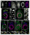
Figure 4
Figure 4. Dll is required for dac and dac RE activation
(A) Late 2nd instar leg disc stained for Dac (red) and Dll (blue). Dac protein is first observed in cells that have lower levels of Dll, consistent with lineage tracing experiments. A′ shows a closeup of the Dac-expressing cells (outlined) with lower levels of Dll. (B) Early 3rd instar leg disc showing that Dll (blue) is no longer observed in most of the Dac (red) expressing cells. Dac and Dll have a 1–2 cell overlap at this stage. (C) Leg disc with clones expressing Dll (green), which activates both dac (blue) and dac RE lacZ (red) in proximal tissue. There is no effect on dac or dac RE activity distally (arrowhead). Larvae heat shocked in early 3rd instar (72–96 hrs AEL) and stained 24 hrs post heat shock. (D) Dll mutant clones (lack of GFP, green). Larvae were heat shocked in early 3rd instar (72–96 hrs AEL) and stained 24 hrs post heat shock. (E–H) Clones are marked by GFP+ expression (green) and discs are stained for dac RE lacZ (red) and Dac (blue). Larvae heat shocked between 24–72 hrs AEL, then fixed and stained 48 hrs post heatshock. (E) UAS-TkvQD clones activate both dac and dac RE lacZ in ventral-proximal tissue. (F) MARCM clones expressing TkvQD and mutant for Dll do not activate the distal leg program or result in ectopic expression of dac or dac RE lacZ. (G) UAS-ArmΔN clones express both dac and dac RE lacZ in dorsal-proximal tissue. (H) MARCM clones expressing ArmΔN and mutant for Dll do not activate dac or dac RE-lacZ.
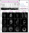
Figure 5
Figure 5. Dll acts through TAAT sequences in dac RE to directly activate expression
(A) EMSAs with Dll protein on TAAT-containing oligos from dac RE. All TAAT sites except t6 show specific binding to Dll that is lost upon mutation. Oligos that contain two TAAT sequences (taat1,2 and taat7,8) show a slower mobility band which is due to the occupancy of both sites. (B) Representative ChIP of 3rd instar leg discs using anti-Dll. Real time PCR of primer sets at dac RE are pulled down specifically relative to IgG controls. Primer sets #1–3 are contained within dac RE, #4 is located just downstream and #5 is located 400 bp further downstream. pdh is a negative control amplicon of the pdh gene on the X chromosome. (C–G) Leg discs at 2nd (D–G), early 3rd (D′–F′), and late 3rd (D″–F″) instar stained for dac RE reporter genes with mutated TAAT sequences. Mutant dac REs are schematized above with the mutant sites represented by open bars. (C) dac RE taatall. RE with all 10 TAAT sequences mutated is nearly inactive. Expression is limited to a few distal cells and in a few proximal cells of the trochanter. (D) dac RE taat1–4. RE with the four 5′ TAAT sequences mutated is expressed in a normal RE ring. In the late 3rd instar there is an ectopic distal ring of expression at the boundary of the pretarsus (see Figure 6). (E) dac RE taat5–10. Mutation of the six 3′ TAAT sequences results in severely limited expression. By the 3rd instar expression is limited to a few cells in a weak ring-like pattern. In the late 3rd instar there is staining in some ventral medial cells but expression is weak elsewhere. Apparent differences in expression in dorsal and ventral regions of the RE domain are largely due to folds in the discs and slight differences in the focal plane. (F,G) dac RE taat5,6. Mutation of TAAT sites 5 and 6 results in a delayed expression of the Dac ring. By the 3rd instar expression is largely normal. See also Table S1.
Figure 6
Figure 6. Late stage distal repression elements in dac RE
(A,B) Leg discs stained for dac RE lacZ (red) and Dac (blue), with Gal4 expressing clones marked by GFP+. Boxed regions are blown up in the right-hand panels. (A) Ectopic Bar clones. Larvae heat shocked 24–72 hrs AEL and stained 64 hrs post heat shock. (B) Clones expressing a constitutive EGFR (λTop, green). Larvae heat shocked 24–72 hrs AEL and stained 48 hrs post heat shock. (C–E) Activity of dac REΔ5 lacZ, a truncated fragment of RE (schematized below). (C) dac REΔ5 lacZ is expressed in cells distal (arrow) to the normal Dac domain in the early 3rd instar. (D) By the late 3rd instar there is a prominent ring (arrow) of expression at the 5th tarsal segment at the boundary with the pretarsus. (E) Late 3rd instar everting leg disc with Bar-Gal4 driving GFP (green) stained for dac REΔ5 lacZ (red) and Dac (blue). The distal ring of REΔ5 is coincident with Bar-Gal4 expression. The distal region of the disc is shown blown up in boxes on right. (F,G) Activity of dac REΔ45 lacZ, a truncated fragment of RE (schematized below). (F) dac REΔ45 lacZ is ectopically expressed in a distal ring (arrow). (G) Clones expressing Bar repress dac (blue) but do not significantly affect dac REΔ45 lacZ expression. Larvae heat shocked 24–72 hrs AEL and stained 64 hrs post heat shock. The boxed region is blown up in the right-hand panels. (H,I) Expression of lacZ from mutant RE constructs in late 3rd instar discs. Mutant REs are schematized at left with the mutant sites represented by open bars. (H) dac RE taat1–4. Mutation of the four 5′ TAAT sequences results in a thin ring of expression at the tarsal/pretarsus boundary (arrow). (I) dac RE taat1,2. Mutating two of the 5′ TAAT sequences results in a partial ring of expression at the tarsal/pretarsus boundary (arrow). See also Figure S3 and Table S1.
Figure 7
Figure 7. Establishment and elaboration of the PD axis of the leg
Shown are schematic diagrams of 2nd instar (left), early 3rd instar (middle) and mid 3rd instar leg imaginal discs. In 2nd instar discs, there is a ventral sector of high Wg signaling (light blue) and a dorsal sector of high Dpp signaling (red). Although not shown, these patterns of Wg and Dpp signaling remain the same throughout the remainder of leg development. Only cells in the center of the leg disc (purple) are receiving high inputs for both Wg and Dpp. The combination of high Wg signaling and high Dpp signaling results in the activation of the Dll LT enhancer element and the repression of the dac RE enhancer element. As the disc grows, some Dll-expressing cells move out of this dac repression domain, allowing Dll to activate dac (Dac+Dll domain; orange). Once dac is activated, it may repress Dll, thus contributing to the establishment of the initial Dac-only domain (blue). By the early 3rd instar, the three primary gene expression domains (Dll-only, Dac+Dll, and Dac-only) become fixed by a maintenance mechanism that is independent of Wg and Dpp signaling. For Dll, this maintenance mechanism involves autoregulation mediated by the M element; for dac, autoregulation may also be involved, but this is not yet known. Also during the early 3rd instar, the EGFR pathway is activated in distal cells, leading to the expression of Bar as well as other downstream transcription factors. Bar continues to repress dac in distal cells, thus helping to maintain the Dll-only domain. High levels of Wg and Dpp signaling, still limited to the center of the disc (purple), may continue to contribute to the repression of dac in distal cells.

Similar articles

Cited by

References

    1. Abu-Shaar M, Mann RS. Generation of multiple antagonistic domains along the proximodistal axis during Drosophila leg development. Development. 1998;125:3821–3830. - PubMed
    1. Ben-Tabou de-Leon S, Davidson EH. Deciphering the underlying mechanism of specification and differentiation: the sea urchin gene regulatory network. Sci STKE. 2006:pe47. - PubMed
    1. Berger MF, Badis G, Gehrke AR, Talukder S, Philippakis AA, Pena-Castillo L, Alleyne TM, Mnaimneh S, Botvinnik OB, Chan ET, et al. Variation in homeodomain DNA binding revealed by high-resolution analysis of sequence preferences. Cell. 2008;133:1266–1276. - PMC - PubMed
    1. Bischof J, Maeda RK, Hediger M, Karch F, Basler K. An optimized transgenesis system for Drosophila using germ-line-specific phiC31 integrases. Proc Natl Acad Sci U S A. 2007;104:3312–3317. - PMC - PubMed
    1. Blauwkamp TA, Chang MV, Cadigan KM. Novel TCF-binding sites specify transcriptional repression by Wnt signalling. EMBO J. 2008;27:1436–1446. - PMC - PubMed

Publication types

MeSH terms

LinkOut - more resources