. 2011:2011:409371.
doi: 10.1155/2011/409371. Epub 2010 Dec 26.

Topography of lipid droplet-associated proteins: insights from freeze-fracture replica immunogold labeling

Affiliations

Topography of lipid droplet-associated proteins: insights from freeze-fracture replica immunogold labeling

Horst Robenek et al. J Lipids. 2011.

Abstract

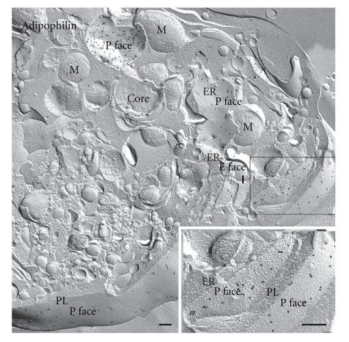
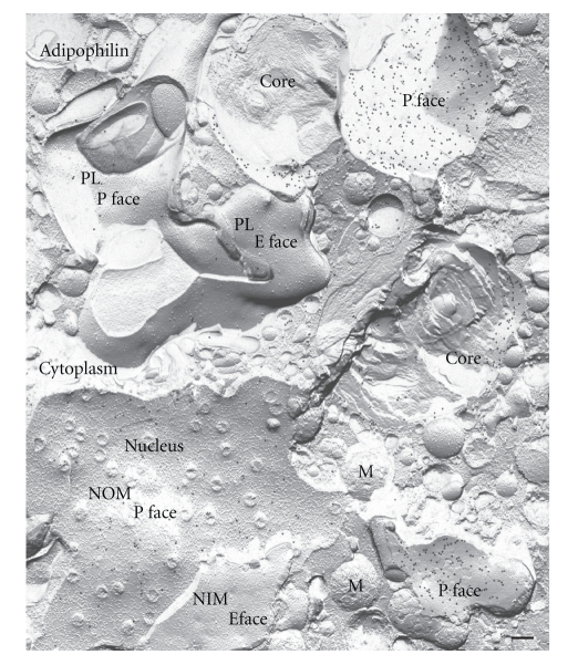
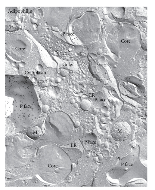
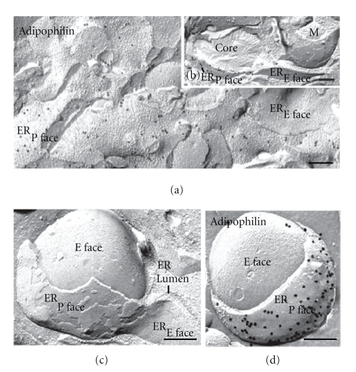
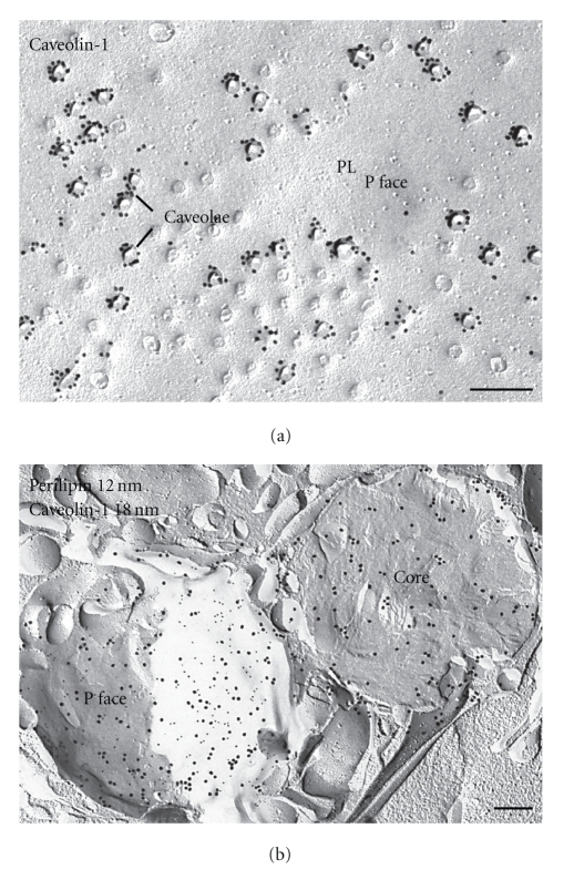
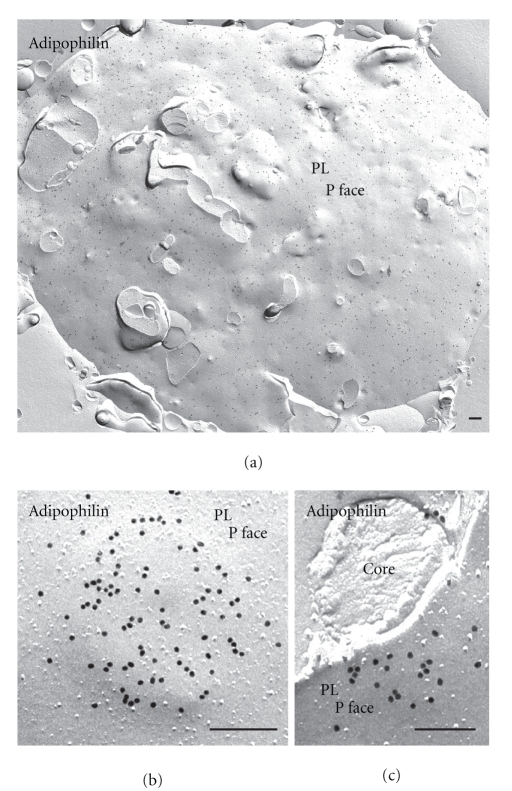
Lipid droplets are not merely storage depots for superfluous intracellular lipids in times of hyperlipidemic stress, but metabolically active organelles involved in cellular homeostasis. Our concepts on the metabolic functions of lipid droplets have come from studies on lipid droplet-associated proteins. This realization has made the study of proteins, such as PAT family proteins, caveolins, and several others that are targeted to lipid droplets, an intriguing and rapidly developing area of intensive inquiry. Our existing understanding of the structure, protein organization, and biogenesis of the lipid droplet has relied heavily on microscopical techniques that lack resolution and the ability to preserve native cellular and protein composition. Freeze-fracture replica immunogold labeling overcomes these disadvantages and can be used to define at high resolution the precise location of lipid droplet-associated proteins. In this paper illustrative examples of how freeze-fracture immunocytochemistry has contributed to our understanding of the spatial organization in the membrane plane and function of PAT family proteins and caveolin-1 are presented. By revisiting the lipid droplet with freeze-fracture immunocytochemistry, new perspectives have emerged which challenge prevailing concepts of lipid droplet biology and may hopefully provide a timely impulse for many ongoing studies.


Figures

Figure 1
Figure 1
Nomenclature for describing the aspects of membranes revealed by freeze-fracture. (a) Cell samples are rapidly frozen and fractured. The freeze-fracture process splits the membrane exposing the fracture faces. The membrane comprises a lipid bilayer with intercalated proteins. The half-membrane leaflet adjacent to the extracellular space is termed the E half; that adjacent to the protoplasm is termed the P half. The term “fracture face” is reserved for the interior views of membranes exposed by freeze fracturing, while the term “surface” is used for the true, natural surfaces of the membrane. The fracture face of the P half is thus termed the P face (or PF), while that of the E half is termed the E face (or EF). The true surfaces of the membrane are correspondingly designated the P surface and the E surface (PS and ES), respectively. (b) Close-up view of the final product in FRIL as viewed in the transmission electron microscope. A platinum-carbon replica is made of the fractured specimen. The replica is treated with SDS to remove the cellular components apart from those attached directly to the replica. Proteins still attached to the replica are then immunogold-labeled. On examination in the electron microscope, the electron dense gold label is clearly visible against the replica, marking the target molecule in the plane of the membrane. The proteins embedded in the replica are detected using a primary antibody followed by a secondary antibody coupled to a colloidal gold marker.
Figure 2
Figure 2
Freeze-fracture of lipid droplets. In freeze-fracture, lipid droplets have a unique smooth appearance enabling their unambiguous discrimination from other organelles. Three different types of view of the droplet are seen with this technique. (1) The fracture may travel upwards and over the droplet to give a convex fracture, (2) downwards and under to give a concave fracture, (3) or the droplet may be cross-fractured to give what is essentially a cross-section of the core. In concave fracture, the enveloping outer phospholipid monolayer is seen en face (P face); convex fractures give mirror image (complementary, E face) views. In practice, the three alternative fracture paths often occur in combination; concavely fractured droplets often include a portion of the core from small regions of cross-fracture, and some fractures skip along successive layers of the lipid revealing a multilayered onion-like appearance. Bar: 0.2 μm.
Figure 3
Figure 3
FRIL demonstrates the presence of adipophilin and perilipin in lipid droplets. Examples in which adipophilin (a) and perilipin (b) label is seen in the outer phospholipid monolayers (P face) of the lipid droplet. The example in (c) illustrates a convexly and a concavely fractured lipid droplet. Perilipin is localized in the P face of the lipid droplet monolayer, whereas the E face is completely devoid of label. ((a) from a lipid-laden macrophage; (b) and (c) from lipid-laden adipocytes). Bars: 0.2 μm.
Figure 4
Figure 4
Survey freeze-fracture view of a lipid-laden macrophage immunogold-labeled for adipophilin. Prominent label is seen in the outer phospholipid monolayer (P face) of the lipid droplets. M: mitochondria. Bar: 0.2 μm.
Figure 5
Figure 5
Adipophilin in cellular membranes. Survey freeze-fracture view of a lipid-laden macrophage immunogold-labeled for adipophilin. Apart from positive labeling in the periphery of lipid droplets prominent label is seen in the P face of the plasma membrane (PL) and ER membranes. The E faces of the ER, mitochondrial and vesicle membranes are devoid of label. Inset: higher magnification of the P face of ER and plasma membrane. M mitochondria. Bar: 0.2 μm.
Figure 6
Figure 6
Freeze-fractured nuclear membrane in a lipid-laden macrophage after immunogold labeling for adipophilin. Apart from positive labeling of lipid droplets, the P face of the outer nuclear membrane (NOM) is prominently labeled. The E face of the outer nuclear membrane and both fracture faces of the inner nuclear membrane (NIM) are typically devoid of label. Bar: 0.2 μm.
Figure 7
Figure 7
Freeze-fracture overview of organelles in a lipid-laden macrophage after labeling for adipophilin. Gold particles marking the presence of adipophilin can be seen in abundance in the outer phospholipid monolayer (P face) of lipid droplets, ER membrane and plasma membrane (PL). The Golgi apparatus (Golgi) is devoid of label. M mitochondria. Bar: 0.2 μm.
Figure 8
Figure 8
Freeze-fracture views of lipid droplet and ER membrane associations in lipid-laden macrophages immunogold-labeled for adipophilin. (a) ER membranes are visible in both P face and E face views. Moderate labeling is seen in the P faces of the ER membranes. (b) Lipid droplet closely associated with ER membranes. Abundant gold label marks the presence of adipophilin in the ER membrane immediately adjacent to the lipid droplet. In contrast, ER membranes adjacent to mitochondria (M) are devoid of label. (c) Lipid droplet situated in a cup formed from ER membranes. The lipid droplet has been convexly fractured and lies beneath (i.e., adjacent to and not within) both ER membranes exposed. (d) Similar view to (c) but with labeling for adipophilin using the FRIL technique. Abundant gold label marks the presence of adipophilin in the ER membrane (P face) immediately adjacent to the lipid droplet. Bars: 0.2 μm.
Figure 9
Figure 9
Distribution of caveolin-1 and perilipin in adipocytes. (a) Caveolae appear as dimples in the P face of the plasma membrane of adipocytes. Gold particles label cavolin-1 in the P face at caveolae. Caveolin-1 labeling is found mainly at the rims of deep caveolae. Apart from positive labeling of caveolae in the plasma membrane, abundant label of caveolin-1 is found in the lipid droplet. (b) Example of immunogold labeling of perilipin (12 nm gold) and caveolin-1 (18 nm gold) in two lipid droplets of the same cell. One droplet shows colocalization of both proteins in the outer monolayer (P face) in almost equal amounts, whereas the other is cross-fractured and the core contains almost exclusively caveolin-1 label. Bars: 0.2 μm.
Figure 10
Figure 10
FRIL images demonstrating that PAT family proteins are present in the plasma membrane. (a) View of the plasma membrane (PL, P face) of a normal cultured macrophage after labeling for adipophilin. The adipophilin is widely distributed throughout the membrane. (b) Upon lipid loading, the adipophilin becomes clustered in elevated domains in the plasma membrane. (c) Fractures that penetrate beneath the plasma membrane demonstrate that lipid droplets lie beneath the elevated adipophilin-rich domains. Bars: 0.2 μm.

Similar articles

Cited by

References

    1. Moor H, Mühlethaler K. Fine structure of frozen-etched yeast cells. The Journal of Cell Biology. 1963;17:p. 609. - PMC - PubMed
    1. Severs NJ. Freeze-fracture electron microscopy. Nature Protocols. 2007;2(3):547–576. - PubMed
    1. Pinto da Silva P, Branton D. Membrane splitting in freeze-ethching. Covalently bound ferritin as a membrane marker. Journal of Cell Biology. 1970;45(3):598–605. - PMC - PubMed
    1. Severs NJ, Robenek H. Detection of microdomains in biomembranes An appraisal of recent developments in freeze-fracture cytochemistry. Biochimica et Biophysica Acta. 1983;737(3-4):373–408. - PubMed
    1. Da Silva PP, Kan FWK. Label-fracture: a method for high resolution labeling of cell surfaces. Journal of Cell Biology. 1984;99(3):1156–1161. - PMC - PubMed

LinkOut - more resources