. 2010 Jul;1(3):192-224.
doi: 10.4161/self.1.3.12794. Epub 2010 Jun 11.

The SCHOOL of nature: III. From mechanistic understanding to novel therapies

Affiliations

The SCHOOL of nature: III. From mechanistic understanding to novel therapies

Alexander B Sigalov. Self Nonself. 2010 Jul.

Abstract

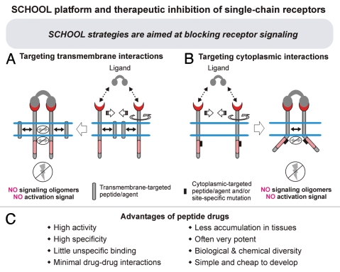
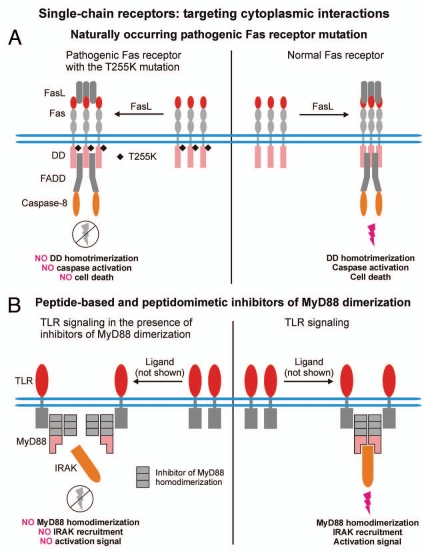
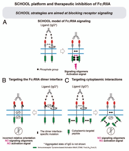
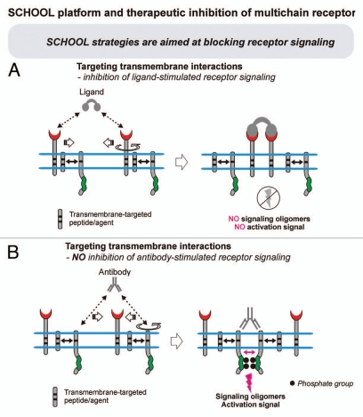
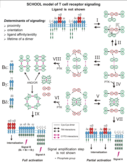
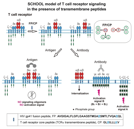
Protein-protein interactions play a central role in biological processes and thus represent an appealing target for innovative drug design and development. They can be targeted by small molecule inhibitors, modulatory peptides and peptidomimetics, which represent a superior alternative to protein therapeutics that carry many disadvantages. Considering that transmembrane signal transduction is an attractive process to therapeutically control multiple diseases, it is fundamentally and clinically important to mechanistically understand how signal transduction occurs. Uncovering specific protein-protein interactions critical for signal transduction, a general platform for receptor-mediated signaling, the signaling chain homooligomerization (SCHOOL) platform, suggests these interactions as universal therapeutic targets. Within the platform, the general principles of signaling are similar for a variety of functionally unrelated receptors. This suggests that global therapeutic strategies targeting key protein-protein interactions involved in receptor triggering and transmembrane signal transduction may be used to treat a diverse set of diseases. This also assumes that clinical knowledge and therapeutic strategies can be transferred between seemingly disparate disorders, such as T cell-mediated skin diseases and platelet disorders or combined to develop novel pharmacological approaches. Intriguingly, human viruses use the SCHOOL-like strategies to modulate and/or escape the host immune response. These viral mechanisms are highly optimized over the millennia, and the lessons learned from viral pathogenesis can be used practically for rational drug design. Proof of the SCHOOL concept in the development of novel therapies for atopic dermatitis, rheumatoid arthritis, cancer, platelet disorders and other multiple indications with unmet needs opens new horizons in therapeutics.


Figures

Figure 1
Figure 1
Therapeutic inhibition of membrane receptors. Due to the lack of knowledge about the molecular mechanism of ligand-induced receptor triggering (A), a vast majority of current inhibitors block the binding of ligand to receptor (B).
Figure 2
Figure 2
SCHOOL platform of single-chain receptor (SR) signaling. Ligand-induced SR clustering and reorientation (in pre-existing SR clusters, ligand binding induces receptor reorientation) results in SR oligomerization mediated by transmembrane interactions. In these oligomers, sufficient proximity and a correct (permissive) relative orientation and geometry of receptors promote homointeractions between cytoplasmic domains. Within the platform, formation of competent signaling oligomers in cytoplasmic milieu is necessary and sufficient to generate the activation signal (for receptor tyrosine kinases, RTKs, this means trans-autophosphorylation of Tyr residues in cytoplasmic signaling sequences), thus triggering downstream signaling pathways. Extracellular ligand-binding domains are shown by red. Intracellular signaling/effector domains are depicted by pink. Protein-protein transmembrane and cytoplasmic interactions are shown by solid black and magenta arrows, respectively. SCHOOL, signaling chain homooligomerization. Adapted from Sigalov AB. The SCHOOL of nature. I. Transmembrane signaling. Self/Nonself 2010; 1:4–39.
Figure 3
Figure 3
SCHOOL platform of multichain receptor signaling. Within the platform, formation of competent signaling oligomers in cytoplasmic milieu is necessary and sufficient to generate the activation signal, thus triggering downstream signaling pathways. Ligand-induced multichain immune recognition receptor (MIRR) clustering and reorientation (in pre-existing MIRR clusters, ligand binding induces receptor reorientation) lead to formation of MIRR oligomers. In these oligomers, sufficient proximity and a correct (permissive) relative orientation and geometry of receptors promote trans-homointeractions between cytoplasmic domains of signaling subunits resulting in formation of competent signaling oligomers. Then, protein tyrosine kinases, PTKs, phosphorylate the tyrosine residues in the cytoplasmic signaling motifs, the immunoreceptor tyrosine-based activation or YxxM motifs (ITAMs/YxxM) that leads to generation of the activation signal. Extracellular ligand-binding domains are shown by red. Intracellular signaling domains (ITAMs/YxxM) are depicted by green. Circular arrows indicate ligand-induced receptor reorientation. Black and magenta arrows indicate specific intersubunit hetero- and homointeractions between transmembrane and cytoplasmic domains, respectively. Curved lines depict intrinsic disorder of the cytoplasmic domains of MIRR signaling subunits. Phosphate groups are shown as dark circles. SCHOOL, signaling chain homooligomerization. Adapted from Sigalov AB. The SCHOOL of nature. I. Transmembrane signaling. Self/Nonself 2010; 1:4–39.
Figure 4
Figure 4
Key protein-protein interactions as new therapeutic targets in receptor signaling. Within the SCHOOL platform, receptor triggering and signaling is an outcome of the ligand-induced interplay between three key protein-protein interactions: (1) ligand-receptor interactions, (2) interreceptor (single-chain receptors, SRs) and intrareceptor (multichain receptors; multichain immune recognition receptors, MIRRs) transmembrane interactions and (3) interreceptor cytoplasmic homointeractions. The last two interactions represent new therapeutic targets. Transmembrane interactions (Target I) between receptors (SRs) or receptor subunits (MIRRs) play an important role in ligand-induced receptor oligomerization (SRs) or in receptor assembly and integrity on resting cells (MIRRs). Cytoplasmic homointeractions (Target II) between receptors (SRs) or receptor signaling subunits (MIRRs) serve as the main driving force of receptor triggering/signaling. Protein-protein transmembrane and cytoplasmic interactions are shown by solid and empty black and magenta arrows, respectively. Curved lines depict intrinsic disorder of the cytoplasmic domains of MIRR signaling subunits.
Figure 5
Figure 5
Single-chain receptors (SRs). The extracellular portion of the receptors is on top and the cytoplasmic portion is on bottom. The lengths of the receptors as shown are only approximately to scale. The inset shows SR domain organization. Extracellular ligand-binding domains are shown by red. Intracellular signaling/effector domains are depicted by pink. EpoR, erythropoietin receptor; G-CSF-R, granulocyte colony-stimulating factor receptor; TGFβ, transforming growth factor-beta; ITAM, immune tyrosine-based activation motif; TNF, tumor necrosis factor; JAK, Janus kinase; EGFR, epidermal growth factor receptor; InsR, insulin receptor; IGF1R, insulin-like growth factor I receptor; IRR, insulin receptor-related receptor; PDGFR, platelet-derived growth factor receptor; CSF1R, colony-stimulating-factor 1 receptor; FGFR, fibroblast growth factor receptor; MuSK, muscle-specific receptor tyrosine kinase; Eph, ephrin; DDR, discoidin domain receptor; Flt1, KDR and Flt4, vascular endothelial growth factor (VEGF) receptors. Adapted from Sigalov AB. The SCHOOL of nature. I. Transmembrane signaling. Self/Nonself 2010; 1:4–39.
Figure 6
Figure 6
Multichain immune recognition receptors (MIRRs). Schematic presentation of the MIRRs expressed on many different immune cells including T and B cells, natural killer cells, mast cells, macrophages, basophils, neutrophils, eosinophils, dendritic cells and platelets. The inset shows MIRR assembly. The extracellular recognition domains and intracellular ITAM-containing signaling domains are located on separate subunits bound together by noncovalent transmembrane interactions (solid arrow). ITAMs/YxxM are shown by green and blue, respectively. Curved lines depict intrinsic disorder of the cytoplasmic domains of MIRR signaling subunits. BCR, B-cell receptor; CLR, C-type lectin receptor; DAP-10 and DAP-12, DNAX adapter proteins of 10 and 12 kD, respectively; DCAR, dendritic cell immunoactivating receptor; GPVI, glycoprotein VI; ILT, Ig-like transcript; ITAM, immunoreceptor tyrosine-based activation motif; KIR, killer cell Ig-like receptor; LIR, leukocyte Ig-like receptor; MAIR-II, myeloid-associated Ig-like receptor; MDL-1, myeloid DAP12-associating lectin 1; NITR, novel immune-type receptor; NK, natural killer cells; SIRP, signal regulatory protein; TCR, T-cell receptor; TREM receptors, triggering receptors expressed on myeloid cells. Adapted from Sigalov AB. The SCHOOL of nature. I. Transmembrane signaling. Self/Nonself 2010; 1:4–39.
Figure 7
Figure 7
SCHOOL platform and therapeutic inhibition of single-chain receptors (SRs). (A) Within the SCHOOL platform, specific blockade of interreceptor transmembrane interactions prevents ligand-induced SR oligomerization. Competent signaling oligomers in cytoplasmic milieu are not formed, thus preventing generation of the activation signal. (B) Specific blockade/prevention of cytoplasmic homointeractions does not affect ligand-induced SR oligomerization but does prevent formation of cytoplasmic competent signaling oligomers resulting in inhibition of receptor-mediated signaling. (C) Peptide inhibitors that block receptor signaling have many competitive advantages over large protein inhibitors that block the binding.
Figure 8
Figure 8
Inhibition of cytoplasmic interactions in single-chain receptor signaling. (A) Fas apoptosis signaling by a normal Fas receptor and the receptor with the Fas T225K mutation that naturally occurs in patients with the autoimmune lymphoproliferative syndrome (ALPS). In contrast to all other ALPS-associated Fas DD mutations, this pathogenic mutation specifically disrupts homooligomerization of the cytoplasmic tails of the receptor but retains the ability to interact with FADD [Siegel RM, Muppidi JR, Sarker M, et al. SPOTS: signaling protein oligomeric transduction structures are early mediators of death receptor-induced apoptosis at the plasma membrane. J Cell Biol 2004; 167:735–44]. As shown, the blockade of the cytoplasmic homointeractions does not allow full caspase-8 activation and apoptosis induction, thus revealing these protein-protein interactions as a therapeutic target. FasL, Fas Ligand; DD, Death Domain; FADD, Fas-associated Death Domain protein. (B) TLR signaling in the absence or presence of peptide-based and peptidomimetics inhibitors of MyD88 dimerization. As reported [Loiarro M, Capolunghi F, Fanto N, et al. Pivotal Advance: Inhibition of MyD88 dimerization and recruitment of IRAK1 and IRAK4 by a novel peptidomimetic compound. J Leukoc Biol 2007; 82:801–10; Loiarro M, Sette C, Gallo G, et al. Peptide-mediated interference of TIR domain dimerization in MyD88 inhibits interleukin-1-dependent activation of NF.B. J Biol Chem 2005; 280:15809–14], cell-permeable analogs of MyD88 peptides derived from the TIR domain of MyD88 as well as a synthetic peptidomimetic compound effectively inhibit homodimerization of MyD88 TIR domains, significantly reducing IL-1 signaling in vitro and dose-dependently inhibiting IL-1β-induced production of IL-6 in treated mice. This suggests that inhibition of MyD88 homodimerization in the cytoplasmic milieu may have therapeutic potential. TLR, toll-like receptor; MyD88, myeloid differentiation factor 88; IRAK, interleukin-1 (IL-1) receptor-associated kinase; TIR, toll/IL-1 receptor domain.
Figure 9
Figure 9
SCHOOL platform and FcγRIIA inhibition. (A) Within the platform, formation of competent signaling oligomers in cytoplasmic milieu is necessary and sufficient to trigger FcγRIIA and generate the activation signal, thus triggering downstream signaling pathways. (B) Mutagenesis of the FcγRIIA dimer interface inhibits receptor signaling but not ligand binding [Powell MS, Barnes NC, Bradford TM, et al. Alteration of the FcγRIIA dimer interface affects receptor signaling but not ligand binding. J Immunol 2006; 176:7489–94]. According to the SCHOOL platform, antibody binding to the FcγRIIA receptor with the altered dimer interface results in incorrect relative orientation in ligand-induced receptor dimers, preventing formation of competent signaling oligomers. (C) In line with the SCHOOL model expectations, specific blockade/prevention of FcγRIIA cytoplasmic homointeractions with a Trojan peptide containing the cytoplasmic tail sequence of FcγRIIA prevents formation of cytoplasmic competent signaling oligomers and results in inhibition of antibody-induced signal transduction and phagolysosome formation [Clark AJ, Petty HR. A cell permeant peptide containing the cytoplasmic tail sequence of Fc receptor type IIA reduces calcium signaling and phagolysosome formation in neutrophils. Cell Immunol 2010; 261:153–8].
Figure 10
Figure 10
SCHOOL platform and therapeutic inhibition of transmembrane interactions in multichain receptor signaling. Within the SCHOOL model of multichain immune recognition receptor (MIRR) signaling, specific blockade or disruption of transmembrane interactions causes a physical and functional disconnection of the MIRR subunits. Ligand/antigen stimulation (A) of these “pre-dissociated” receptors leads to reorientation and clustering of the recognition but not signaling subunits. As a result, signaling oligomers are not formed, immunoreceptor tyrosine-based activation motif (ITAM) Tyr residues do not become phosphorylated and the signaling cascade is not initiated. In contrast, the “pre-dissociation” does not prevent the formation of signaling oligomers when signaling subunits are clustered by specific antibodies that trigger cell activation (B). The proposed transmembrane-targeted strategies can be used not only to inhibit but also to modulate receptor-mediated signal transduction, thus modulating the immune response. Extracellular ligand-binding domains are shown by red. Intracellular signaling domains (ITAMs/YxxM) are denoted by green. Curved lines depict intrinsic disorder of the cytoplasmic domains of MIRR signaling subunits.
Figure 11
Figure 11
SCHOOL model of T-cell receptor (TCR) signaling. Interaction with multivalent ligand (not shown) clusters the receptors and pushes them to reorientate (I), to bring signaling subunits into a correct (permissive) relative orientation and in sufficient proximity in the formed receptor oligomer (for illustrative purposes, receptor dimer is shown), and thus to promote the trans-homointeractions between ζ molecules (II). Then, two alternative pathways can take a place depending on the nature of activating stimuli. First is going through a stage IV resulting in formation of ζ2 dimer (dimer of dimers) and phosphorylation of the ζ ITAM tyrosines, thus triggering the activation signal A. Then, the signaling ζ oligomers formed subsequently dissociate from the TCR-CD3 complex, resulting in internalization of the remaining engaged TCR-CD3 complexes (VII). This pathway leads to partial (or incomplete) T-cell activation. Alternatively, the intermediate complex formed at the stage II can undergo further rearrangements, starting trans-homointeractions between CD3 proteins (III) and resulting in formation of an oligomeric intermediate. The stages I, II and III can be reversible or irreversible depending on interreceptor proximity and relative orientation of the receptors in TCR dimers/oligomers as well as on time duration of the TCR-ligand contact and lifetime of the receptor in TCR dimers/oligomers that generally correlate with the nature of the stimulus and its specificity and affinity/avidity. Next, in the signaling oligomers formed (III), the ITAM tyrosines undergo phosphorylation by PTKs that leads to generation of the activation signal, dissociation of signaling oligomers and internalization of the remaining engaged TCRαβ chains (VIII, XI). This pathway provides at least two different activation signals from the ζ and CD3 signaling oligomers (signals A and B), respectively, and results in full T-cell activation. The distinct signaling through . and CD3 oligomers (or through various combinations of signaling chains in CD3 oligomeric structures) might be also responsible for distinct functions such as T-cell proliferation, effector functions, T-cell survival, pathogen clearance, TCR anergy, etc. In addition, the signaling oligomers formed can sequentially interact with the signaling subunits of nonengaged TCRs resulting in formation of higher-order signaling oligomers, thus amplifying and propagating the activation signal (not shown). Also, this leads to the release and subsequent internalization of the remaining nonengaged TCR complexes and/or TCRαβ chains (not shown). Immunoreceptor tyrosine-based activation motifs (ITAMs) are shown as green rectangles. TCR-CD3-ζ components are represented as whole polypeptides and as a simplified axial view. Circular arrows indicate ligand-induced receptor reorientation. Black and magenta arrows indicate specific intersubunit hetero- and homointeractions between transmembrane and cytoplasmic domains, respectively. All interchain interactions in intermediate complexes are shown by dotted arrows reflecting their transition state. Phosphate groups are shown as filled gray circles. In an axial view, one solid small black line depicts one phosphorylated ITAM domain. PTK, protein tyrosine kinase; SCHOOL, signaling chain homooligomerization. Adapted from Sigalov AB. The SCHOOL of nature. I. Transmembrane signaling. Self/Nonself 2010; 1:4–39.
Figure 12
Figure 12
A schematic representation of the SCHOOL-based mechanisms of action of T cell receptor transmembrane inhibitors such as the T cell receptor core peptide (CP) and HIV-1 gp41 fusion peptide (FP). Considering the close similarity in patterns of inhibition of T cell activation and immunosuppressive activity observed for CP and FP, the SCHOOL model reasonably suggests a similar molecular mechanism of action for both peptides. Within the model, these peptides compete with the TCRα chain for binding to the CD3δε and ζ signaling subunits, thus disrupting the transmembrane (TM) interactions between these subunits and resulting in disconnection and pre-dissociation of the relevant signaling subunits from the remaining receptor complex (also shown in the inset as a simplified axial view). This prevents formation of signaling oligomers upon multivalent antigen stimulation, thus inhibiting antigen-mediated T-cell activation. In contrast, stimulation of these “pre-dissociated” MIRRs with cross-linking antibodies to signaling subunit(s) should still lead to receptor triggering and cell activation. The model predicts that the same mechanisms of inhibitory action can be applied to TCR TM peptides corresponding to the TM regions of not only the TCRαβ recognition subunits but the corresponding CD3ε, CD3δ, CD3γ and ζ signaling subunits as well. Adapted from Sigalov AB. New therapeutic strategies targeting transmembrane signal transduction in the immune system. Cell Adh Migr 2010; 4:255–67.
Figure 13
Figure 13
A schematic representation of the SCHOOL-based mechanisms of action of different T-cell receptor transmembrane inhibitors. Within the SCHOOL model, upon antigen stimulation of T cells, T-cell receptor α-chain (TCRα) transmembrane peptide (TMP) prevents formation of all signaling oligomers, including ζ, CD3ε, CD3δ and CD3γ. This inhibits T-cell activation in both in vitro and in vivo. In contrast, other TMPs prevent formation of signaling oligomers (and therefore signaling) of selected signaling subunits. This inhibits T-cell activation in vivo whereas inhibition in vitro depends on the evaluation method used. For example, antigen-stimulated induction of cytokine secretion and T-cell proliferation in T cells lacking CD3γ and/or CD3δ cytoplasmic domains are intact [Luton F, Buferne M, Legendre V, et al. Role of CD3gamma and CD3delta cytoplasmic domains in cytolytic T lymphocyte functions and TCR/CD3 down-modulation. J Immunol 1997; 158:4162–70], thus explaining the absence of inhibitory effect of the CD3γ and CD3δ TM peptides in the in vitro IL-2 production and T-cell proliferation assays used as markers of T-cell activation [Collier S, Bolte A, Manolios N. Discrepancy in CD3-transmembrane peptide activity between in vitro and in vivo T-cell inhibition. Scand J Immunol 2006; 64:388–91]. Abbreviations: AD, atopic dermatitis; AIA, adjuvant-induced arthritis; IL-2, interleukin 2. Adapted from Sigalov AB. New therapeutic strategies targeting transmembrane signal transduction in the immune system. Cell Adh Migr 2010; 4:255–67.
Figure 14
Figure 14
SCHOOL platform and therapeutic inhibition of cytoplasmic interactions in multichain receptor signaling. Within the SCHOOL model of multichain immune recognition receptor (MIRR) signaling, specific blockade of cytoplasmic homointeractions between signaling subunits prevents formation of signaling oligomers, thus inhibiting ligand/antigen-dependent immune cell activation (A). In contrast to transmembrane-targeted agents, stimulation of MIRRs with cross-linking antibodies to the signaling subunit in the presence of cytoplasmic-targeted agents and/or site-specific mutations should not result in receptor triggering and cell activation (B). The proposed cytoplasmic-targeted strategies can be used not only to inhibit but also to modulate receptor-mediated signal transduction, thus modulating the immune response. Extracellular ligand-binding domains are shown by red. Intracellular signaling domains (ITAMs/YxxM) are denoted by green. Curved lines depict intrinsic disorder of the cytoplasmic domains of MIRR signaling subunits. ITAM, immunoreceptor tyrosine-based activation motif.
Figure 15
Figure 15
Adhesion of normal and GPVI-deficient platelets under flow conditions. Blood from a normal individual (A) and GPVI-deficient blood (B) were flowed over denuded rabbit vessel for 5 min and the adhered platelets were fixed with glutaraldehyde after washing with phosphate-buffered saline. The adhered platelets were cut and observed by light microscopy (magnification of ×800). The normal platelets form large aggregates on the subendothelium, but the GPVI-deficient patient's platelets adhere as single cells or form only small aggregates. Reprinted with permission from Moroi M, Jung SM. Platelet glycoprotein VI: its structure and function. Thrombosis Research 2004; 114:221–33.
Figure 16
Figure 16
Novel concept of platelet inhibition. (A) The SCHOOL model of collagen-stimulated GPVI-FcRγ transmembrane (TM) signaling, proposing that the homooligomerization of the FcRγ signaling subunit plays a central role in triggering the GPVI-FcRγ receptor complex. The model also assumes that not only is sufficient proximity of the receptor units in formed and/or preformed receptor dimers/oligomers required to trigger MIRRs but also a correct interunit relative orientation and geometry. Extracellular ligand-binding domains are shown by red. Intracellular signaling ITAM domain is denoted by green. Small solid black arrows indicate specific intersubunit hetero- and homointeractions between TM and cytoplasmic domains, respectively. Circular arrows indicate collagen-induced receptor reorientation. All interchain interactions in a dimeric intermediate are shown by large white arrows reflecting their transition state. Immunoreceptor tyrosine-based activation motifs are shown as dark gray rectangles. Phosphate groups are shown as black circles. (B) Specific disruption of the GPVI-FcRγ TM interactions results in “pre-dissociation” of the GPVI-FcRγ receptor complex, thus preventing formation of FcRγ signaling oligomers and inhibiting collagen-dependent platelet activation and aggregation. Reproduced with permission from [Sigalov AB. SCHOOL model and new targeting strategies. Adv Exp Med Biol 2008; 640:268–311].

Similar articles

Cited by

References

    1. Fry DC. Protein-protein interactions as targets for small molecule drug discovery. Biopolymers. 2006;84:535–552. - PubMed
    1. Ryan DP, Matthews JM. Protein-protein interactions in human disease. Curr Opin Struct Biol. 2005;15:441–446. - PubMed
    1. Toogood PL. Inhibition of protein-protein association by small molecules: approaches and progress. J Med Chem. 2002;45:1543–1558. - PubMed
    1. Fletcher S, Hamilton AD. Targeting protein-protein interactions by rational design: mimicry of protein surfaces. J R Soc Interface. 2006;3:215–233. - PMC - PubMed
    1. Hershberger SJ, Lee SG, Chmielewski J. Scaffolds for blocking protein-protein interactions. Curr Top Med Chem. 2007;7:928–942. - PubMed

LinkOut - more resources