. 2010;1(6):e50.
doi: 10.1038/cddis.2010.28.

IGF1 activates cell cycle arrest following irradiation by reducing binding of ΔNp63 to the p21 promoter

Affiliations

IGF1 activates cell cycle arrest following irradiation by reducing binding of ΔNp63 to the p21 promoter

G C Mitchell et al. Cell Death Dis. 2010.

Abstract

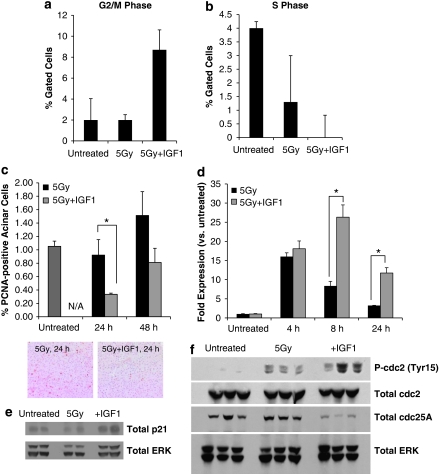
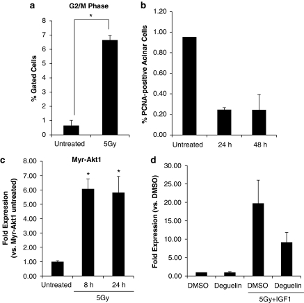
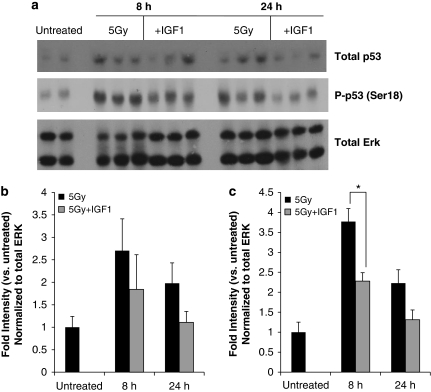
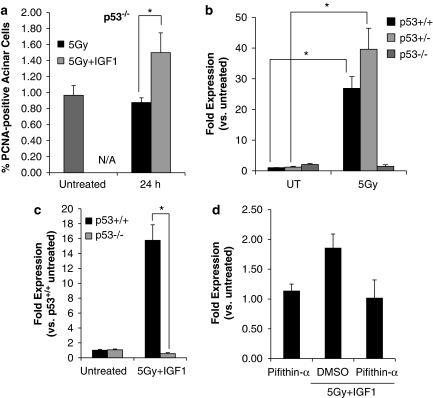
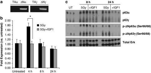
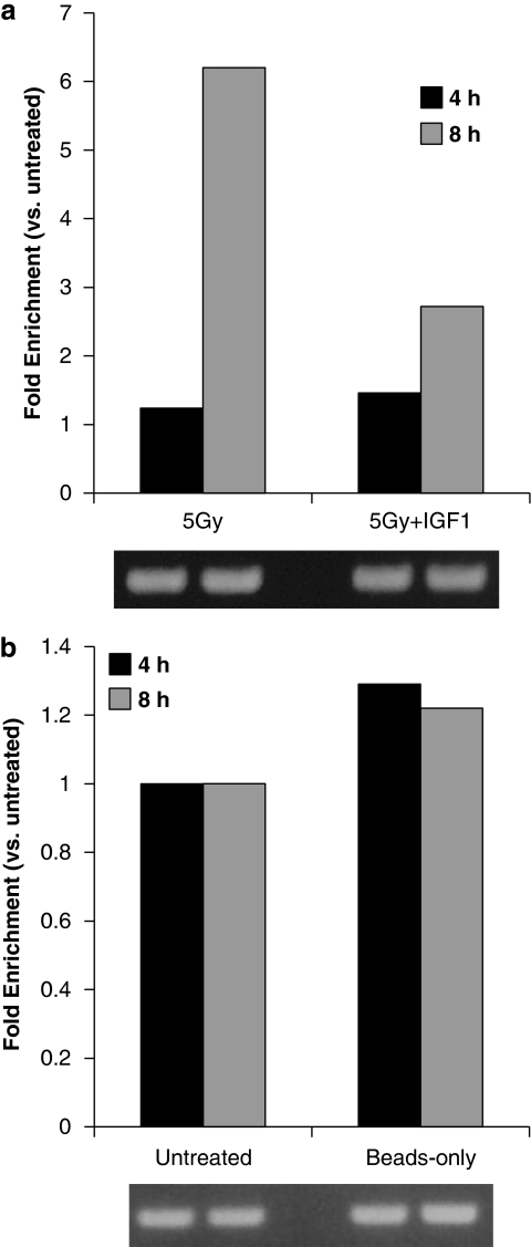
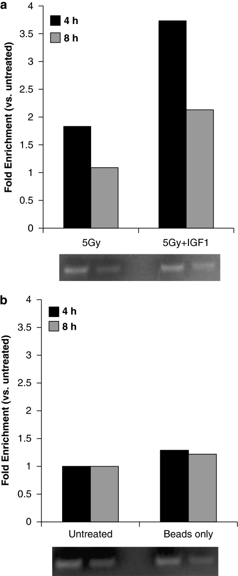
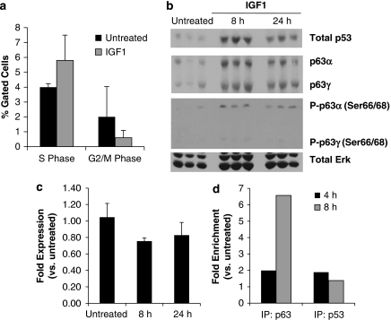
Radiotherapy for head and neck tumors often results in persistent loss of function in salivary glands. Patients suffering from impaired salivary function frequently terminate treatment prematurely because of reduced quality of life caused by malnutrition and other debilitating side-effects. It has been previously shown in mice expressing a constitutively active form of Akt (myr-Akt1), or in mice pretreated with IGF1, apoptosis is suppressed, which correlates with maintained salivary gland function measured by stimulated salivary flow. Induction of cell cycle arrest may be important for this protection by allowing cells time for DNA repair. We have observed increased accumulation of cells in G2/M at acute time-points after irradiation in parotid glands of mice receiving pretreatment with IGF1. As p21, a transcriptional target of the p53 family, is necessary for maintaining G2/M arrest, we analyzed the roles of p53 and p63 in modulating IGF1-stimulated p21 expression. Pretreatment with IGF1 reduces binding of ΔNp63 to the p21 promoter after irradiation, which coincides with increased p53 binding and sustained p21 transcription. Our data indicate a role for ΔNp63 in modulating p53-dependent gene expression and influencing whether a cell death or cell cycle arrest program is initiated.

Keywords: IGF1; cell cycle arrest; p53; p63; radiation; xerostomia.


Figures

Figure 1
Figure 1
Pretreatment with IGF1 induces cycle arrest in irradiated parotid glands. The head and neck regions of wild-type mice were irradiated ±IGF1 pretreatment. Parotid glands were removed 4, 8, 24 and 48 h after treatment. (a, b) In all, 8 h tissues were dispersed, stained with propidium iodide and analyzed by flow cytometry. The data are shown as the mean percentage of gated cells in G2/M (a) or S phase (b) +S.E.M. of ⩾3 mice per treatment. (c) In total, 24 and 48 h tissues were embedded in paraffin and stained for PCNA. The graphs represent the number of PCNA-positive acinar cells as a percentage of total acinar cells counted. The data are shown as the mean+S.E.M. of ⩾3 mice per treatment. Representative PCNA images are shown below the graph. (d) RNA was isolated from 4, 8 and 24 h tissues, and real-time RT-PCR was run with primers to amplify total p63. Results were calculated using the 2−ΔΔCt method, normalized to untreated and shown as the mean+S.E.M. of ⩾3 mice per treatment. (e, f) Protein lysates were prepared from 8 (e) and 6 h (f) tissues, and western blotting was performed as described in Materials and methods. Total ERK was used to confirm equal loading of lanes. *A significant difference (P⩽0.05) between irradiated glands±IGF1 pretreatment as measured by a Student's t-test
Figure 2
Figure 2
Radiation induces cell cycle arrest in myr-Akt1 mice. The head and neck regions of myr-Akt1 mice were irradiated (ac). Parotid glands were removed 8, 24 and 48 h after treatment. (a) In all, 8 h tissues were dispersed, stained with propidium iodide and analyzed by flow cytometry. The data are shown as the mean percentage of gated cells in G2/M+S.E.M. of ⩾3 mice per treatment. (b) In total, 24 and 48 h tissues were embedded in paraffin and stained for PCNA. The graphs represent the number of PCNA-positive acinar cells as a percentage of total acinar cells counted. The data are shown as the mean+S.E.M. (c) RNA was isolated from 8 and 24 h tissues, and real-time RT-PCR was run with primers to amplify p21. Results were calculated using the 2−ΔΔCt method, normalized to wild-type untreated and shown as the mean+S.E.M. of ⩾3 mice per treatment. *A significant difference (P⩽0.05) between untreated and irradiated glands. (d) Wild-type mice were treated intraperitoneally with deguelin (4 mg/kg) immediately before IGF1 injections and head and neck irradiation. Parotid glands were removed after 8 h, RNA was isolated, and real-time RT-PCR was run with primers to amplify p21. Results were calculated using the 2−ΔΔCt method, normalized to vehicle control (DMSO alone) and shown as the mean+S.D.
Figure 3
Figure 3
Parotid glands of irradiated mice pretreated with IGF1 have reduced levels of total and phosphorylated p53 protein. The head and neck regions of wild-type mice were irradiated ±IGF1 pretreatment. Parotid glands were removed, protein lysates were prepared, and western blotting was performed (a) as described in Materials and methods. Total ERK was used to confirm equal loading of lanes. (b, c) Blots were analyzed by densitometry, normalized to total Erk and shown as mean intensity relative to untreated+S.E.M. *A significant difference (P⩽0.05) between irradiated glands ±IGF1 pretreatment as measured by a Student's t-test
Figure 4
Figure 4
IGF1-induced cell cycle arrest in irradiated parotid glands is p53 dependent. The head and neck regions of wild-type (p53+/+), p53+/− and p53−/− mice were irradiated (a–c). After 4, 8 and 24 h, parotid glands were removed. (a) In all, 24 h tissues were embedded in paraffin and stained for PCNA. The graphs represent the number of PCNA-positive acinar cells as a percentage of total acinar cells counted. The data are shown as the mean+S.E.M. of ⩾3 mice per treatment. *A significant difference (P⩽0.05) between p53+/+ and p53−/− glands as measured by a Student's t-test. (b, c) RNA was isolated after 4 (b) and 8 h (c), and real-time RT-PCR was run with primers to amplify p21. Results were calculated using the 2−ΔΔCt method, normalized to wild-type untreated and shown as the mean+S.E.M. of ⩾3 mice per treatment. *A significant difference (P⩽0.05) between untreated and irradiated glands of the same genotype (b) or irradiated glands ±IGF1 pretreatment (c). (d) Wild-type mice were treated interperitoneally with pifithrin-α (0.25 mg) for 12 h, then again immediately before IGF1 injection and head and neck irradiation. Parotid glands were removed after 8 h, RNA was isolated, and real-time RT-PCR was run with primers to amplify p21. Results were calculated using the 2−ΔΔCt method, normalized to pifithrin-α alone and shown as the mean+S.D.
Figure 5
Figure 5
ΔNp63 mRNA and protein expression increases in irradiated parotid glands. (a) RNA was isolated from untreated parotid glands, and RT-PCR was run with primers to amplify p63 isoforms (TAα, ΔNα, TAγ and ΔNγ). PCR products were electrophoresed on a 1% agarose gel. (b) The head and neck regions of wild-type mice were irradiated ±IGF1 pretreatment. Parotid glands were removed, RNA was isolated and real-time RT-PCR was run with primers to amplify total p63. Results were calculated using the 2−ΔΔCt method, normalized to untreated and shown as the mean+S.E.M. of ⩾3 mice per treatment. *A significant difference (P⩽0.05) between irradiated glands ±IGF1 pretreatment as measured by a Student's t-test. (c) The head and neck regions of wild-type mice were irradiated ±IGF1 pretreatment. Parotid glands were removed, protein lysates were prepared and western blotting was performed as described in Materials and methods. Total ERK was used to confirm equal loading of lanes
Figure 6
Figure 6
Binding of ΔNp63 to the p21 promoter is reduced in parotid glands of irradiated mice pretreated with IGF1. The head and neck regions of wild-type mice were irradiated ±IGF1 pretreatment. (a) ChIP was performed on parotid glands removed after 4 and 8 h with an antibody recognizing total p63. (b) ChIP was performed utilizing beads-only as a control. Precipitated DNA was analyzed by real-time PCR using primers for a region of the p21 promoter ∼1390 bases upstream of the transcription start site. Results were calculated by normalizing the Ct value for each IP sample with the Ct value for its corresponding input (pre-IP chromatin) and calculating a ΔCt. Normalized values are shown as fold versus untreated and represent trends that were consistent between multiple independent experiments. Agarose gels shown with each graph were loaded with PCR-amplified input samples to show equal starting material for each IP
Figure 7
Figure 7
Binding of p53 to the p21 promoter is increased in parotid glands of irradiated mice pretreated with IGF1. The head and neck regions of wild-type mice were irradiated ±IGF1 pretreatment. (a) ChIP was performed on parotid glands removed after 4 and 8 h with an antibody recognizing total p53. (b) ChIP was performed utilizing beads-only as a control. Precipitated DNA was analyzed and graphed as described for Figure 6. Normalized values are shown as fold versus untreated and represent trends that were consistent between multiple independent experiments
Figure 8
Figure 8
IGF1-induced effects after irradiation are not detected in unstressed salivary glands. Wild-type mice were treated with IGF1, and parotid glands were removed after 4, 8, and 24 h. (a) Cell cycle distribution was analyzed for 8 h tissues as described for Figure 1a. The data are shown as the mean percentage of gated cells in G2/M phase+S.E.M. of ⩾3 mice per treatment. (b) In all, 8 and 24 h protein lysates were analyzed as described for Figure 3. (c) p21 mRNA expression was measured after 8 and 24 h as described for Figure 4. (d) ChIP was performed on 4 and 8 h tissues with an antibody recognizing total p63 or total p53. Precipitated DNA was analyzed and graphed as described for Figure 6. Normalized values are shown as fold versus untreated and represent trends that were consistent between multiple independent experiments

References

    1. Jemal A, Siegel R, Ward E, Hao Y, Xu J, Thun MJ. Cancer statistics, 2009. CA Cancer J Clin. 2009;59:225–249. - PubMed
    1. Grundmann O, Mitchell GC, Limesand KH. Sensitivity of salivary glands to radiation: from animal models to therapies. J Dent Res. 2009;88:894–903. - PMC - PubMed
    1. Eisbruch A, Ten Haken RK, Kim HM, Marsh LH, Ship JA. Dose, volume, and function relationships in parotid salivary glands following conformal and intensity-modulated irradiation of head and neck cancer. Int J Radiat Oncol Biol Phys. 1999;45:577–587. - PubMed
    1. Banin S, Moyal L, Shieh S, Taya Y, Anderson CW, Chessa L, et al. Enhanced phosphorylation of p53 by ATM in response to DNA damage. Science. 1998;281:1674–1677. - PubMed
    1. Canman CE, Lim DS, Cimprich KA, Taya Y, Tamai K, Sakaguchi K, et al. Activation of the ATM kinase by ionizing radiation and phosphorylation of p53. Science. 1998;281:1677–1679. - PubMed

Publication types

MeSH terms