. 2011 May 13;286(19):17069-78.
doi: 10.1074/jbc.M111.230961. Epub 2011 Mar 22.

Citrullination of inhibitor of growth 4 (ING4) by peptidylarginine deminase 4 (PAD4) disrupts the interaction between ING4 and p53

Affiliations

Citrullination of inhibitor of growth 4 (ING4) by peptidylarginine deminase 4 (PAD4) disrupts the interaction between ING4 and p53

Qin Guo et al. J Biol Chem. .

Abstract

Gene expression is regulated by a number of interrelated posttranslational modifications of histones, including citrullination. For example, peptidylarginine deminase 4 (PAD4) converts peptidyl arginine to citrulline in histone H3 and can repress gene expression. However, regulation of gene expression through citrullination of non-histone proteins is less well defined. Herein, we identify a tumor suppressor protein, inhibitor of growth 4 (ING4), as a novel non-histone substrate of PAD4. ING4 is known to bind p53 via its nuclear localization signal (NLS) region and to enhance transcriptional activity of p53. We show that PAD4 preferentially citrullinates ING4 in the same NLS region and thereby disrupts the interaction between ING4 and p53. A citrulline-mimicking Arg-NLS-Gln ING4 mutant, which has all Arg residues in the NLS mutated to Gln, loses its affinity for p53, can no longer promote p53 acetylation, and results in repression of downstream p21 expression. In addition, we found that citrullination leads to increased susceptibility of ING4 to degradation, likely impacting p53-independent pathways as well. These findings elucidate an interaction between posttranslational citrullination, acetylation, and methylation and highlight an unusual mechanism whereby citrullination of a non-histone protein impacts gene regulation.


Figures
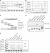
FIGURE 1.
FIGURE 1.
PAD4 citrullinates ING4. A, PAD4 citrullinates ING4 in vitro. MBP-ING4 is treated with GST-PAD4 (30 min) with or without calcium at 25 °C. Samples were Western-blotted (WB) using an anti-modified citrulline (α MC) antibody. A duplicate Coomassie-stained gel was run to indicate equal loading. B, PAD4 citrullinates ING4 in HEK 293T cells. PAD4 (or PAD4-C645A) and FLAG-ING4 plasmids were cotransfected into HEK 293T cells. Half of the cells were treated with calcium ionophore A23187 followed by immunoprecipitation using M2-agarose beads. The total amount of immunoprecipitated ING4 was probed by an anti-FLAG antibody (α FLAG) to indicate equal amounts per lane, and the corresponding citrulline content was probed by an anti-modified citrulline. They were detected on the same membrane using the two-color imaging system from Li-COR. C, shown is a schematic of ING4 deletion mutants. D, mapping the citrullinated domain of ING4 is shown. MBP-tagged deletion mutants were treated with GST-PAD4 followed by Western blot, as in A. E, PAD4 preferentially citrullinates ING4 at Arg-166. MBP-tagged ING4 and its mutants were treated with GST-PAD4. At the indicated time points, the reactions were stopped and Western-blotted as in A.
FIGURE 2.
FIGURE 2.
PAD4 binds ING4, independent of added calcium. A, PAD4 coimmunoprecipitates (IP) with FLAG-ING4 from HEK 293T cells co-expressing PAD4 and FLAG-ING4. B, ING4 coimmunoprecipitates with FLAG-PAD4 from HEK 293T cells co-expressing FLAG-PAD4 and ING4. C, shown is a schematic of PAD4 deletion mutants. D, the N-terminal IgL of PAD4 was sufficient for the interaction with ING4. Immobilized on the glutathione resin, the full-length and truncations of PAD4 were used to pull down MBP-ING4. The bound MBP-ING4 was detected by Western blot (WB) using an anti-MBP antibody (α MBP). E, the PHD domain of ING4 is the major region involved in PAD4 binding. PAD4 were immobilized on the glutathione resin and incubated with full-length ING4 or its deletion mutants (Fig. 1C). The assay was performed using the same methods as D. F, the PAD4 IgL domains directly interact with the PHD domain of ING4 in a calcium-independent manner. Immobilized on the glutathione resin, GST or GST-IgL was incubated with MBP-PHD in the absence or presence of 2 mm CaCl2. The assay was performed as in D and E.
FIGURE 3.
FIGURE 3.
Interactions among PAD4, ING4, and p53. A, shown is a schematic diagram of MBP-p53 and its truncations used in the pulldown experiments. B, the RD of p53 is necessary and sufficient for the interaction with ING4. MBP-tagged domain deletions of p53 were expressed, purified, and bound to glutathione resin. GST pulldown assays were performed as in Fig. 2D. WB, Western blot. C, shown is a schematic of domain interactions among PAD4, ING4, and p53.
FIGURE 4.
FIGURE 4.
Citrullination of ING4 disrupts p53 binding. A, ING4 citrullination disrupts p53 binding in vitro. Pretreated with combinations of PAD4 and CaCl2 as indicated at top, MBP-ING4 was incubated with resin-bound GST-p53. The input, flow-through (FT), and eluted samples were analyzed by Western blots using anti-PAD4 antibody (α PAD4), anti-modified citrulline (α MC) to detect the citrulline content of ING4, anti-MBP (α MBP) to detect MBP-ING4, and anti-GST antibody (α GST) to detect GST-p53. B, citrullination of ING4 disrupts p53 binding in HEK 293T cell lysates. FLAG-ING4 together with p53 and PAD4 was immunoprecipitated from 293T cells coexpressing FLAG-ING4 and PAD4. Half of the resulting resin was treated with CaCl2 and washed, and the retained proteins were eluted and analyzed by Western blots using anti-modified citrulline, anti-FLAG antibody (α FLAG) to detect FLAG-ING4 and anti-p53 antibody (α p53). C, loss of ING4-p53 interaction in cells with activated PAD4 is shown. HEK 293T cells coexpressing PAD4 and FLAG-ING4 were treated with or without calcium ionophore A23187. FLAG-ING4 was immunoprecipitated (IP), and the coimmunoprecipitated endogenous p53 was detected by anti-p53. Anti-FLAG (α FLAG) was also probed to indicate equal loading of FLAG-ING4. D, citrulline-mimicking mutants of ING4 disrupt binding to p53. FLAG-ING4 and single (R166Q), double (R133Q/R166Q), and tetra (Arg-NLS-Gln) Arg to Gln mutants were generated and expressed in HEK 293T cells. The FLAG epitope immunoprecipitates were analyzed for endogenous p53 binding by Western blot. The ratio of band densities for p53 (α p53) and FLAG-ING4 (α FLAG) are shown and represent three independent experiments.
FIGURE 5.
FIGURE 5.
Citrulline-mimicking mutant of ING4 (Arg-NLS-Gln) inhibits p53 acetylation and p21 expression. A, mutant Arg-NLS-Gln blocks ING4-induced p53 acetylation. Endogenous p53 was immunoprecipitated from RKO cells expressing FLAG-ING4, Arg-NLS-Gln, or empty vector. The immunoprecipitates (IP) were probed by Western blot using an anti-acetyl Lys-382 p53 antibody (α Ac-p53 (Lys-382)) to detect p53 acetylation on Lys-382 and anti-p53 (α p53) to detect the total amount of immunoprecipitated p53. Expression of FLAG-ING4 and Arg-NLS-Gln were also tested in cell lysates using anti-FLAG (α FLAG). Anti-tubulin antibody was used for a loading control. B, citrulline-mimicking ING4 mutations block ING4 promotion of p21/waf1 expression. RKO cells expressing FLAG-ING4, Arg-NLS-Gln, or empty vector were lysed and Western-blotted using an anti-p21/WAF1 antibody (α p21), anti-FLAG (α FLAG), and anti-tubulin (α tubulin). The ratio of band densities for p21 and tubulin are shown and represent three independent experiments.
FIGURE 6.
FIGURE 6.
Inactive PAD4 stabilizes ING4 and citrullination of ING4 promotes its degradation in HEK 293T cells. A, cells expressing FLAG-ING4 and wild-type or C645A PAD4 (transfected at two concentrations, shown above) were analyzed by Western blot using anti-FLAG (FLAG-ING4), anti- tubulin (α tubulin), and anti-PAD4 (α PAD4). Relative band densities for FLAG-ING4 and tubulin were determined as in Fig. 5B and are shown at the top. B, inactive PAD4 slows degradation of ING4 in cells. Cells expressing FLAG-ING4 and C645A PAD4, empty vector, or wild-type PAD4 in the absence of a calcium ionophore, were treated with cycloheximide and harvested at later time points for Western blot using anti-FLAG (FLAG-ING4), anti-tubulin, and anti- PAD4. C, active PAD4 promotes ING4 degradation in cells. Cells expressing FLAG-ING4 and wild-type or C645A PAD4 were treated with 5 μm A23187, 2 mm CaCl2, and 100 μg/ml cycloheximide. Cells were harvested at later time points, and the expression of FLAG-ING4 was examined using Western blot as described in B. Data represent three independent experiments and are relative values based on the density at time zero. D, citrullinated ING4 was preferentially degraded. Cells expressing FLAG-ING4 and wild type or C645A PAD4 were treated with 5 μm A23187 in Locke's solution at 37 °C for 30 min and then changed to fresh medium containing 100 μg/ml cycloheximide. Cells were harvested at later time points, and FLAG-ING4 was immunoprecipitated. Citrullination content of ING4 was detected by anti-modified citrulline (α MC). α FLAG was also probed as the loading control.

Similar articles

Cited by

References

    1. Tarcsa E., Marekov L. N., Mei G., Melino G., Lee S. C., Steinert P. M. (1996) J. Biol. Chem. 271, 30709–30716 - PubMed
    1. Pritzker L. B., Joshi S., Gowan J. J., Harauz G., Moscarello M. A. (2000) Biochemistry 39, 5374–5381 - PubMed
    1. Klose R. J., Zhang Y. (2007) Nat. Rev. Mol. Cell Biol. 8, 307–318 - PubMed
    1. Jones J. E., Causey C. P., Knuckley B., Slack-Noyes J. L., Thompson P. R. (2009) Curr. Opin. Drug Discov. Devel. 12, 616–627 - PMC - PubMed
    1. Slack J. L., Causey C. P., Thompson P. R. (2011) Cell. Mol. Life Sci. 68, 709–720 - PMC - PubMed

Publication types

MeSH terms