Review
. 2011 May;12(5):295-307.
doi: 10.1038/nrm3099. Epub 2011 Mar 30.

The spatial and temporal organization of ubiquitin networks

Affiliations
Review

The spatial and temporal organization of ubiquitin networks

Caroline Grabbe et al. Nat Rev Mol Cell Biol. 2011 May.

Abstract

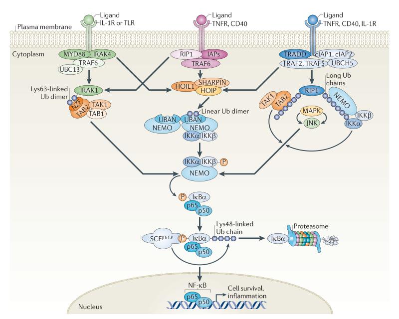
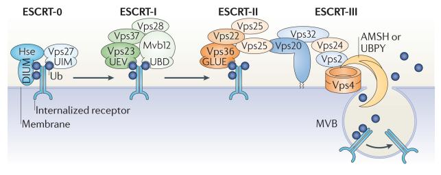
In the past decade, the diversity of signals generated by the ubiquitin system has emerged as a dominant regulator of biological processes and propagation of information in the eukaryotic cell. A wealth of information has been gained about the crucial role of spatial and temporal regulation of ubiquitin species of different lengths and linkages in the nuclear factor-κB (NF-κB) pathway, endocytic trafficking, protein degradation and DNA repair. This spatiotemporal regulation is achieved through sophisticated mechanisms of compartmentalization and sequential series of ubiquitylation events and signal decoding, which control diverse biological processes not only in the cell but also during the development of tissues and entire organisms.


Figures

Figure 1
Figure 1. Ubiquitylation — bringing temporal and spatial order to the NF-κB pathway
Activation of the nuclear factor-κB (NF-κB) pathway downstream of multiple inflammatory cell surface receptors, such as interleukin-1 receptor (IL-1R), Toll-like receptor (TLR), tumour necrosis factor receptor (TNFR) and CD40, triggers the recruitment and ubiquitylation of many molecules with scaffolding and catalytic activities. Recent evidence suggests that multiple ubiquitin species join forces to bring spatial and temporal organization to the different branches of the NF-κB pathway. Distinct ubiquitin signals seem to be spatially restricted close to specific ubiquitin-binding proteins such as TAK1-binding 2 (TAB2) and NF-κB essential modulator (NEMO; also known as IKKγ). TAB2 can bind Lys63-linked chains, which are produced by the E2 complex of ubiquitin-conjugating enzyme 13 (UBC13; also known as UBE2N) and UBE2 variant 1A (UEV1A) and E3 ligases such as TNFR-associated factor 6 (TRAF6). NEMO can bind linear diubiquitin chains, produced by the linear ubiquitin chain assembly complex (LUBAC), which is composed of HOIL1-interacting protein (HOIP), haeme-oxidized IRP2 ubiquitin ligase 1 (HOIL1; also known as RBCK1) and SHANK-associated RH domain-interacting protein (SHARPIN). NEMO can also bind longer ubiquitin chains with variable linkages, including Lys11, which is generated by the E2 UBCH5 and E3 ligase cellular inhibitor of apoptosis protein 1 (cIAP1). Regardless of which pathway is activated and by what type of ubiquitin chain, all scenarios result in the activation of the inhibitor of NF-κB (IκB) kinase (IKK) complex. Following activation, the IKKs phosphorylate the inhibitory IκBα, thus creating a binding site for the Skp–cullin–F-box–βTrCP (SCFβTrCP) ubiquitin E3 ligase complex. This triggers the Lys48-linked ubiquitylation and subsequent proteasomal degradation of IκBα. NF-κB (made up of p50 and p65) can then translocate to the nucleus and activate the transcription of genes promoting cell survival and inflammation. IRAK, IL-1R-associated kinase; JNK, Jun N-terminal kinase; MAPK, mitogen-activated protein kinase; MYD88, myeloid differentiation primary response 88; NZF, neural zinc-finger factor; RIP1, receptor-interacting protein 1; TAK1, TGFβ-activated kinase 1; TRADD, TNFR1-associated DEATH domain; Ub, ubiquitin; UBAN, ubiquitin binding in ABIN and NEMO.
Figure 2
Figure 2. Ubiquitin-mediated coordination of receptor endocytosis
The sequential recognition of ubiquitin signals by components of the endosomal sorting complexes required for transport (ESCRTs) is essential for orchestrating the transportation and sorting of internalized plasma membrane proteins along the endocytic pathway. Upon ligand binding, many plasma membrane proteins are marked by monoubiquitylation or Lys63-linked polyubiquitylation and internalized into early endosomes. At early endosomes, the internalized cargo is captured by ESCRT-0, which can simultaneously interact with three ubiquitin moieties through the diubiquitin motif (DIUM) of Hse (the yeast homologue of mammalian hepatocyte growth factor-regulated Tyr kinase substrate (HRS)) and the ubiquitin-interacting motif (UIM) of vacuolar protein sorting 27 (Vps27; the yeast homologue of signal-transducing adaptor molecule (STAM)). Following the ESCRT-0-mediated concentration of ubiquitylated cargo at early endosomes, ESCRT-I and ESCRT-II are sequentially recruited to the endosomal membrane by the ubiquitin-conjugating enzyme E2 variant (UEV) domain in Vps23 (the yeast homologue of tumour susceptibility gene 101 (TSG101)), the newly identified ubiquitin-binding domain (UBD) of MVB sorting factor 12 (Mvb12) and the GRAM-like ubiquitin-binding in EAP45 (GLUE) domain of Vps36, as well as by a direct interaction between the ESCRTs. Together with ESCRT-III, ESCRT-I and ESCRT-II facilitate the maturation of early endosomes into late endosomes, from which multivesicular bodies (MVBs) pinch-off to fuse with lysosomes, where the internalized cargo is ultimately degraded. In addition to the ESCRT components, several E3 ubiquitin ligases and deubiquitylating enzymes (DUBs) associate with the ESCRTs along the endocytic pathway, initially for directing the sorting events that separate cargo destined for either recycling or degradation and in the final stages to dissociate ubiquitin species from the internalized cargo before MVB formation. Dissociation of ubiquitin relies on the activity of the DUBs associated molecule with the SH3 domain of STAM (AMSH) and ubiquitin isopeptidase Y (UBPY), which, by directly interacting with ESCRT-III components, are suitably positioned at the membrane of late endosomes. Ub, ubiquitin.
Figure 3
Figure 3. Polarized functions of ubiquitylation in neurons
Stringent spatial control of the ubiquitin network is crucial for many aspects in the life, death and functionality of the nervous system. a | The ubiquitin E3 ligase anaphase-promoting complex (APC) is considered to be a key regulator of presynaptic axonal differentiation. APC and its co-activator CD20 (APCCD20) induce the degradation of the proneuronal transcription factor neurogenic differentiation factor 2 (NEUROD2), which regulates the transcription of complexin II (CPLX2) and thereby suppresses presynaptic differentiation specifically at presynaptic sites. b | The spatially restricted ubiquitylation and subsequent proteasomal degradation of AKT in dendrites underlies the generation of neuronal polarity, during which axonal and dendritic fates are distinguished by high and low levels of phosphoinositide 3-kinase (PI3K) signalling, respectively. To further reinforce high PI3K activation in axons, phosphatase and tensin homologue (PTEN; which antagonizes PI3K signalling) was recently found to be reciprocally downregulated by NEDD4-mediated ubiquitylation in axonal growth cones. c,d | A spatially controlled ubiquitin–proteasome system network is essential for maintaining optimal protein levels and synaptic balance. At the presynaptic side, the turnover of proteins regulating vesicle priming and release, such as DUNC13 in Drosophila melanogaster and RAB3-interacting molecule 1 (RIM1) in mice, are strictly controlled by ubiquitylation. In mice, the E3 responsible for RIM1 ubiquitylation is SCRAPPER (c). In response to neuronal stimulation, active proteasomes are highly enriched in the postsynaptic compartment, where they are crucial for controlling spine size, receptor trafficking, synapse plasticity and signalling downstream of neurotransmitter receptors. In Caenorhabditis elegans, Glu receptor 1 (GLR-1) is regulated by ubiquitylation on many levels, including receptor internalization and trafficking (by the APC) and receptor degradation (by a cullin–RING ubiquitin ligase (CRL) complex made up of cullin 3 and the BTB-Kelch protein KEL-8). Moreover, GLR-1 is also transcriptionally controlled by WNT signalling, which in turn is regulated by the Skp–cullin–F-box–βTrCP (SCFβTrCP)-mediated ubiquitylation and degradation of β-catenin (d). Ub, ubiquitin.
Figure 4
Figure 4. Control of development by localized ubiquitylation events
a | The developmental morphogen Decapentaplegic (DPP) is responsible for mediating dorsoventral polarity in Drosophila melanogaster. When bound to its receptor, DPP activates a transcriptional programme mediated by the SMAD transcriptional activators MAD and MEDEA, which translocate to the nucleus as dimers. The SMAD ubiquitylation regulatory factor (SMURF) family of HECT E3 ligases targets MAD for proteasomal degradation, and sumoylation of MEDEA in the nucleus triggers its nuclear export, thus negatively regulating its activity. b | Miranda is a multidomain scaffold protein that plays an important part in neuroblast asymmetric division in D. melanogaster. Its central domain binds cargo proteins, such as Prospero, Staufen and Brain tumour (BRAT). It has been shown that the localization of Miranda is regulated by the multidomain E3 ligase anaphase-promoting complex (APC), which probably directly monoubiquitylates Miranda, leading to its asymmetric localization and retention at the neuroblast cortex through binding to an unknown ubiquitin receptor. c | The migration of glial cells along motor axons in the D. melanogaster peripheral nervous system relies on the homophilic interaction between two distinct isoforms of the immunoglobulin superfamily cell adhesion molecule Fasciclin 2 (FAS2), which is expressed on the cell surface of motor neurons and glial cells, respectively. Recently, the ubiquitin E3 ligase APCFZR was reported to promote, in a graded manner, the ubiquitylation and subsequent degradation of FAS2 in motor neurons, thereby forming a gradient of cell surface-bound FAS2 along the length of the axon. This allows glial cells to migrate and encapsulate motor neuron axons as they grow. SUMO, small ubiquitin-related modifier; Ub, ubiquitin.

Similar articles

Cited by

References

    1. Weissman AM. Themes and variations on ubiquitylation. Nature Rev. Mol. Cell Biol. 2001;2:169–178. - PubMed
    1. Hershko A, Ciechanover A. The ubiquitin system. Annu. Rev. Biochem. 1998;67:425–479. - PubMed
    1. Varshavsky A. Regulated protein degradation. Trends Biochem. Sci. 2005;30:283–286. - PubMed
    1. Deshaies RJ, Joazeiro CA. RING domain E3 ubiquitin ligases. Annu. Rev. Biochem. 2009;78:399–434. - PubMed
    1. Ye Y, Rape M. Building ubiquitin chains: E2 enzymes at work. Nature Rev. Mol. Cell Biol. 2009;10:755–764. - PMC - PubMed

Publication types